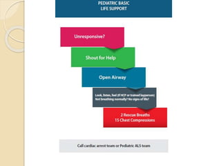
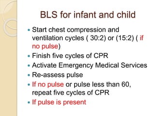
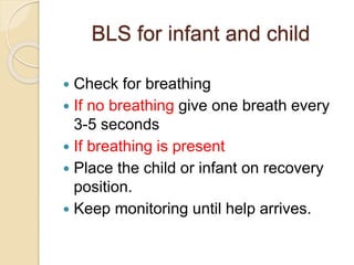
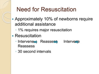
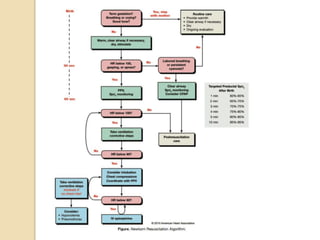
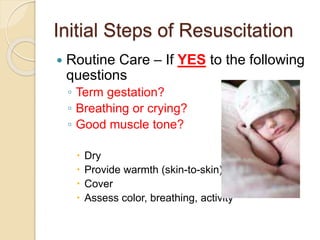
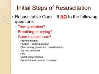
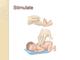
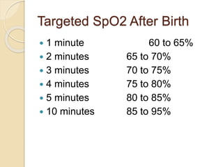
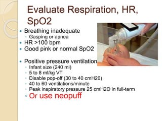
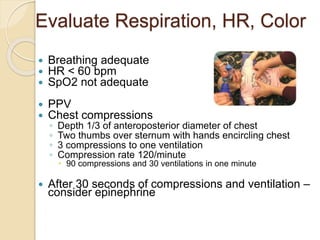
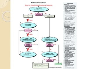
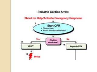
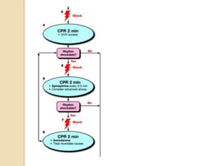
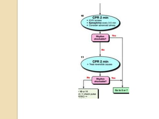
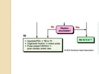
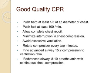
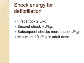
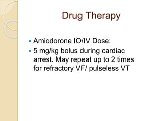
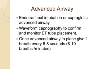
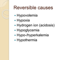
Neonatal and
Pediatric
Resuscitation
Objectives:
BLS for infant and child
Neonatal resuscitation
Pediatric advanced life support
BLS for infant and child
A – B – C
 Assess for responsiveness
 Shout for help
 Open the airway
 Check for breathing ( look, listen and
feel)
 Give two breaths ( if no breathing)
 Check for pulse ( carotid or brachial)
BLS for infant and child
 Start chest compression and
ventilation cycles ( 30:2) or (15:2) ( if
no pulse)
 Finish five cycles of CPR
 Activate Emergency Medical Services
 Re-assess pulse
 If no pulse or pulse less than 60,
repeat five cycles of CPR
 If pulse is present
BLS for infant and child
 Check for breathing
 If no breathing give one breath every
3-5 seconds
 If breathing is present
 Place the child or infant on recovery
position.
 Keep monitoring until help arrives.
Newborn Resuscitation
 Recommendations are primarily for
neonates transitioning to extrauterine
life
 Also applicable to neonates and
infants during the first few weeks to
months following birth
2010 American Heart Association Guidelines for Cardiopulmonary
Resuscitation and Emergency Cardiovascular Care
Need for Resuscitation
 Approximately 10% of newborns require
additional assistance
◦ 1% requires major resuscitation
 Resuscitation
◦ Intervene Reassess Intervene
Reassess
◦ 30 second intervals
Initial Steps of Resuscitation
 Routine Care – If YES to the following
questions
◦ Term gestation?
◦ Breathing or crying?
◦ Good muscle tone?
 Dry
 Provide warmth (skin-to-skin)
 Cover
 Assess color, breathing, activity
Initial Steps of Resuscitation
 Resuscitative Care – If NO to the following
questions
◦ Term gestation?
◦ Breathing or crying?
◦ Good muscle tone?
 Provide warmth
 Position – sniffing position
 Clear airway (meconium consideration)
 Dry and stimulate
 PPV
 Chest compressions
 Epinephrine or volume expansion
Stimulate
Initial Steps (Golden Minute)
 Approximately 60 seconds to
complete, reevaluate, and ventilate if
necessary
◦ Provide warmth
◦ Clear airway
◦ Dry
◦ Stimulate
◦ Position - sniffing
Initial Steps (Golden
Minute)
 Decision to proceed beyond initial steps is
based on evaluation of:
◦ Respirations
 Apnea
 Gasping
 Labored breathing
◦ Heart rate
 Less than 100 bpm
 Auscultation of heart sounds
 Palpation of umbilical pulse
Assessment After PPV or
Supplemental Oxygenation
 Evaluate
◦ Heart rate
◦ Respirations
◦ Oxygenation
 Most sensitive indicator of successful
response is an increase in heart rate
Assessment of Oxygen Need
and Oxygen Administration
 Blood oxygen levels do not reach
extrauterine values in uncompromised
babies until approximately 10 minutes after
birth
 Cyanosis may appear until that point (10
minutes)
 Skin color is very poor indicator of oxygen
saturation immediately after birth
 Lack of cyanosis is a very poor indicator
state of oxygenation in uncompromised
Neonatal Pulse Oximetry
 New pulse oximeters with neonatal
probes
◦ Provide reliable readings within 1 to 2
minutes following birth
◦ Must have sufficient cardiac output to skin
 SpO2 recommended
◦ Resuscitation anticipated
◦ PPV for more than a few breaths
◦ Persistent cyanosis
◦ Supplemental oxygen is administered
Neonatal Pulse Oximetry
 Probe location
◦ Right upper extremity
 Medial surface of the palm
 Wrist
 Attach probe to baby prior to device
◦ More rapid acquisition of signal
PPV and Supplemental
Oxygen
 100% oxygen administration is not
recommended
 Titrate oxygen to SpO2 range
 Initiate resuscitation with air if blended
oxygen is not available
◦ If bradycardia persists (HR <60 bpm) after
90 seconds, increase oxygen to 100%
until HR > 100 bpm
Targeted SpO2 After Birth
 1 minute 60 to 65%
 2 minutes 65 to 70%
 3 minutes 70 to 75%
 4 minutes 75 to 80%
 5 minutes 80 to 85%
 10 minutes 85 to 95%
Newborn Intervention Triggers
 Secretions = suction
 Apnea or gasping respirations = PPV
 Labored breathing or low SpO2 =
oxygen or CPAP
 HR< 100 bpm = PPV
 HR< 60 = Chest compressions and
PPV
 Persistent HR< 60 = epinephrine
Evaluate Respiration, HR,
Oxygenation
 Breathing adequate (rate and effort)
◦ No apnea
◦ No gasping
◦ No labored breathing
 HR >100 bpm
 SpO2 in normal range
 Observe and suction only to keep
airway clear
Evaluate Respiration, HR,
Color
 Breathing adequate
 HR >100 bpm
 Core cyanosis is persistent
 Low SpO2 reading
 Provide blow by oxygen
◦ Warm and humidify oxygen
◦ 5 lpm
◦ Do not blow directly in eyes
Evaluate Respiration, HR,
Color
 Breathing adequate
 HR >100 bpm
 Acrocyanosis with normal SpO2
 No intervention
 If acrocyanosis with poor SpO2
provide blow-by O2
Evaluate Respiration, HR,
SpO2
 Breathing inadequate
◦ Gasping or apnea
 HR >100 bpm
 Good pink or normal SpO2
 Positive pressure ventilation
◦ Infant size (240 ml)
◦ 5 to 8 ml/kg VT
◦ Disable pop-off (30 to 40 cmH20)
◦ 40 to 60 ventilations/minute
◦ Peak inspiratory pressure 25 cmH2O in full-term
◦ Or use neopuff
CPAP
 Breathing spontaneously but labored
 HR> 100 bpm
 SpO2 normal or low
 Research lacking – only studied in
preterm babies
Evaluate Respiration, HR,
Color
 Breathing adequate
 HR <100 bpm
 SpO2 normal
 Positive pressure ventilation
◦ Infant size (240 ml)
◦ 5 to 8 ml/kg VT
◦ Disable pop-off (30 to 40 cmH20)
◦ 40 to 60 ventilations/minute
◦ Peak inspiratory pressure 25 mmHg in full-term
Evaluate Respiration, HR, Color
 Breathing adequate
 HR < 60 bpm
 SpO2 not adequate
 PPV
 Chest compressions
◦ Depth 1/3 of anteroposterior diameter of chest
◦ Two thumbs over sternum with hands encircling chest
◦ 3 compressions to one ventilation
◦ Compression rate 120/minute
 90 compressions and 30 ventilations in one minute
 After 30 seconds of compressions and ventilation –
consider epinephrine
Persistent Bradycardia
 Usually due to
◦ Inadequate lung inflation
◦ Profound hypoxemia
 Primary emergency intervention
◦ Adequate ventilation
 HR remains < 60 bpm with 100%
oxygen
 Consider epinephrine
Epinephrine Administration
 Intravenous route is recommended
only
◦ 0.01 to 0.03 mg/kg
◦ 1:10,000 dilution
 If ET route is used
◦ 0.05 to 0.1 mg/kg
◦ 1:10,000 dilution
Volume Expansion
 Blood loss known or suspected
◦ Pale skin
◦ Poor perfusion
◦ Weak pulse
◦ HR not responding to other interventions
 Isotonic crystalloid
◦ 10 mL/kg
 Avoid rapid infusion in premature
infants
Oral Airways
 Rarely used for neonates
 Use tongue depressor to insert airway
Respiratory Distress or
Inadequacy
 HR < 100 bpm = hypoxia
 Periodic breathing (20 second or
longer period of apnea)
 Intercostal retractions
 Nasal flaring
 Grunting
Meconium Stained Amniotic Fluid
(MSAF)
 10 to 15% of deliveries
 High risk of morbidity
 Passage may occur before or during
delivery
 More common in post-term infants and
neonates small for the gestational age
 Fetus normally does not pass stool prior to
brith
Meconium Stained Amniotic
Fluid
 Complications if aspirated –
Meconium Aspiration Syndrome
(MAS)
◦ Atelectasis
◦ Persistent pulmonary hypertension
◦ Pneumonitis
◦ Pneumothorax
Meconium Stained Amniotic
Fluid
 Determine if fluid is thin and green or thick
and particulate
 If baby is crying vigorously – use standard
resuscitation criteria
 If baby is depressed
◦ DO NOT dry or stimulate
◦ Intubate trachea
◦ Attach a meconium aspirator
◦ Apply suction to endotracheal tube
◦ Dry and stimulate
◦ Continue with standard resuscitation
Apnea
 Common in infants delivered before 32
weeks of gestation
 Risk factors
◦ Prematurity
◦ Infection
◦ Prolonged or difficult labor and delivery
◦ Drug exposure
◦ CNS abnormalities
◦ Seizures
◦ Metabolic disorders
◦ Gastroesophageal reflux
Apnea
 Pathophysiology
◦ Prematurity due to underdeveloped CNS
◦ Gastroesophageal reflux can trigger a
vagal response
◦ Drug-induced from CNS depression
 Bradycardia is key assessment finding
Premature and Low Birth Weight
Infants
 Delivered before 37th week of gestation
 Less than 5.5 lbs or 2,500 grams
Premature and Low Birth Weight
Infants
 Low birth weight
◦ Chronic maternal HTN
◦ Smoking
◦ Placental anomalies
◦ Chromosomal abnormalities
 Born <24 weeks and less than 1 lb –
poor chance of survival
PALS
Good Quality CPR
• Push hard at least 1/3 of ap diameter of chest.
• Push fast at least 100 /min.
• Allow complete chest recoil.
• Minimize interruption in chest compression.
• Avoid excessive ventilation.
• Rotate compressor every two minutes.
• If no advanced airway 15:2 compression to
ventilation ratio.
• If advanced airway, 8-10 breaths /min with
continuous chest compression.
Shock energy for
defibrillation
 First shock 2 J/kg.
 Second shock 4 J/kg.
 Subsequent shocks more than 4 J/kg
 Maximum 10 J/kg or adult dose.
Drug Therapy
 Epinephrine IO/IV Dose:
 0.01 mg/kg (0.1 ml/kg of 1:10 000
concentration)
 Repeat every 3-5 minutes.
 If no IV/IO access, may give
endotracheal dose: 0.01 mg/kg (0.1
ml/kg of 1:1000 concentration)
Drug Therapy
 Amiodorone IO/IV Dose:
 5 mg/kg bolus during cardiac
arrest. May repeat up to 2 times
for refractory VF/ pulseless VT
Advanced Airway
 Endotracheal intubation or supraglotic
advanced airway.
 Waveform capnography to confirm
and monitor ET tube placement.
 Once advanced airway in place give 1
breath every 6-8 seconds (8-10
breaths /minutes)
Return of spontaneous
circulation (ROSC)
 Pulse and blood pressure
 Spontaneous arterial pressure waves
with intra-arterial monitoring
Reversible causes
 Hypovolemia
 Hypoxia
 Hydrogen ion (acidosis)
 Hypoglycemia
 Hypo-/hyperkalemia
 Hypothermia
Reversible Causes
 Tension pneumothorax.
 Temponade, cardiac
 Toxins
 Thrombosis, pulmonary
 Thrombosis, coronary
16- Neonatal Resuscitation.ppt

16- Neonatal Resuscitation.ppt

  • 1.
  • 2.
    Objectives: BLS for infantand child Neonatal resuscitation Pediatric advanced life support
  • 4.
    BLS for infantand child A – B – C  Assess for responsiveness  Shout for help  Open the airway  Check for breathing ( look, listen and feel)  Give two breaths ( if no breathing)  Check for pulse ( carotid or brachial)
  • 5.
    BLS for infantand child  Start chest compression and ventilation cycles ( 30:2) or (15:2) ( if no pulse)  Finish five cycles of CPR  Activate Emergency Medical Services  Re-assess pulse  If no pulse or pulse less than 60, repeat five cycles of CPR  If pulse is present
  • 6.
    BLS for infantand child  Check for breathing  If no breathing give one breath every 3-5 seconds  If breathing is present  Place the child or infant on recovery position.  Keep monitoring until help arrives.
  • 8.
    Newborn Resuscitation  Recommendationsare primarily for neonates transitioning to extrauterine life  Also applicable to neonates and infants during the first few weeks to months following birth 2010 American Heart Association Guidelines for Cardiopulmonary Resuscitation and Emergency Cardiovascular Care
  • 9.
    Need for Resuscitation Approximately 10% of newborns require additional assistance ◦ 1% requires major resuscitation  Resuscitation ◦ Intervene Reassess Intervene Reassess ◦ 30 second intervals
  • 11.
    Initial Steps ofResuscitation  Routine Care – If YES to the following questions ◦ Term gestation? ◦ Breathing or crying? ◦ Good muscle tone?  Dry  Provide warmth (skin-to-skin)  Cover  Assess color, breathing, activity
  • 12.
    Initial Steps ofResuscitation  Resuscitative Care – If NO to the following questions ◦ Term gestation? ◦ Breathing or crying? ◦ Good muscle tone?  Provide warmth  Position – sniffing position  Clear airway (meconium consideration)  Dry and stimulate  PPV  Chest compressions  Epinephrine or volume expansion
  • 13.
  • 14.
    Initial Steps (GoldenMinute)  Approximately 60 seconds to complete, reevaluate, and ventilate if necessary ◦ Provide warmth ◦ Clear airway ◦ Dry ◦ Stimulate ◦ Position - sniffing
  • 15.
    Initial Steps (Golden Minute) Decision to proceed beyond initial steps is based on evaluation of: ◦ Respirations  Apnea  Gasping  Labored breathing ◦ Heart rate  Less than 100 bpm  Auscultation of heart sounds  Palpation of umbilical pulse
  • 16.
    Assessment After PPVor Supplemental Oxygenation  Evaluate ◦ Heart rate ◦ Respirations ◦ Oxygenation  Most sensitive indicator of successful response is an increase in heart rate
  • 17.
    Assessment of OxygenNeed and Oxygen Administration  Blood oxygen levels do not reach extrauterine values in uncompromised babies until approximately 10 minutes after birth  Cyanosis may appear until that point (10 minutes)  Skin color is very poor indicator of oxygen saturation immediately after birth  Lack of cyanosis is a very poor indicator state of oxygenation in uncompromised
  • 19.
    Neonatal Pulse Oximetry New pulse oximeters with neonatal probes ◦ Provide reliable readings within 1 to 2 minutes following birth ◦ Must have sufficient cardiac output to skin  SpO2 recommended ◦ Resuscitation anticipated ◦ PPV for more than a few breaths ◦ Persistent cyanosis ◦ Supplemental oxygen is administered
  • 20.
    Neonatal Pulse Oximetry Probe location ◦ Right upper extremity  Medial surface of the palm  Wrist  Attach probe to baby prior to device ◦ More rapid acquisition of signal
  • 21.
    PPV and Supplemental Oxygen 100% oxygen administration is not recommended  Titrate oxygen to SpO2 range  Initiate resuscitation with air if blended oxygen is not available ◦ If bradycardia persists (HR <60 bpm) after 90 seconds, increase oxygen to 100% until HR > 100 bpm
  • 22.
    Targeted SpO2 AfterBirth  1 minute 60 to 65%  2 minutes 65 to 70%  3 minutes 70 to 75%  4 minutes 75 to 80%  5 minutes 80 to 85%  10 minutes 85 to 95%
  • 23.
    Newborn Intervention Triggers Secretions = suction  Apnea or gasping respirations = PPV  Labored breathing or low SpO2 = oxygen or CPAP  HR< 100 bpm = PPV  HR< 60 = Chest compressions and PPV  Persistent HR< 60 = epinephrine
  • 24.
    Evaluate Respiration, HR, Oxygenation Breathing adequate (rate and effort) ◦ No apnea ◦ No gasping ◦ No labored breathing  HR >100 bpm  SpO2 in normal range  Observe and suction only to keep airway clear
  • 25.
    Evaluate Respiration, HR, Color Breathing adequate  HR >100 bpm  Core cyanosis is persistent  Low SpO2 reading  Provide blow by oxygen ◦ Warm and humidify oxygen ◦ 5 lpm ◦ Do not blow directly in eyes
  • 26.
    Evaluate Respiration, HR, Color Breathing adequate  HR >100 bpm  Acrocyanosis with normal SpO2  No intervention  If acrocyanosis with poor SpO2 provide blow-by O2
  • 28.
    Evaluate Respiration, HR, SpO2 Breathing inadequate ◦ Gasping or apnea  HR >100 bpm  Good pink or normal SpO2  Positive pressure ventilation ◦ Infant size (240 ml) ◦ 5 to 8 ml/kg VT ◦ Disable pop-off (30 to 40 cmH20) ◦ 40 to 60 ventilations/minute ◦ Peak inspiratory pressure 25 cmH2O in full-term ◦ Or use neopuff
  • 29.
    CPAP  Breathing spontaneouslybut labored  HR> 100 bpm  SpO2 normal or low  Research lacking – only studied in preterm babies
  • 30.
    Evaluate Respiration, HR, Color Breathing adequate  HR <100 bpm  SpO2 normal  Positive pressure ventilation ◦ Infant size (240 ml) ◦ 5 to 8 ml/kg VT ◦ Disable pop-off (30 to 40 cmH20) ◦ 40 to 60 ventilations/minute ◦ Peak inspiratory pressure 25 mmHg in full-term
  • 31.
    Evaluate Respiration, HR,Color  Breathing adequate  HR < 60 bpm  SpO2 not adequate  PPV  Chest compressions ◦ Depth 1/3 of anteroposterior diameter of chest ◦ Two thumbs over sternum with hands encircling chest ◦ 3 compressions to one ventilation ◦ Compression rate 120/minute  90 compressions and 30 ventilations in one minute  After 30 seconds of compressions and ventilation – consider epinephrine
  • 32.
    Persistent Bradycardia  Usuallydue to ◦ Inadequate lung inflation ◦ Profound hypoxemia  Primary emergency intervention ◦ Adequate ventilation  HR remains < 60 bpm with 100% oxygen  Consider epinephrine
  • 33.
    Epinephrine Administration  Intravenousroute is recommended only ◦ 0.01 to 0.03 mg/kg ◦ 1:10,000 dilution  If ET route is used ◦ 0.05 to 0.1 mg/kg ◦ 1:10,000 dilution
  • 34.
    Volume Expansion  Bloodloss known or suspected ◦ Pale skin ◦ Poor perfusion ◦ Weak pulse ◦ HR not responding to other interventions  Isotonic crystalloid ◦ 10 mL/kg  Avoid rapid infusion in premature infants
  • 36.
    Oral Airways  Rarelyused for neonates  Use tongue depressor to insert airway
  • 37.
    Respiratory Distress or Inadequacy HR < 100 bpm = hypoxia  Periodic breathing (20 second or longer period of apnea)  Intercostal retractions  Nasal flaring  Grunting
  • 38.
    Meconium Stained AmnioticFluid (MSAF)  10 to 15% of deliveries  High risk of morbidity  Passage may occur before or during delivery  More common in post-term infants and neonates small for the gestational age  Fetus normally does not pass stool prior to brith
  • 39.
    Meconium Stained Amniotic Fluid Complications if aspirated – Meconium Aspiration Syndrome (MAS) ◦ Atelectasis ◦ Persistent pulmonary hypertension ◦ Pneumonitis ◦ Pneumothorax
  • 40.
    Meconium Stained Amniotic Fluid Determine if fluid is thin and green or thick and particulate  If baby is crying vigorously – use standard resuscitation criteria  If baby is depressed ◦ DO NOT dry or stimulate ◦ Intubate trachea ◦ Attach a meconium aspirator ◦ Apply suction to endotracheal tube ◦ Dry and stimulate ◦ Continue with standard resuscitation
  • 41.
    Apnea  Common ininfants delivered before 32 weeks of gestation  Risk factors ◦ Prematurity ◦ Infection ◦ Prolonged or difficult labor and delivery ◦ Drug exposure ◦ CNS abnormalities ◦ Seizures ◦ Metabolic disorders ◦ Gastroesophageal reflux
  • 42.
    Apnea  Pathophysiology ◦ Prematuritydue to underdeveloped CNS ◦ Gastroesophageal reflux can trigger a vagal response ◦ Drug-induced from CNS depression  Bradycardia is key assessment finding
  • 43.
    Premature and LowBirth Weight Infants  Delivered before 37th week of gestation  Less than 5.5 lbs or 2,500 grams
  • 44.
    Premature and LowBirth Weight Infants  Low birth weight ◦ Chronic maternal HTN ◦ Smoking ◦ Placental anomalies ◦ Chromosomal abnormalities  Born <24 weeks and less than 1 lb – poor chance of survival
  • 45.
  • 51.
    Good Quality CPR •Push hard at least 1/3 of ap diameter of chest. • Push fast at least 100 /min. • Allow complete chest recoil. • Minimize interruption in chest compression. • Avoid excessive ventilation. • Rotate compressor every two minutes. • If no advanced airway 15:2 compression to ventilation ratio. • If advanced airway, 8-10 breaths /min with continuous chest compression.
  • 52.
    Shock energy for defibrillation First shock 2 J/kg.  Second shock 4 J/kg.  Subsequent shocks more than 4 J/kg  Maximum 10 J/kg or adult dose.
  • 53.
    Drug Therapy  EpinephrineIO/IV Dose:  0.01 mg/kg (0.1 ml/kg of 1:10 000 concentration)  Repeat every 3-5 minutes.  If no IV/IO access, may give endotracheal dose: 0.01 mg/kg (0.1 ml/kg of 1:1000 concentration)
  • 54.
    Drug Therapy  AmiodoroneIO/IV Dose:  5 mg/kg bolus during cardiac arrest. May repeat up to 2 times for refractory VF/ pulseless VT
  • 55.
    Advanced Airway  Endotrachealintubation or supraglotic advanced airway.  Waveform capnography to confirm and monitor ET tube placement.  Once advanced airway in place give 1 breath every 6-8 seconds (8-10 breaths /minutes)
  • 56.
    Return of spontaneous circulation(ROSC)  Pulse and blood pressure  Spontaneous arterial pressure waves with intra-arterial monitoring
  • 57.
    Reversible causes  Hypovolemia Hypoxia  Hydrogen ion (acidosis)  Hypoglycemia  Hypo-/hyperkalemia  Hypothermia
  • 58.
    Reversible Causes  Tensionpneumothorax.  Temponade, cardiac  Toxins  Thrombosis, pulmonary  Thrombosis, coronary