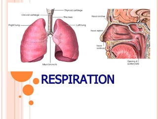
RESPIRATION
HUMAN RESPIRATORY SYSTEM
UPPER RESPIRATORY TRACT LOWER RESPIRATORY TRACT
1) NOSTRILS
2) NASAL
CAVITIES
3) PHARYNX
4) LARYNX
CONDUCTING ZONE RESPIRATORY ZONE
1) TRACHEA
2) PRIMARY BRONCHI
3) SECONDARY BRONCHI
4) TRETIARY BRONCHI
5) BRONCHIOLES
6) TERMINAL BRONCHIOLE
1) RESPIRATORY
BRONCHIOLES
2) ALVEOLI
LOWER RESPIRATORY TRACT
CONDUCTING ZONE RESPIRATORY ZONE
1) THICKER WALL 1) THINNER WALL
2) LARGER DIAMETER 2) SMALLER DIAMETER
3) SMOOTH MUSCLES IN WALLS 3) ABSENT
4) TOTAL AREA OCCUPIED 4) MORE
WITHIN THE LUNG IS LESS
5) VELOCITY OF AIR MOVEMENT* 5) SLOW
IS FASTER
RESPIRATORY OR BRONCHIAL TREE
INFUNDIBULUM
• Atria are
also called
infundibulum.
ALVEOLI
• The number of alveoli present in each lung is 300
million.
• An average diameter of each alveolus is 0.2mm.
• The alveoli of both lungs provide a combined surface
area of 80 sq.meters.
• The trachea undergoes 23 levels of branching to produce
the bronchial tree or respiratory tree.
LUNGS
LEFT LUNG
1) Smaller
2) Weighing 550 grams
3) Divided into two lobes namely
superior & inferior by an oblique
fissure
4) Has a cardiac notch to
accommodate the tip of the heart
RIGHT LUNG
1) Larger
2) Weighing 650 grams
3) Divided into three lobes namely
superior, middle & inferior lobes
by oblique and horizontal fissures
4) It is absent
PLEURA
• Lungs are surrounded by two pleural membranes. They
are
● Inner visceral/pulmonary
● Outer parietal
PLEURISY
• It is the painful infection involving inflammation of
pleura and overproduction of pleural fluid.
RESPIRATORY MECHANISMS
CELL SURFACE RESPIRATION
• It is the direct exchange of gases between cells and the
environment.
Example; Sponges, coelenterates and protistans
CUTANEOUS RESPIRATION
• Skin functions as respiratory surface Example;
Earthworm, leech and frog
TRACHEAL RESPIRATION
• This system consists of network of white shining tubes
called tracheae. Tracheae communicate to the outside
through lateral holes called spiracles or stigmata.
It occurs in insects and myriapods.
BRANCHIAL/GILL RESPIRATION
• It is the exchange of gases with the help of gills. They
occur in aquatic animals.
• There are two kinds of gills
● External
● Internal
PULMONARY RESPIRATION
• It occurs with the help of lungs and
respiratory tract
• Examples; All land vertebrates
including birds and mammals. (Even
whale)
PULMONARY SAC RESPIRATION
• It is seen in molluscs.
• Here the mantle or glandular membrane of molluscs is
modified in many cases to function as respiratory sac or
pulmonary sac.
BUCCOPHARYNGEAL RESPIRATION
• It is seen in frog and some related animals where the
buccopharyngeal surface functions as respiratory
surface.
CLOACAL RESPIRATION
• It is seen in some turtles.
• Some turtles, especially those specialized in diving,
are highly reliant on cloacal respiration during dives.
They accomplish this by having a pair of accessory air
bladders connected to the cloaca, which can absorb
oxygen from the water.
• In zoological anatomy, a cloaca is the posterior
opening that serves as the only such opening for
the intestinal, urinary, and genital tracts of certain
animal species. The word comes from Latin, and means
"sewer". All birds, reptiles, and amphibians possess
this orifice, by which they simultaneously evacuate both
urine and feces
SURFACTANT
• The walls of the alveoli are extremely thin, being composed
primarily of a single layer of squamous epithelial cells called type
I cells. Scattered among them are cuboidal cells called type II
cells or Septal cells or surfactant cells. In addition to these cells,
there are roaming phagocytic cells called alveolar macrophages
or dust cells. They are involved in defence.
• These type II cells secrete a fluid called surfactant.
• It forms a coating to the inner surface of an alveolus.
• It lowers the surface tension of alveoli. If the surface tension is
not kept low, the alveoli become smaller during expiration and
collapses.
SURFACTANT
Dust cells
EUSTACHIAN TUBES
• They are also known as auditory tubes and
pharyngotympanic tubes
• This tube connect the pharynx with the cavity of the
middle ear.
CHOANAE
• They are the internal nostrils or nares
• During swallowing of food, it is closed by Uvula.
GUARDIAN OF AIRWAYS
• Epiglottis
is called
guardian of
airways
NASAL CHAMBER
• Nasal chamber is differentiated into three regions
namely vestibular, respiratory and olfactory parts.
● Vestibular part; It is the anterior most region, lined
by pseudostratified ciliated epithelium, meant for
filtration.
● Respiratory tract; It is the middle region, lined
by glandular and vascular epithelium, meant for
warming up of air.
● Olfactory part; It is the posterior region, lined by
sensory epithelium, meant for olfaction.
BRONCHIAL TREE
• The bronchial tree divides 23 times.
• The first 16 generations form the conducting zone.
It includes bronchi, bronchioles and terminal
bronchioles.
• The remaining 7 generations form the transitional
and respiratory zones. They are made up of
respiratory bronchioles, alveolar ducts and alveoli.
PHRENIC MUSCLES
• The muscles of diaphragm are called phrenic muscles.
THE TONSILS
• The tonsils are the 2 oval masses of lymphoid tissue
lying in the side walls of the oral pharynx. The tonsils
forms part of a ring of lymphoid tissue guarding the
alimentary and respiratory tracts against bacteria.
CARTILAGES OF LARYNX
• The wall of larynx is supported by 9 pieces of cartilages.
● 3 of them are unpaired and 3 of them are paired.
● The three unpaired pieces are thyroid cartilage (Adams
apple), epiglottic cartilage (epiglottis) and cricoid cartilage.
● Paired ones are the arytenoids, corniculate and cuneiform
cartilages.
PRESSURES INVOLVED IN
BREATHING
Atmospheric
pressure
It is the pressure
exerted by
surrounding air on
the body.
It is 760 mm Hg.
It is a positive
pressure.
Intrapulmonary
pressure
It is the pressure
exerted within the
lung.
It is 760 mm Hg.
It is a positive
pressure.
Intrapleural
pressure
It is the pressure
exerted within the
pleural space.
It is 756 mm Hg.
It is a negative
pressure.
RESPIROMETER OR SPIROMETER
• It is a device to measure the amount of air exchanged
during breathing. Its recording is called a “spirogram”.
• Spirometer is also called pulmometer.
• Measurement of lung capacity with the help of
spirometer is called spirometry.
SPIROMETER
• The instrument used in
measuring and recording
the volume of air exchanged
during breathing is called
spirometer or respirometer.
BREATHING RATE
▪ Normal breathing rate in a new-born baby is 32/ minute.
▪ In a child of 6 years of age , it is about 26/minute.
▪ It is 12-15/minute in a man of 25-30 years of age.
▪ It is 18/minute in a man of 50 years of age.
RESPIRATORY VOLUMES
• Tidal volume: - It is the amount of air move in
and out of lungs during normal breathing. It is
approximately 500 ml of air for an average person.
• Inspiratory reserve volume: - It is the amount of air
inspired forcefully over and above the tidal volume.
It is about 3000ml for a normal person.
• Expiratory reserve volume: - It is the amount of air
that can be forcefully expired over and above the
tidal volume. It is about 1200 ml on the average.
RESPIRATORY VOLUMES
• Vital capacity: - It is the maximum amount of
air a person can move in and out of his lungs.
It is equivalent to the sum of the tidal volume +
inspiratory and expiratory reserve volumes.
The vital capacity of an average person is about
4500-5500 ml.
• Residual volume: - Even after a maximum
expiration about 1200 ml of air remains in the
lungs. This is known as the residual volume.
RESPIRATORY VOLUMES
• Tidal volume; 500mL
• Inspiratory reserve volume (Inspiratory capacity); 3000-
3500ml
• Expiratory reserve volume; 1200ml
• Vital capacity; 4500-5500ml
(TV+IRV+ERV=500+3000+1200= 4700ml)
• Residual volume; 1200ml
TIDAL VOLUME
• It is 500ml
• It consists of 150ml of dead space volume and 350 ml of
alveolar volume
CARRYING CAPACITY OF BLOOD
• 20ml of oxygen and 3-7ml of CO2/100 ml of blood
OXYGEN CARRYING CAPACITY OF
HAEMOGLOBIN
• There is about 15gm of haemoglobin in 100 ml of blood,
which can transport about 20 ml of oxygen.
RESPIRATORY MINUTE VOLUME
(RMV)
• It is the product of tidal volume and normal breathing
rate per minute i.e. 500 x 12 =6000ml/minute
RESPIRATORY VOLUMES;
• Functional residual capacity; It is sum total of ERV+
residual volume i.e. 1200+1200= 2400
• Total lung capacity; Maximum amount of air the lungs
can hold after forceful inspiration. It is 6000ml. It is the
sum of IRV+ERV+TV+RV
ANATOMICAL DEAD SPACE AIR
• It is the volume of air, present in the conducting zone of
human respiratory system.
• It is not used for exchange of gases
• It is 150 ml
RESPIRATORY CYCLE
• A respiratory cycle includes one inspiration and one
expiration
• An average period taken by one respiratory cycle is 5
seconds
• Out of 5 seconds, 2 seconds are for inspiration and 3
seconds for expiration. It is calculated by multiplying
tidal volume and respiratory rate per minute. (500ml x
12= 6000ml/minute)
INSPIRATION
EXPIRATION
PHONATION
• It is the production of sound. It is the non-respiratory
function of larynx
MOUNTAIN SICKNESS
• When a person living on plains ascends and stays on a
mountain about 8000 feet from sea level, he develops
mountain sickness. Its symptoms are nausea, headache,
breathlessness, dizziness, mental fatigue and a bluish
tinge on skin, nails and lips.
• Rate of breathing increases with altitude. Hill people
have more RBC’s.
MECHANISM OF RESPIRATION
• External respiration
• Transport of gases by blood
● Transport of CO2
• In the form of carbonic acid
• In the form of ions and bicarbonates
• In the form of carbaminohaemoglobin
● Transport of O2
• As solution in plasma
• Through RBC as oxyhaemoglobin
• Internal respiration and cellular oxidation
AIR-BLOOD BARRIER
• The alveolar-capillary membrane acts as the air-blood
barrier. This is due to the membrane with air on the
alveolar side and blood on the capillary side
LUDWIG (1872);
• He described gaseous exchange in blood.
CHRISTIAN BOHR (1904)
• He put forth Bohr’s effect in 1909
• According to this” the affinity of haemoglobin to O2
reduces with increased concentration of CO2”
HALDANE EFFECT
• Carbaminohaemoglobin is produced more readily when
the haemoglobin is deoxygenated. Therefore binding of
O2 to Hb reduces its affinity for CO2. This effect is called
Haldane effect.
ROSE FEVER
• Hay fever is even known by the name rose fever or
Rhinitis or Pollenosis.
EMPHYSEMA
▪ It is one of the diseases along with bronchitis and asthma,
coming under COPD (chronic obstructive pulmonary
diseases).
▪ The meaning of emphysema is fully blown out.
▪ It is a disorder of the pulmonary system characterized by
destructive changes of alveolar walls resulting in loss of
lung elasticity and reduces the ventilation of alveoli.
BARREL CHEST
• Emphysema and smoke inhalation makes the lungs to
loose their elasticity. The lungs become permanently
inflated. To adjust to the increased lung size, size of
thoracic cavity also increases. This condition is called as
Barrel chest.
BIOPSY
• It is a technique of detecting abnormal malignant cells in
a tissue.
METASTASIS
• It is a state in which malignant tumour cells move from
the site of formation to other parts through transport
system (blood and lymph)
HYPOXIA
• Reduced supply of oxygen to the tissues.
• Types of hypoxia;
● Hypoxic hypoxia; It is a state where PO2 of the arterial blood is
extremely low.
● Anemic hypoxia; It is a state where the arterial PO2 is normal but
the amount of haemoglobin available to carry O2 is reduced.
● Stagnant or Ischemic hypoxia; It is a state where the blood flow to
a tissue is so low, that adequate O2 is not delivered to it inspite of
normal PO2 and haemoglobin concentration.
● Histotoxic hypoxia; It is a state where the amount of O2 delivered to
a tissue is adequate but because of the action of toxic agents, cells
cannot make use of the O2. Cyanide poisoning leads to histotoxic
hypoxia, as the poison blocks electron transport chain.
ANOXIA
• An absence of oxygen supply to the tissues is known as
anoxia
HYPERCAPNIA
• Increased concentration of CO2 in blood is known as
hypercapnia
APNOEA
• The complete cessation of breathing is known as apnoea
DYSPNOEA
• The forceful breathing (i.e. difficulty in breathing) is
known as dyspnoea.
EUPNOEA
• It is the process of normal breathing
HYPOPNOEA
• Slower breathing rate
HYPERPNOEA
• Rapid i.e. increased breathing caused by emotions,
exercise etc.
ASPHIXIA (BREATHING ARREST)
• Paralysis of respiratory centre due to excessive CO2.
• It is commonly due to irreversible combination of CO
with haemoglobin to form carboxyhaemoglobin. It
results in death.
• It is common in closed rooms with coal burning, kerosine
lamp etc.
EPISTAXIS
• Nose bleeding
• It may be due to scratching of nasal membranes or
hypertension.
BOOK LUNG
• It is a terrestrial respiratory organ similar to the purse
with a number of compartments.
• It occurs in spiders and scorpions
BOOK GILL
• It is an aquatic respiratory organ found in Limulus. It
consists of a number of leaf-like structures , between
which water circulates.
THE LYMPH
• The lymph flows in lymphatic vessels very slowly. Only
about 10-20 liters of lymph passes into the blood in a
day.
HICCOUGH (HICCUP)
• Inspiratory spasm caused by sudden contraction of
diaphragm accompanied by loud closure of glottis.
PHARYNGITIS
• Inflammation of pharynx.
LARYNGITIS
• Inflammation of larynx.
SNEEZING
• An involuntary, sudden, violent and audible expulsion of
air through mouth and nose.
YAWNING
• A deep involuntary inspiration with mouth open, often
accompanied by act of stretching.
SARS
• It is a killer typical pneumonia called “SEVERE ACUTE
RESPIRATORY SYNDROME”.
• It is caused by a variant of common cold Corona virus.

123

  • 1.
  • 2.
    HUMAN RESPIRATORY SYSTEM UPPERRESPIRATORY TRACT LOWER RESPIRATORY TRACT 1) NOSTRILS 2) NASAL CAVITIES 3) PHARYNX 4) LARYNX CONDUCTING ZONE RESPIRATORY ZONE 1) TRACHEA 2) PRIMARY BRONCHI 3) SECONDARY BRONCHI 4) TRETIARY BRONCHI 5) BRONCHIOLES 6) TERMINAL BRONCHIOLE 1) RESPIRATORY BRONCHIOLES 2) ALVEOLI
  • 3.
    LOWER RESPIRATORY TRACT CONDUCTINGZONE RESPIRATORY ZONE 1) THICKER WALL 1) THINNER WALL 2) LARGER DIAMETER 2) SMALLER DIAMETER 3) SMOOTH MUSCLES IN WALLS 3) ABSENT 4) TOTAL AREA OCCUPIED 4) MORE WITHIN THE LUNG IS LESS 5) VELOCITY OF AIR MOVEMENT* 5) SLOW IS FASTER
  • 4.
  • 5.
    INFUNDIBULUM • Atria are alsocalled infundibulum.
  • 6.
    ALVEOLI • The numberof alveoli present in each lung is 300 million. • An average diameter of each alveolus is 0.2mm. • The alveoli of both lungs provide a combined surface area of 80 sq.meters. • The trachea undergoes 23 levels of branching to produce the bronchial tree or respiratory tree.
  • 7.
    LUNGS LEFT LUNG 1) Smaller 2)Weighing 550 grams 3) Divided into two lobes namely superior & inferior by an oblique fissure 4) Has a cardiac notch to accommodate the tip of the heart RIGHT LUNG 1) Larger 2) Weighing 650 grams 3) Divided into three lobes namely superior, middle & inferior lobes by oblique and horizontal fissures 4) It is absent
  • 9.
    PLEURA • Lungs aresurrounded by two pleural membranes. They are ● Inner visceral/pulmonary ● Outer parietal
  • 11.
    PLEURISY • It isthe painful infection involving inflammation of pleura and overproduction of pleural fluid.
  • 12.
  • 13.
    CELL SURFACE RESPIRATION •It is the direct exchange of gases between cells and the environment. Example; Sponges, coelenterates and protistans
  • 14.
    CUTANEOUS RESPIRATION • Skinfunctions as respiratory surface Example; Earthworm, leech and frog
  • 15.
    TRACHEAL RESPIRATION • Thissystem consists of network of white shining tubes called tracheae. Tracheae communicate to the outside through lateral holes called spiracles or stigmata. It occurs in insects and myriapods.
  • 16.
    BRANCHIAL/GILL RESPIRATION • Itis the exchange of gases with the help of gills. They occur in aquatic animals. • There are two kinds of gills ● External ● Internal
  • 17.
    PULMONARY RESPIRATION • Itoccurs with the help of lungs and respiratory tract • Examples; All land vertebrates including birds and mammals. (Even whale)
  • 18.
    PULMONARY SAC RESPIRATION •It is seen in molluscs. • Here the mantle or glandular membrane of molluscs is modified in many cases to function as respiratory sac or pulmonary sac.
  • 19.
    BUCCOPHARYNGEAL RESPIRATION • Itis seen in frog and some related animals where the buccopharyngeal surface functions as respiratory surface.
  • 20.
    CLOACAL RESPIRATION • Itis seen in some turtles. • Some turtles, especially those specialized in diving, are highly reliant on cloacal respiration during dives. They accomplish this by having a pair of accessory air bladders connected to the cloaca, which can absorb oxygen from the water. • In zoological anatomy, a cloaca is the posterior opening that serves as the only such opening for the intestinal, urinary, and genital tracts of certain animal species. The word comes from Latin, and means "sewer". All birds, reptiles, and amphibians possess this orifice, by which they simultaneously evacuate both urine and feces
  • 21.
    SURFACTANT • The wallsof the alveoli are extremely thin, being composed primarily of a single layer of squamous epithelial cells called type I cells. Scattered among them are cuboidal cells called type II cells or Septal cells or surfactant cells. In addition to these cells, there are roaming phagocytic cells called alveolar macrophages or dust cells. They are involved in defence. • These type II cells secrete a fluid called surfactant. • It forms a coating to the inner surface of an alveolus. • It lowers the surface tension of alveoli. If the surface tension is not kept low, the alveoli become smaller during expiration and collapses.
  • 22.
  • 24.
    EUSTACHIAN TUBES • Theyare also known as auditory tubes and pharyngotympanic tubes • This tube connect the pharynx with the cavity of the middle ear.
  • 28.
    CHOANAE • They arethe internal nostrils or nares • During swallowing of food, it is closed by Uvula.
  • 29.
    GUARDIAN OF AIRWAYS •Epiglottis is called guardian of airways
  • 31.
    NASAL CHAMBER • Nasalchamber is differentiated into three regions namely vestibular, respiratory and olfactory parts. ● Vestibular part; It is the anterior most region, lined by pseudostratified ciliated epithelium, meant for filtration. ● Respiratory tract; It is the middle region, lined by glandular and vascular epithelium, meant for warming up of air. ● Olfactory part; It is the posterior region, lined by sensory epithelium, meant for olfaction.
  • 32.
    BRONCHIAL TREE • Thebronchial tree divides 23 times. • The first 16 generations form the conducting zone. It includes bronchi, bronchioles and terminal bronchioles. • The remaining 7 generations form the transitional and respiratory zones. They are made up of respiratory bronchioles, alveolar ducts and alveoli.
  • 33.
    PHRENIC MUSCLES • Themuscles of diaphragm are called phrenic muscles.
  • 34.
    THE TONSILS • Thetonsils are the 2 oval masses of lymphoid tissue lying in the side walls of the oral pharynx. The tonsils forms part of a ring of lymphoid tissue guarding the alimentary and respiratory tracts against bacteria.
  • 35.
    CARTILAGES OF LARYNX •The wall of larynx is supported by 9 pieces of cartilages. ● 3 of them are unpaired and 3 of them are paired. ● The three unpaired pieces are thyroid cartilage (Adams apple), epiglottic cartilage (epiglottis) and cricoid cartilage. ● Paired ones are the arytenoids, corniculate and cuneiform cartilages.
  • 37.
    PRESSURES INVOLVED IN BREATHING Atmospheric pressure Itis the pressure exerted by surrounding air on the body. It is 760 mm Hg. It is a positive pressure. Intrapulmonary pressure It is the pressure exerted within the lung. It is 760 mm Hg. It is a positive pressure. Intrapleural pressure It is the pressure exerted within the pleural space. It is 756 mm Hg. It is a negative pressure.
  • 38.
    RESPIROMETER OR SPIROMETER •It is a device to measure the amount of air exchanged during breathing. Its recording is called a “spirogram”. • Spirometer is also called pulmometer. • Measurement of lung capacity with the help of spirometer is called spirometry.
  • 39.
    SPIROMETER • The instrumentused in measuring and recording the volume of air exchanged during breathing is called spirometer or respirometer.
  • 40.
    BREATHING RATE ▪ Normalbreathing rate in a new-born baby is 32/ minute. ▪ In a child of 6 years of age , it is about 26/minute. ▪ It is 12-15/minute in a man of 25-30 years of age. ▪ It is 18/minute in a man of 50 years of age.
  • 41.
    RESPIRATORY VOLUMES • Tidalvolume: - It is the amount of air move in and out of lungs during normal breathing. It is approximately 500 ml of air for an average person. • Inspiratory reserve volume: - It is the amount of air inspired forcefully over and above the tidal volume. It is about 3000ml for a normal person. • Expiratory reserve volume: - It is the amount of air that can be forcefully expired over and above the tidal volume. It is about 1200 ml on the average.
  • 42.
    RESPIRATORY VOLUMES • Vitalcapacity: - It is the maximum amount of air a person can move in and out of his lungs. It is equivalent to the sum of the tidal volume + inspiratory and expiratory reserve volumes. The vital capacity of an average person is about 4500-5500 ml. • Residual volume: - Even after a maximum expiration about 1200 ml of air remains in the lungs. This is known as the residual volume.
  • 43.
    RESPIRATORY VOLUMES • Tidalvolume; 500mL • Inspiratory reserve volume (Inspiratory capacity); 3000- 3500ml • Expiratory reserve volume; 1200ml • Vital capacity; 4500-5500ml (TV+IRV+ERV=500+3000+1200= 4700ml) • Residual volume; 1200ml
  • 44.
    TIDAL VOLUME • Itis 500ml • It consists of 150ml of dead space volume and 350 ml of alveolar volume
  • 45.
    CARRYING CAPACITY OFBLOOD • 20ml of oxygen and 3-7ml of CO2/100 ml of blood
  • 46.
    OXYGEN CARRYING CAPACITYOF HAEMOGLOBIN • There is about 15gm of haemoglobin in 100 ml of blood, which can transport about 20 ml of oxygen.
  • 47.
    RESPIRATORY MINUTE VOLUME (RMV) •It is the product of tidal volume and normal breathing rate per minute i.e. 500 x 12 =6000ml/minute
  • 48.
    RESPIRATORY VOLUMES; • Functionalresidual capacity; It is sum total of ERV+ residual volume i.e. 1200+1200= 2400 • Total lung capacity; Maximum amount of air the lungs can hold after forceful inspiration. It is 6000ml. It is the sum of IRV+ERV+TV+RV
  • 49.
    ANATOMICAL DEAD SPACEAIR • It is the volume of air, present in the conducting zone of human respiratory system. • It is not used for exchange of gases • It is 150 ml
  • 50.
    RESPIRATORY CYCLE • Arespiratory cycle includes one inspiration and one expiration • An average period taken by one respiratory cycle is 5 seconds • Out of 5 seconds, 2 seconds are for inspiration and 3 seconds for expiration. It is calculated by multiplying tidal volume and respiratory rate per minute. (500ml x 12= 6000ml/minute)
  • 52.
  • 53.
  • 54.
    PHONATION • It isthe production of sound. It is the non-respiratory function of larynx
  • 55.
    MOUNTAIN SICKNESS • Whena person living on plains ascends and stays on a mountain about 8000 feet from sea level, he develops mountain sickness. Its symptoms are nausea, headache, breathlessness, dizziness, mental fatigue and a bluish tinge on skin, nails and lips. • Rate of breathing increases with altitude. Hill people have more RBC’s.
  • 56.
    MECHANISM OF RESPIRATION •External respiration • Transport of gases by blood ● Transport of CO2 • In the form of carbonic acid • In the form of ions and bicarbonates • In the form of carbaminohaemoglobin ● Transport of O2 • As solution in plasma • Through RBC as oxyhaemoglobin • Internal respiration and cellular oxidation
  • 57.
    AIR-BLOOD BARRIER • Thealveolar-capillary membrane acts as the air-blood barrier. This is due to the membrane with air on the alveolar side and blood on the capillary side
  • 58.
    LUDWIG (1872); • Hedescribed gaseous exchange in blood.
  • 59.
    CHRISTIAN BOHR (1904) •He put forth Bohr’s effect in 1909 • According to this” the affinity of haemoglobin to O2 reduces with increased concentration of CO2”
  • 60.
    HALDANE EFFECT • Carbaminohaemoglobinis produced more readily when the haemoglobin is deoxygenated. Therefore binding of O2 to Hb reduces its affinity for CO2. This effect is called Haldane effect.
  • 61.
    ROSE FEVER • Hayfever is even known by the name rose fever or Rhinitis or Pollenosis.
  • 62.
    EMPHYSEMA ▪ It isone of the diseases along with bronchitis and asthma, coming under COPD (chronic obstructive pulmonary diseases). ▪ The meaning of emphysema is fully blown out. ▪ It is a disorder of the pulmonary system characterized by destructive changes of alveolar walls resulting in loss of lung elasticity and reduces the ventilation of alveoli.
  • 63.
    BARREL CHEST • Emphysemaand smoke inhalation makes the lungs to loose their elasticity. The lungs become permanently inflated. To adjust to the increased lung size, size of thoracic cavity also increases. This condition is called as Barrel chest.
  • 64.
    BIOPSY • It isa technique of detecting abnormal malignant cells in a tissue.
  • 65.
    METASTASIS • It isa state in which malignant tumour cells move from the site of formation to other parts through transport system (blood and lymph)
  • 66.
    HYPOXIA • Reduced supplyof oxygen to the tissues. • Types of hypoxia; ● Hypoxic hypoxia; It is a state where PO2 of the arterial blood is extremely low. ● Anemic hypoxia; It is a state where the arterial PO2 is normal but the amount of haemoglobin available to carry O2 is reduced. ● Stagnant or Ischemic hypoxia; It is a state where the blood flow to a tissue is so low, that adequate O2 is not delivered to it inspite of normal PO2 and haemoglobin concentration. ● Histotoxic hypoxia; It is a state where the amount of O2 delivered to a tissue is adequate but because of the action of toxic agents, cells cannot make use of the O2. Cyanide poisoning leads to histotoxic hypoxia, as the poison blocks electron transport chain.
  • 67.
    ANOXIA • An absenceof oxygen supply to the tissues is known as anoxia
  • 68.
    HYPERCAPNIA • Increased concentrationof CO2 in blood is known as hypercapnia
  • 69.
    APNOEA • The completecessation of breathing is known as apnoea
  • 70.
    DYSPNOEA • The forcefulbreathing (i.e. difficulty in breathing) is known as dyspnoea.
  • 71.
    EUPNOEA • It isthe process of normal breathing
  • 72.
  • 73.
    HYPERPNOEA • Rapid i.e.increased breathing caused by emotions, exercise etc.
  • 74.
    ASPHIXIA (BREATHING ARREST) •Paralysis of respiratory centre due to excessive CO2. • It is commonly due to irreversible combination of CO with haemoglobin to form carboxyhaemoglobin. It results in death. • It is common in closed rooms with coal burning, kerosine lamp etc.
  • 75.
    EPISTAXIS • Nose bleeding •It may be due to scratching of nasal membranes or hypertension.
  • 76.
    BOOK LUNG • Itis a terrestrial respiratory organ similar to the purse with a number of compartments. • It occurs in spiders and scorpions
  • 77.
    BOOK GILL • Itis an aquatic respiratory organ found in Limulus. It consists of a number of leaf-like structures , between which water circulates.
  • 78.
    THE LYMPH • Thelymph flows in lymphatic vessels very slowly. Only about 10-20 liters of lymph passes into the blood in a day.
  • 79.
    HICCOUGH (HICCUP) • Inspiratoryspasm caused by sudden contraction of diaphragm accompanied by loud closure of glottis.
  • 80.
  • 81.
  • 82.
    SNEEZING • An involuntary,sudden, violent and audible expulsion of air through mouth and nose.
  • 83.
    YAWNING • A deepinvoluntary inspiration with mouth open, often accompanied by act of stretching.
  • 85.
    SARS • It isa killer typical pneumonia called “SEVERE ACUTE RESPIRATORY SYNDROME”. • It is caused by a variant of common cold Corona virus.