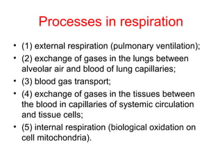
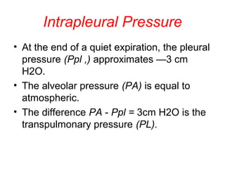
EXTERNAL RESPIRATION
Processes in respiration
• (1) external respiration (pulmonary ventilation);
• (2) exchange of gases in the lungs between
alveolar air and blood of lung capillaries;
• (3) blood gas transport;
• (4) exchange of gases in the tissues between
the blood in capillaries of systemic circulation
and tissue cells;
• (5) internal respiration (biological oxidation on
cell mitochondria).
Changes in the position of
diaphragm & chest during quiet
breathing.
Diaphragm & thoracis cavity
movements
inhalation exhalation
Р
Abdominal cavity
Р intrapleural
-6 mm Hg
Р intrapleural
-3 mm Hg
Sponsored
Medical Lecture Notes – All Subjects
USMLE Exam (America) – Practice
Inspiratory muscles
• muscular part of the diaphragm
• external intercostals & intercartilarginous
parts of the internal intercostal muscles
• the scalenus, the major and minor
pectoralis , the serratus anterior,
trapezius, rhomboidei & the levatores
scapulae muscles.
Mechanics of costal movements
Expiratory muscles
• Peaceful expiration is passive
• abdominal muscles & internal
intercostal muscles
Pleural cavity
Visceral pleura
Parietal pleura Pleural cavity
Intrapleural Pressure
• At the end of a quiet expiration, the pleural
pressure (Ppl ,) approximates —3 cm
H2O.
• The alveolar pressure (PA) is equal to
atmospheric.
• The difference PA - Ppl = 3cm H2O is the
transpulmonary pressure (PL).
Factors forming negative pressure
in the pleural cavity
• Hermeticized cavity (closed volume)
• In embrio thorasic cavity grows faster than
lung parenhime
• Sucking force of parietal & visceral pleura
• Elastic recoil force
Elastic recoil force of the lungs
• (1) surface tension in the alveolar
fluid film lining the alveoli
• (2) tissue elasticity of the alveolar
walls due to the elastic fiber`s.
Surfactant effects
•Fats, mainly lecithin
•Produced by pneumocytes of II type
Surfactant effects
Surfactant effects
Lung volumes are measured by a spirometer. A cross-section of the spirometer is shown
on the left. With inspiration, the pen shows an upward deflection on the spirogram, and
with expiration, a downward deflection. On the right, a spirogram illustrating the different
lung volumes is depicted. Note that the functional residual capacity (FRC), residual
volume (RV), and total lung capacity (TLC) cannot be measured directly with a spirometer.
Lung Volumes
Lung Volumes
• The volume of air breathed in and out during quiet breathing is
about 500 ml (from 300 to 800 ml) and is known as the tidal volume.
• The maximal volume of air that can be additionally inspired in deep
inspiration is about 3000 ml. This is the inspiratory_reserve volume.
• The maximal volume of air that can be expired after quiet expiration
is about 1300 ml and is called the expiratory reserve volume.
• The sum of these volumes is the vital capacity of the lungs (VC):
500 + 3000 + 1300 = 4800 ml The VC is slightly higher in the male
(4000-5500 ml) than in the female (3000-4500 ml).
• A considerable volume of air (about 1200 ml) remains in the lungs
after maximal expiration. It is called the residual volume.
Pneumothorax
• closed pneumothorax is the
presence of small volume of air
within the pleural cavity
• In open pneumothorax the pleural
cavity has a direct communication
with atmospheric air (the thorax is
open by wound or during
intrathoracic operations)
Resistance to respiration
• ELASTIC (evaluated by – VC/PVC х
100%)
• NON-ELASTIC
А) VISCOUS
B) AERODYNAMIC(evaluated by
Votchal-Tiffno index VFE/VC х 100%)
DEAD SPACE
• ANATOMICAL (150 ml)
• PHYSIOLOGICAL
Bronchial tree
Functions of airconducting
pathways
• Warm up the air
• Cleanse the air
• Air is becoming more humid
External Respiration

External Respiration

  • 1.
  • 2.
    Processes in respiration •(1) external respiration (pulmonary ventilation); • (2) exchange of gases in the lungs between alveolar air and blood of lung capillaries; • (3) blood gas transport; • (4) exchange of gases in the tissues between the blood in capillaries of systemic circulation and tissue cells; • (5) internal respiration (biological oxidation on cell mitochondria).
  • 3.
    Changes in theposition of diaphragm & chest during quiet breathing.
  • 4.
    Diaphragm & thoraciscavity movements inhalation exhalation Р Abdominal cavity Р intrapleural -6 mm Hg Р intrapleural -3 mm Hg
  • 5.
    Sponsored Medical Lecture Notes– All Subjects USMLE Exam (America) – Practice
  • 6.
    Inspiratory muscles • muscularpart of the diaphragm • external intercostals & intercartilarginous parts of the internal intercostal muscles • the scalenus, the major and minor pectoralis , the serratus anterior, trapezius, rhomboidei & the levatores scapulae muscles.
  • 7.
  • 8.
    Expiratory muscles • Peacefulexpiration is passive • abdominal muscles & internal intercostal muscles
  • 9.
  • 10.
    Intrapleural Pressure • Atthe end of a quiet expiration, the pleural pressure (Ppl ,) approximates —3 cm H2O. • The alveolar pressure (PA) is equal to atmospheric. • The difference PA - Ppl = 3cm H2O is the transpulmonary pressure (PL).
  • 11.
    Factors forming negativepressure in the pleural cavity • Hermeticized cavity (closed volume) • In embrio thorasic cavity grows faster than lung parenhime • Sucking force of parietal & visceral pleura • Elastic recoil force
  • 12.
    Elastic recoil forceof the lungs • (1) surface tension in the alveolar fluid film lining the alveoli • (2) tissue elasticity of the alveolar walls due to the elastic fiber`s.
  • 13.
    Surfactant effects •Fats, mainlylecithin •Produced by pneumocytes of II type
  • 15.
  • 16.
  • 18.
    Lung volumes aremeasured by a spirometer. A cross-section of the spirometer is shown on the left. With inspiration, the pen shows an upward deflection on the spirogram, and with expiration, a downward deflection. On the right, a spirogram illustrating the different lung volumes is depicted. Note that the functional residual capacity (FRC), residual volume (RV), and total lung capacity (TLC) cannot be measured directly with a spirometer.
  • 19.
  • 20.
    Lung Volumes • Thevolume of air breathed in and out during quiet breathing is about 500 ml (from 300 to 800 ml) and is known as the tidal volume. • The maximal volume of air that can be additionally inspired in deep inspiration is about 3000 ml. This is the inspiratory_reserve volume. • The maximal volume of air that can be expired after quiet expiration is about 1300 ml and is called the expiratory reserve volume. • The sum of these volumes is the vital capacity of the lungs (VC): 500 + 3000 + 1300 = 4800 ml The VC is slightly higher in the male (4000-5500 ml) than in the female (3000-4500 ml). • A considerable volume of air (about 1200 ml) remains in the lungs after maximal expiration. It is called the residual volume.
  • 22.
    Pneumothorax • closed pneumothoraxis the presence of small volume of air within the pleural cavity • In open pneumothorax the pleural cavity has a direct communication with atmospheric air (the thorax is open by wound or during intrathoracic operations)
  • 25.
    Resistance to respiration •ELASTIC (evaluated by – VC/PVC х 100%) • NON-ELASTIC А) VISCOUS B) AERODYNAMIC(evaluated by Votchal-Tiffno index VFE/VC х 100%)
  • 26.
    DEAD SPACE • ANATOMICAL(150 ml) • PHYSIOLOGICAL
  • 27.
  • 28.
    Functions of airconducting pathways •Warm up the air • Cleanse the air • Air is becoming more humid