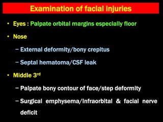
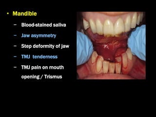
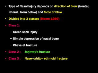
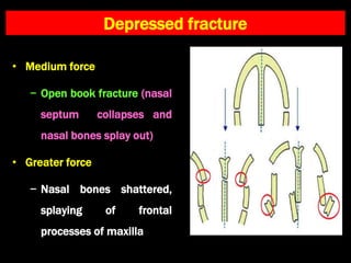
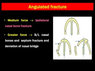
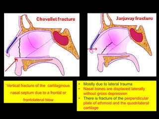
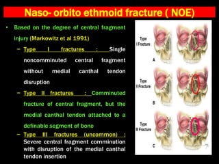
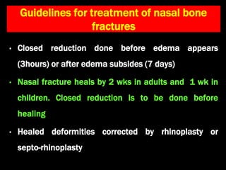
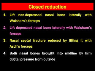
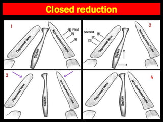
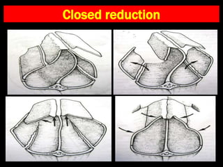
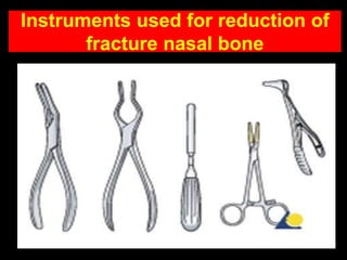
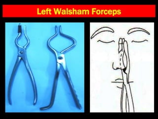
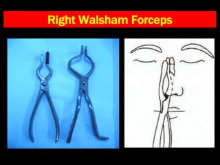
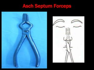
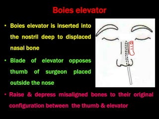
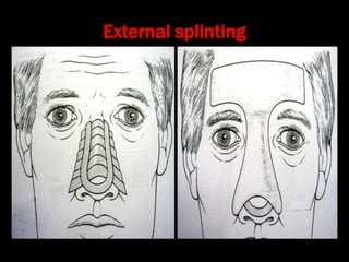
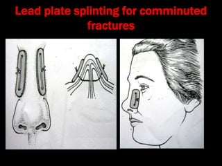
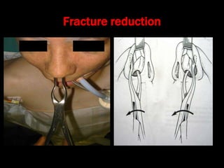
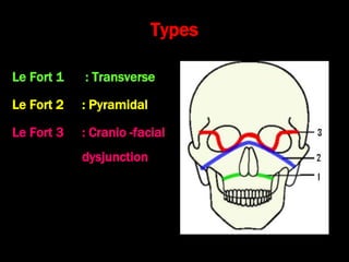
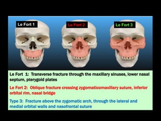
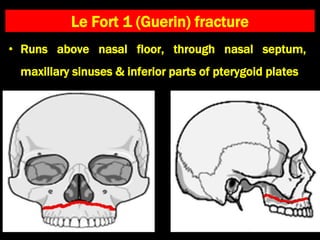
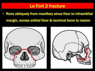
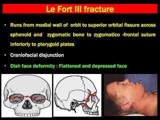
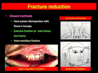
Facial Bone Fractures
Dr. Krishna Koirala
• Nasal bones are the most common types of
facial fractures ( ~ 50% of all facial fractures)
–Face is specifically targeted during assaults
–Nose is centrally placed and most anteriorly projecting part
• Others:
• Frontal bone, zygomatic, maxilla, mandible
Etiology
• Road Traffic Accident
• Fall injury
• Assault
• Sports injury
• In children
–‘Fall’ while playing
–Child abuse
Management of facial trauma
• Manage airway, breathing and circulation
• Control hemorrhage
• Treat associated injuries of head, neck, cervical spine,
chest, abdomen, pelvis and limbs
• Wound debridement
• Treatment of maxillo -facial bone injury
Examination of facial injuries
• Eyes : Palpate orbital margins especially floor
• Nose
− External deformity/bony crepitus
− Septal hematoma/CSF leak
• Middle 3rd
− Palpate bony contour of face/step deformity
− Surgical emphysema/Infraorbital & facial nerve
deficit
• Mandible
− Blood-stained saliva
− Jaw asymmetry
− Step deformity of jaw
− TMJ tenderness
− TMJ pain on mouth
opening / Trismus
Nasal bone fracture
• Type of Nasal Injury depends on direction of blow (frontal,
lateral, from below) and force of blow
• Divided into 3 classes (Moore 1989)
• Class 1:
− Green stick injury
− Simple depression of nasal bone
− Chevalet fracture
• Class 2 : Jarjavey's fracture
• Class 3 : Naso- orbito - ethmoid fracture
Depressed fracture
• Medium force
− Open book fracture (nasal
septum collapses and
nasal bones splay out)
• Greater force
− Nasal bones shattered,
splaying of frontal
processes of maxilla
Angulated fracture
• Medium force  Ipsilateral
nasal bone fracture
• Greater force  B/L nasal
bones and septum fracture and
deviation of nasal bridge
Vertical fracture of the cartilaginous
nasal septum due to a frontal or
frontolateral blow
• Mostly due to lateral trauma
• Nasal bones are displaced laterally
without gross depression
• There is fracture of the perpendicular
plate of ethmoid and the quadrilateral
cartilage
Naso- orbito ethmoid fracture ( NOE)
• Based on the degree of central fragment
injury (Markowitz et al 1991)
− Type I fractures : Single
noncomminuted central fragment
without medial canthal tendon
disruption
− Type II fractures : Comminuted
fracture of central fragment, but the
medial canthal tendon attached to a
definable segment of bone
− Type III fractures (uncommon) :
Severe central fragment comminution
with disruption of the medial canthal
tendon insertion
• Clinical signs
− Telecanthus
− Telescoping of the orbit
− Pig snout deformity
− Positive bowstring sign
Clinical features
• External nasal deformity
• Epistaxis
• Laceration of skin of nose
• Edema over nasal bridge
(within few hours of injury )
• Peri -orbital ecchymosis
• Nasal bone tenderness
• Nasal bone crepitus
Plain X-ray soft tissue nasal bone lateral view
Treatment
• Fractures without displacement : no treatment
• Fractures with displacement : closed reduction
• Open reduction required rarely for
• Infection
• Comminuted fracture
• Failed closed reduction
Guidelines for treatment of nasal bone
fractures
• Closed reduction done before edema appears
(3hours) or after edema subsides (7 days)
• Nasal fracture heals by 2 wks in adults and 1 wk in
children. Closed reduction is to be done before
healing
• Healed deformities corrected by rhinoplasty or
septo-rhinoplasty
Closed reduction
1. Lift non-depressed nasal bone laterally with
Walsham’s forceps
2. Lift depressed nasal bone laterally with Walsham’s
forceps
3. Nasal septal fracture reduced by lifting it with
Asch’s forceps
4. Both nasal bones brought into midline by firm
digital pressure from outside
Closed reduction
Closed reduction
Instruments used for reduction of
fracture nasal bone
Left Walsham Forceps
Right Walsham Forceps
Asch Septum Forceps
Boies elevator
• Boies elevator is inserted into
the nostril deep to displaced
nasal bone
• Blade of elevator opposes
thumb of surgeon placed
outside the nose
• Raise & depress misaligned bones to their original
configuration between the thumb & elevator
External splinting
Lead plate splinting for comminuted
fractures
Rhinoplasty for healed deformity
Nasal Septum Fracture
Fracture reduction
Le Fort fractures
Types
Le Fort 1 : Transverse
Le Fort 2 : Pyramidal
Le Fort 3 : Cranio -facial
dysjunction
Le Fort 1: Transverse fracture through the maxillary sinuses, lower nasal
septum, pterygoid plates
Le Fort 2: Oblique fracture crossing zygomaticomaxillary suture, inferior
orbital rim, nasal bridge
Type 3: Fracture above the zygomatic arch, through the lateral and
medial orbital walls and nasofrontal suture
Le Fort 3
Le Fort 2
Le Fort 1
Le Fort 1 (Guerin) fracture
• Runs above nasal floor, through nasal septum,
maxillary sinuses & inferior parts of pterygoid plates
Le Fort 2 fracture
• Runs obliquely from maxillary sinus floor to infraorbital
margin, across orbital floor & lacrimal bone to nasion
Le Fort III fracture
• Runs from medial wall of orbit to superior orbital fissure across
sphenoid and zygomatic bone to zygomatico -frontal suture
inferiorly to pterygoid plates
• Craniofacial disjunction
• Dish face deformity : Flattened and depressed face
Fracture reduction
• Closed methods
− Hard palate disimpaction with
Rowe’s forceps
− External fixation on halo frame,
box frame
− Inter-maxillary fixation
External Fixation
Inter-maxillary fixation
• Open methods
− Inter-osseous
wiring
− Compression
plates & screws
Orbital blowout fracture
Mechanism of injury : Medium strength blunt trauma to
eyeball
• Clinical Features
− Enophthalmos
− Orbital emphysema
− Inferior rectus & orbital tissue get
trapped in # site  prevents upward
eye movement  diplopia
− Orbital hemorrhage
− Ruptured eyeball
• Forced duction Test ?
• CT Nose and PNS
− Tear drop sign
Treatment Protocol
• Approaches : Transconjunctival , transantral
• Principles of surgery
− Release of entrapped orbital soft tissue & inferior rectus
muscle
− Removal of small bony fragments & repositioning of large
bony fragments
− Repair of bony defect with micro-plating & biocompatible
implants
Zygomatic bone fracture
Tripod zygomatic fracture
Gillie’s approach
• Incision made 4 cm superior to
zygomatic arch & posterior to
temporal hairline
• Periosteal elevator carried
forward, between the temporalis
fascia & temporalis muscle &
positioned beneath zygomatic
arch
• Lateral traction placed on
zygomatic arch
Mandible fractures
Sites and Frequency
Clinical features and investigations
• Blood-stained saliva
• Trismus
• Malocclusion of teeth
• Ecchymosis / hematoma on the
floor of mouth
• Tenderness at site of fracture
• Crepitus at site of fracture
• Step-deformity on palpation
CT Scan of mandible
Fracture reduction
• Closed methods (Inter-maxillary
fixation)
− Inter-dental wiring
− Arch bars and rubber bands
• Open methods
− Inter-osseous wiring
− Lag screws
− Compression plates & screws

11. Facial Bone fractures.ppt

  • 1.
  • 2.
    • Nasal bonesare the most common types of facial fractures ( ~ 50% of all facial fractures) –Face is specifically targeted during assaults –Nose is centrally placed and most anteriorly projecting part • Others: • Frontal bone, zygomatic, maxilla, mandible
  • 3.
    Etiology • Road TrafficAccident • Fall injury • Assault • Sports injury • In children –‘Fall’ while playing –Child abuse
  • 4.
    Management of facialtrauma • Manage airway, breathing and circulation • Control hemorrhage • Treat associated injuries of head, neck, cervical spine, chest, abdomen, pelvis and limbs • Wound debridement • Treatment of maxillo -facial bone injury
  • 5.
    Examination of facialinjuries • Eyes : Palpate orbital margins especially floor • Nose − External deformity/bony crepitus − Septal hematoma/CSF leak • Middle 3rd − Palpate bony contour of face/step deformity − Surgical emphysema/Infraorbital & facial nerve deficit
  • 6.
    • Mandible − Blood-stainedsaliva − Jaw asymmetry − Step deformity of jaw − TMJ tenderness − TMJ pain on mouth opening / Trismus
  • 7.
  • 8.
    • Type ofNasal Injury depends on direction of blow (frontal, lateral, from below) and force of blow • Divided into 3 classes (Moore 1989) • Class 1: − Green stick injury − Simple depression of nasal bone − Chevalet fracture • Class 2 : Jarjavey's fracture • Class 3 : Naso- orbito - ethmoid fracture
  • 9.
    Depressed fracture • Mediumforce − Open book fracture (nasal septum collapses and nasal bones splay out) • Greater force − Nasal bones shattered, splaying of frontal processes of maxilla
  • 10.
    Angulated fracture • Mediumforce  Ipsilateral nasal bone fracture • Greater force  B/L nasal bones and septum fracture and deviation of nasal bridge
  • 11.
    Vertical fracture ofthe cartilaginous nasal septum due to a frontal or frontolateral blow • Mostly due to lateral trauma • Nasal bones are displaced laterally without gross depression • There is fracture of the perpendicular plate of ethmoid and the quadrilateral cartilage
  • 12.
    Naso- orbito ethmoidfracture ( NOE) • Based on the degree of central fragment injury (Markowitz et al 1991) − Type I fractures : Single noncomminuted central fragment without medial canthal tendon disruption − Type II fractures : Comminuted fracture of central fragment, but the medial canthal tendon attached to a definable segment of bone − Type III fractures (uncommon) : Severe central fragment comminution with disruption of the medial canthal tendon insertion
  • 13.
    • Clinical signs −Telecanthus − Telescoping of the orbit − Pig snout deformity − Positive bowstring sign
  • 14.
    Clinical features • Externalnasal deformity • Epistaxis • Laceration of skin of nose • Edema over nasal bridge (within few hours of injury ) • Peri -orbital ecchymosis • Nasal bone tenderness • Nasal bone crepitus
  • 15.
    Plain X-ray softtissue nasal bone lateral view
  • 16.
    Treatment • Fractures withoutdisplacement : no treatment • Fractures with displacement : closed reduction • Open reduction required rarely for • Infection • Comminuted fracture • Failed closed reduction
  • 17.
    Guidelines for treatmentof nasal bone fractures • Closed reduction done before edema appears (3hours) or after edema subsides (7 days) • Nasal fracture heals by 2 wks in adults and 1 wk in children. Closed reduction is to be done before healing • Healed deformities corrected by rhinoplasty or septo-rhinoplasty
  • 18.
    Closed reduction 1. Liftnon-depressed nasal bone laterally with Walsham’s forceps 2. Lift depressed nasal bone laterally with Walsham’s forceps 3. Nasal septal fracture reduced by lifting it with Asch’s forceps 4. Both nasal bones brought into midline by firm digital pressure from outside
  • 19.
  • 20.
  • 21.
    Instruments used forreduction of fracture nasal bone
  • 22.
  • 23.
  • 24.
  • 25.
    Boies elevator • Boieselevator is inserted into the nostril deep to displaced nasal bone • Blade of elevator opposes thumb of surgeon placed outside the nose • Raise & depress misaligned bones to their original configuration between the thumb & elevator
  • 26.
  • 27.
    Lead plate splintingfor comminuted fractures
  • 28.
  • 29.
  • 30.
  • 31.
  • 32.
    Types Le Fort 1: Transverse Le Fort 2 : Pyramidal Le Fort 3 : Cranio -facial dysjunction
  • 33.
    Le Fort 1:Transverse fracture through the maxillary sinuses, lower nasal septum, pterygoid plates Le Fort 2: Oblique fracture crossing zygomaticomaxillary suture, inferior orbital rim, nasal bridge Type 3: Fracture above the zygomatic arch, through the lateral and medial orbital walls and nasofrontal suture Le Fort 3 Le Fort 2 Le Fort 1
  • 34.
    Le Fort 1(Guerin) fracture • Runs above nasal floor, through nasal septum, maxillary sinuses & inferior parts of pterygoid plates
  • 35.
    Le Fort 2fracture • Runs obliquely from maxillary sinus floor to infraorbital margin, across orbital floor & lacrimal bone to nasion
  • 36.
    Le Fort IIIfracture • Runs from medial wall of orbit to superior orbital fissure across sphenoid and zygomatic bone to zygomatico -frontal suture inferiorly to pterygoid plates • Craniofacial disjunction • Dish face deformity : Flattened and depressed face
  • 37.
    Fracture reduction • Closedmethods − Hard palate disimpaction with Rowe’s forceps − External fixation on halo frame, box frame − Inter-maxillary fixation External Fixation Inter-maxillary fixation
  • 38.
    • Open methods −Inter-osseous wiring − Compression plates & screws
  • 39.
  • 40.
    Mechanism of injury: Medium strength blunt trauma to eyeball
  • 41.
    • Clinical Features −Enophthalmos − Orbital emphysema − Inferior rectus & orbital tissue get trapped in # site  prevents upward eye movement  diplopia − Orbital hemorrhage − Ruptured eyeball • Forced duction Test ? • CT Nose and PNS − Tear drop sign
  • 42.
    Treatment Protocol • Approaches: Transconjunctival , transantral • Principles of surgery − Release of entrapped orbital soft tissue & inferior rectus muscle − Removal of small bony fragments & repositioning of large bony fragments − Repair of bony defect with micro-plating & biocompatible implants
  • 44.
  • 45.
  • 46.
    Gillie’s approach • Incisionmade 4 cm superior to zygomatic arch & posterior to temporal hairline • Periosteal elevator carried forward, between the temporalis fascia & temporalis muscle & positioned beneath zygomatic arch • Lateral traction placed on zygomatic arch
  • 47.
  • 48.
  • 49.
    Clinical features andinvestigations • Blood-stained saliva • Trismus • Malocclusion of teeth • Ecchymosis / hematoma on the floor of mouth • Tenderness at site of fracture • Crepitus at site of fracture • Step-deformity on palpation CT Scan of mandible
  • 50.
    Fracture reduction • Closedmethods (Inter-maxillary fixation) − Inter-dental wiring − Arch bars and rubber bands • Open methods − Inter-osseous wiring − Lag screws − Compression plates & screws

Editor's Notes

  • #50 CT Scan of mandible