Download to read offline

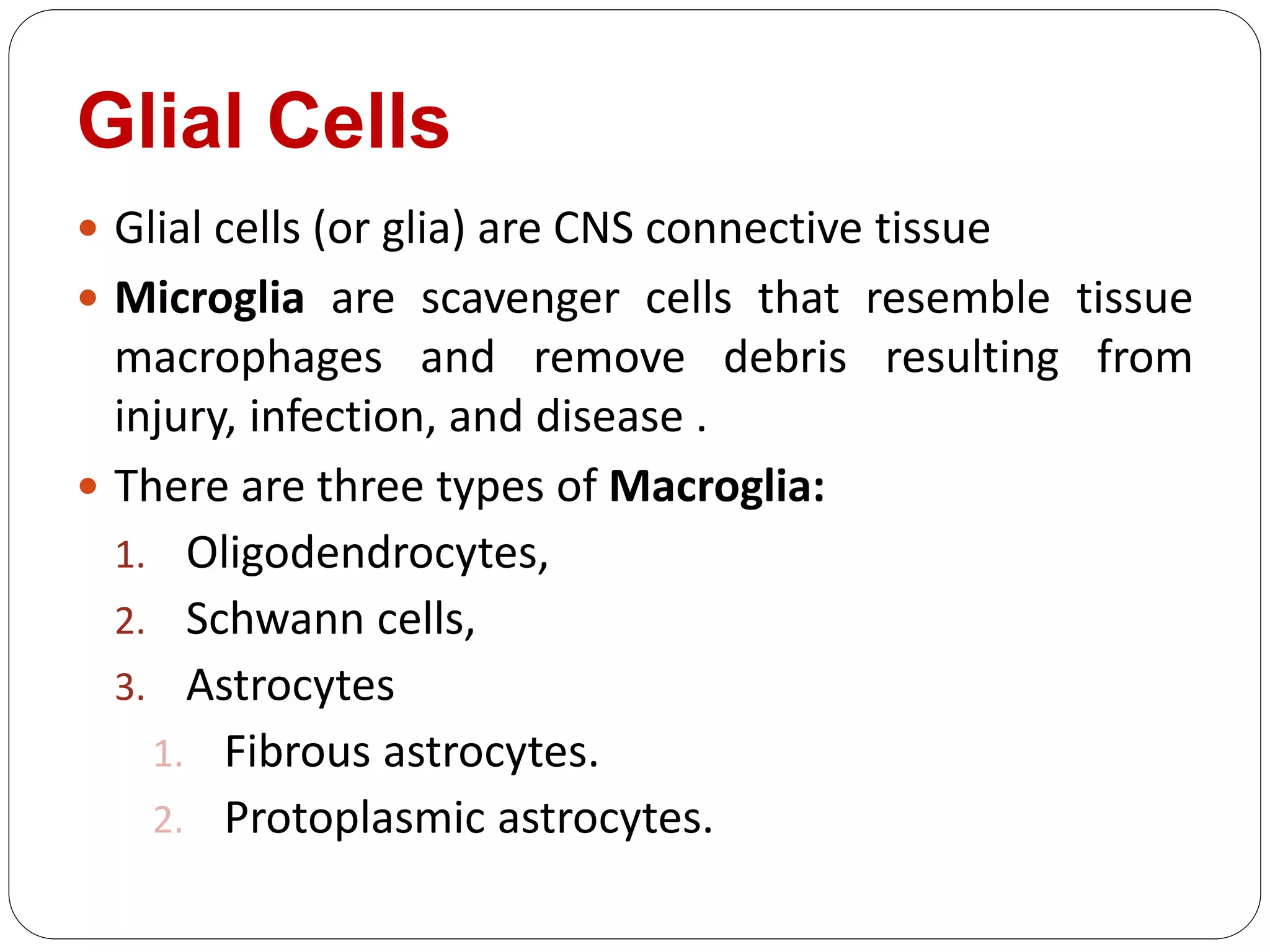
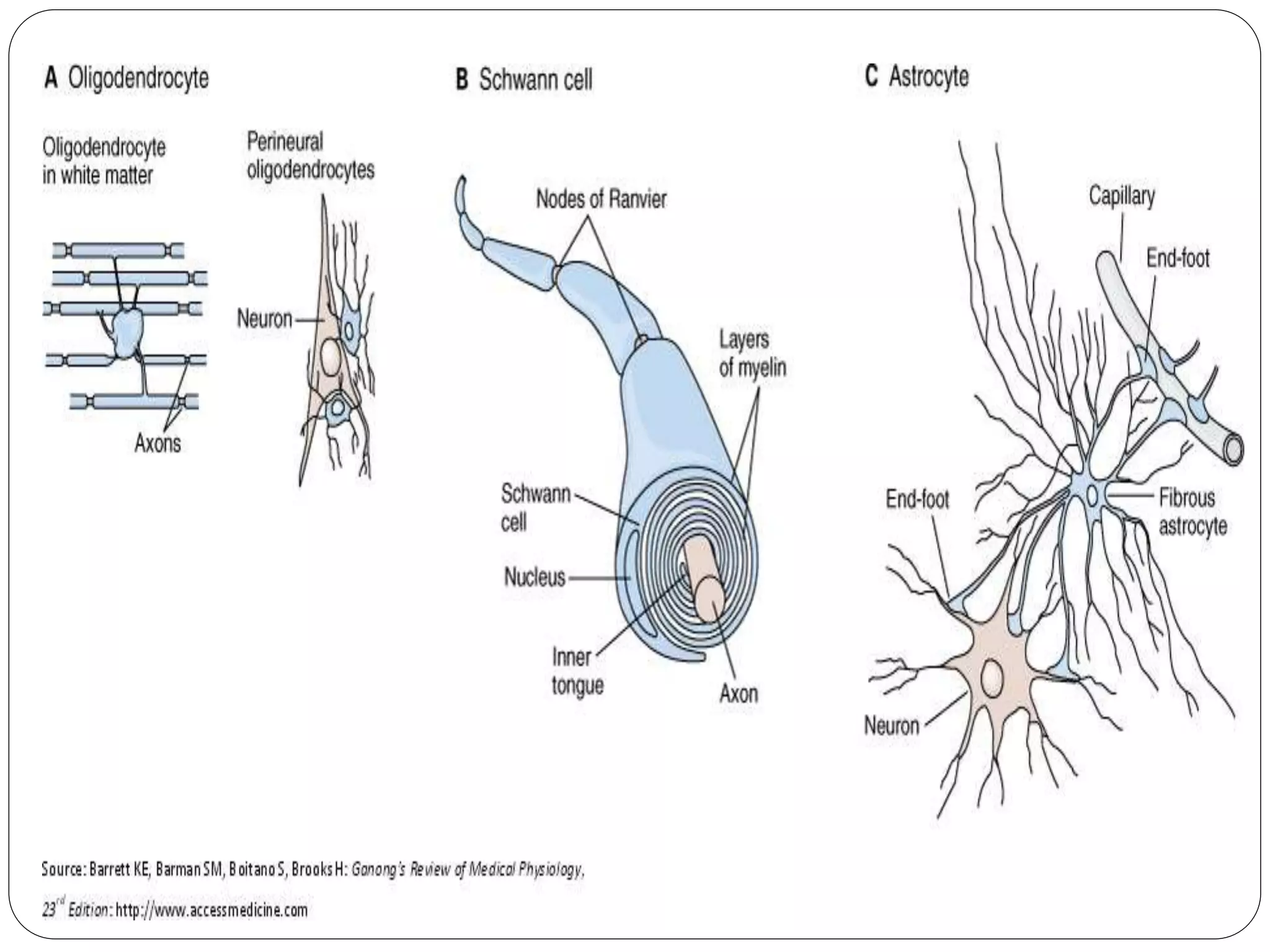


Here are the answers to your questions: 1. Glial cells are the connective tissue of the CNS. There are three main types of glial cells: astrocytes, oligodendrocytes, and microglia. Astrocytes help form the blood-brain barrier and provide nutrients. Oligodendrocytes create myelin sheaths around axons. Microglia are immune cells that remove cellular debris. 2. The main classifications of nerve fibers are: - Erlanger and Gasser classification (Aα, Aβ, Aγ, Aδ, B, C, etc.) based on diameter and conduction velocity. - Numerical classification (Ia, Ib


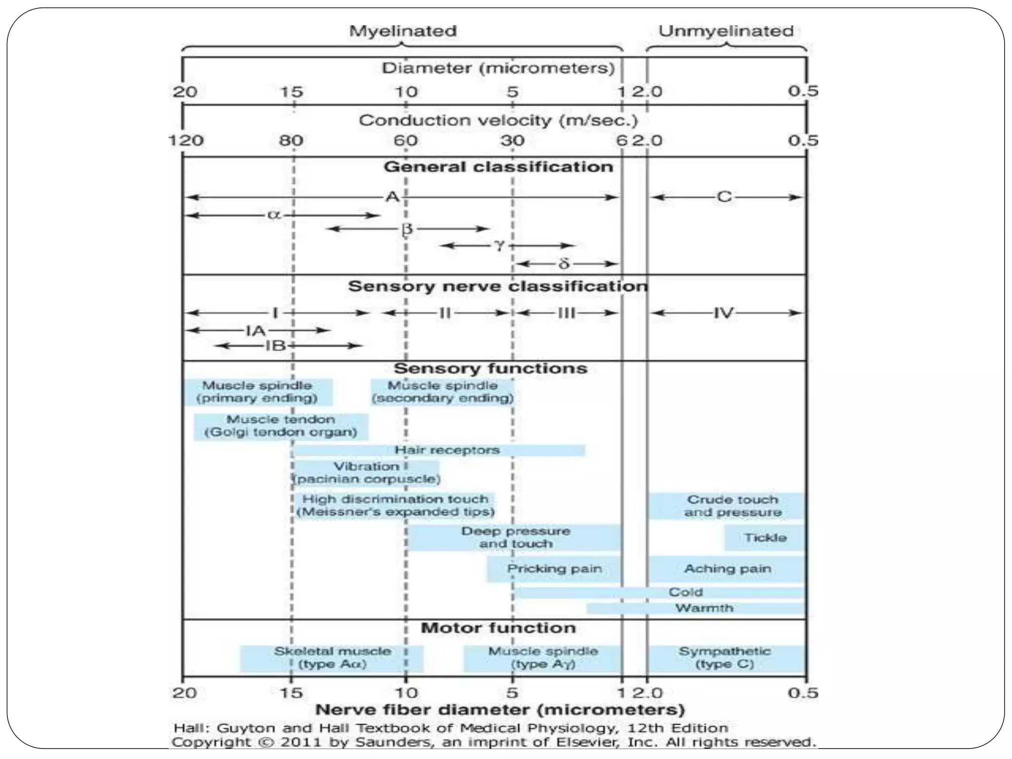
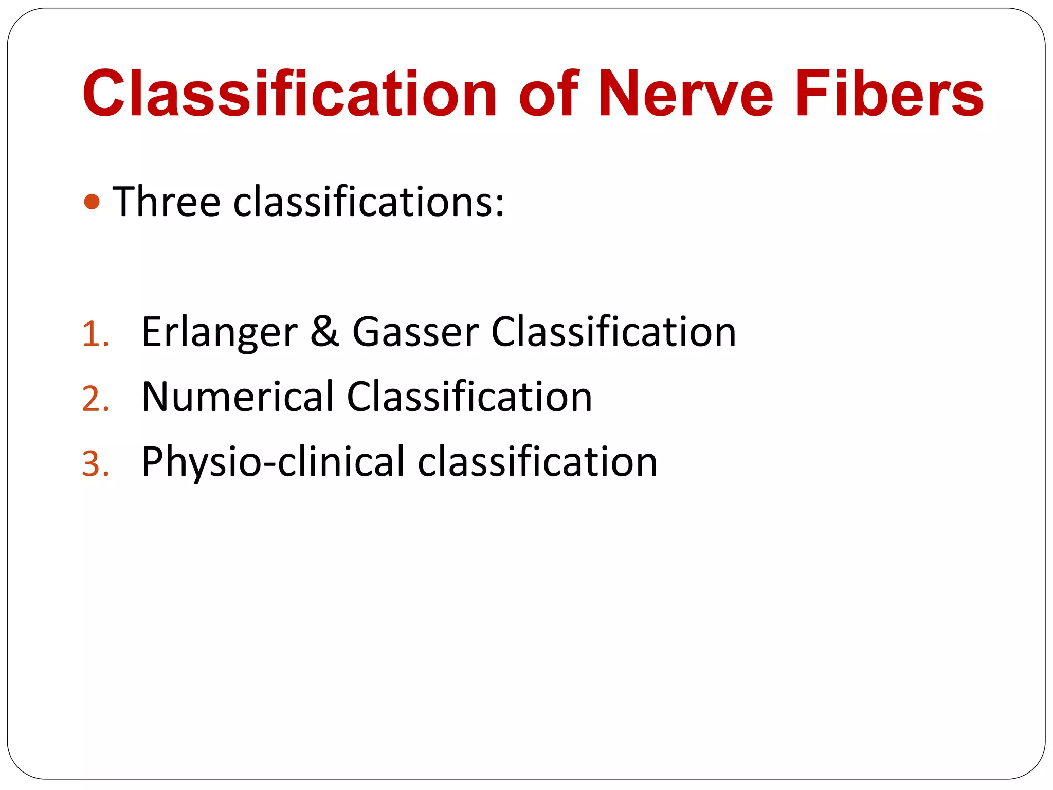
![16 [chapter 16 sensory, motor, and integrative systems]](https://cdn.slidesharecdn.com/ss_thumbnails/16chapter16sensorymotorandintegrativesystems-170828041940-thumbnail.jpg?width=640&height=640&fit=bounds)


