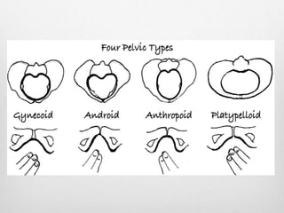
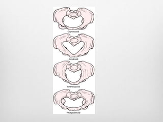
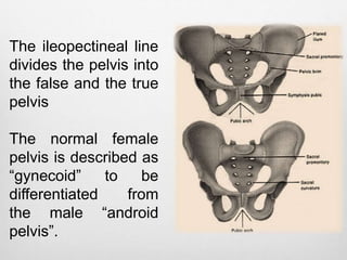
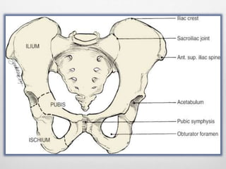
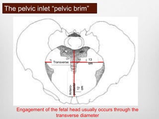
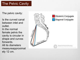
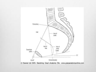
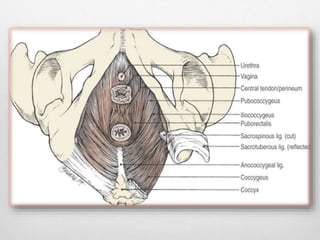
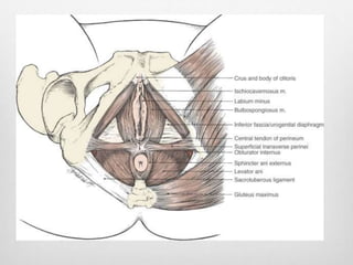
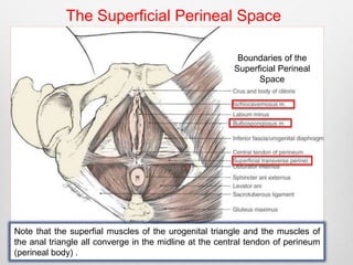
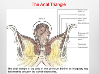
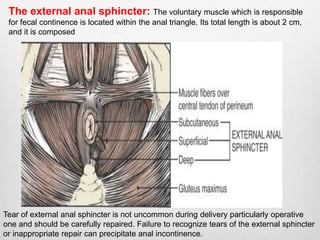
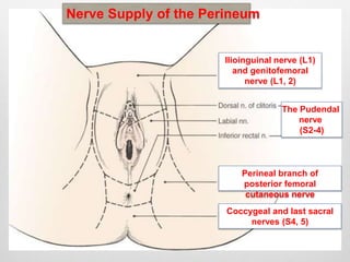
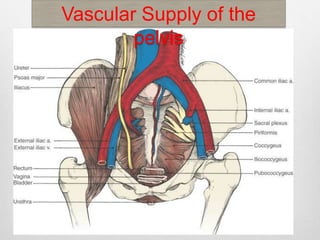
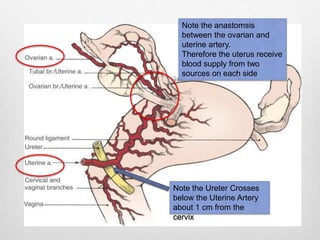
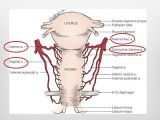
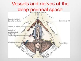
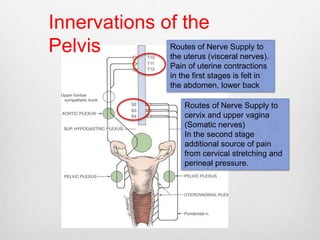
Clinical Anatomy of 
the Female Pelvis 
For the Obstetrician 
Professor Hassan Nasrat
The Bony Pelvis
The ileopectineal line 
divides the pelvis into 
the false and the true 
pelvis 
The normal female 
pelvis is described as 
“gynecoid” to be 
differentiated from 
the male “android 
pelvis”.
The pelvic inlet “pelvic brim” 
13 
cm 
Antero-posterior 
Transverse 
12 
cm 
Engagement of the fetal head usually occurs through the 
transverse diameter
The Pelvic Cavity 
The pelvic cavity: 
Is the curved canal 
between inlet and 
outlet. 
In the normal 
female pelvis the 
cavity is circular in 
shape and curves 
forwards. 
All its diameters 
measureapproximat 
ely 12 cm.
The Pelvic Outlet 
the two pubic bones make the pubic arch, which in the normal female pelvis forms an 
angle not less than 90°. A narrow angle will force the fetal head at delivery posteriorly 
and
Pelvic ligaments and 
Diaphragm
The pudendal neurovascular bundle exits out of the greater sciatic foramen and 
reenters the pelvis through the lesser sciatic foramen. 
This is the site for administration of pudenal block for local anesthesia.
The two main muscles: 
The levator ani muscle group: 
Pubococcygeus, puborectalis, and iliococcygeus. 
They muscles extend from the lateral pelvic walls 
downward and medially to fuse with each other 
posteriorly. 
The levator hiatus lies anteriorly and accommodates the 
urethra, vagina, and anus. 
The coccygeus muscles 
A triangular muscle arises from the ischial spine and 
inserts onto the sacrum and coccyx
The Perineum
The Perineum 
The perineum is divided into two parts (or 
triangles): 
Anterior or urogenital triangle: 
Subdivided into: 
A superficial and deep perineal spaces by a 
fibromuscular septum called the urogenital 
diaphragm 
Posterior or anal triangle: 
The midline attachment forms the fibromuscular 
perineal body. between the anal canal and the 
vagina
The Superficial Perineal Space 
Boundaries of the 
Superficial Perineal 
Space 
Note that the superfial muscles of the urogenital triangle and the muscles of 
the anal triangle all converge in the midline at the central tendon of perineum 
(perineal body) .
During episiotomy :It is important to 
recognize superficial transverse perineil-muscle 
in order to ensure proper cooptation. 
Is bounded by three sets of muscles: 
•The ischiocavernosus: 
•The bulbocavernosus (the sphincter of the Vagina): 
•The superficial transverse perinei: 
It also includes the Bartholin’s glands and the 
vestibular bulbs. 
The superfial muscles of the urogenital triangle and 
the muscles of the anal triangle all converge in the 
midline.
The Deep Perineal Space
The Anal Triangle 
The anal triangle is the area of the perineum behind an imaginary line 
that extends between the ischial tuberosities.
The ischiorectal fossae : 
•A potential space that allows distention of the 
rectum during defecation and the vaginal wall during 
second stage of labor. 
•It is also a potential space for huge (up to one liter) 
hematoma collection and abscess formation. 
•The obturator nerve and internal pudendal 
vessels: run alongside the lateral wall of the 
ischiorectal fossa in the pudendal or Alcock’s canal. 
This canal is formed from the splitting of the fascia 
on the lateral wall of the ischiorectal fossa together 
with the obturator fascia itself.
The external anal sphincter: The voluntary muscle which is responsible 
for fecal continence is located within the anal triangle. Its total length is about 2 cm, 
and it is composed 
Tear of external anal sphincter is not uncommon during delivery particularly operative 
one and should be carefully repaired. Failure to recognize tears of the external sphincter 
or inappropriate repair can precipitate anal incontinence.
Nerve Supply of the Perineum 
Ilioinguinal nerve (L1) 
and genitofemoral 
The Pudendal 
nerve 
(S2-4) 
nerve (L1, 2) 
Perineal branch of 
posterior femoral 
cutaneous nerve 
Coccygeal and last sacral 
nerves (S4, 5)
The Uterus 
In 75% the uterus is in the anteverted, anteflexed position. 
On rare occasion a retro-verted gravid uterus may get entrapped within the pelvis and 
beneath the sacral promontory, giving rise to anterior sacculatoin of the uterus. 
Clinically this presents with acute retention of urine.
The Isthmus is the short constricted area that marks the junction of the uterine 
body with the cervix.
The body of the uterus: 
It has three layers: The endometrium, the myometrium and the perimetrium: 
The myometrium: Has longitudinal, circular and oblique muscle fibers 
and is very expansile. The oblique muscle fibers run “criss-cross” and compress 
the blood vessels when the uterus is well contracted. 
It is found mostly in the upper segment of the uterus, where the placenta normally 
embeds. 
The richness in muscle fibers and its criss-cross important to ensure proper 
hemostasis following placental delivery. 
In contrast to that is the lower uterine segment which is poor hemostasis following 
placental delivery. 
This explains why bleeding in the third stage is more difficult to control if the 
placenta is implanted in the lower uterine segment as in cases of placenta 
praevia. 
The Endometrium: During pregnancy and childbirth, the endometrium 
is referred to as the decidua. 
The perimetrium: Is a layer of peritoneum that covers the uterus except 
at the sides where It extends to form the broad ligaments. 
Significant bleeding and hematoma can extend whithin the layers of the broad 
ligament into the extra peritoneal space with serious consequences
The Cervix: 
Consists predominantly of collagenous connective tissue and 
mucopolysaccaride ground substance. 
It communicates with the uterine cavity through the internal os 
and with the vaginal canal through the external os. 
The endocervical canal is about 2.5 to 3 cm in length. It is lined 
by a single layer of specialized columnar epithelium and 
secretes mucus to facilitate sperm transport. 
During pregnancy the glands secretion forms a plug of mucus 
which helps protect against infection. 
This plug of mucous comes away stained with some blood just 
before labor commences. Many women refer to this as the 
“show”.
Vascular Supply of the 
pelvis
Note the anastomsis 
between the ovarian and 
uterine artery. 
Therefore the uterus receive 
blood supply from two 
sources on each side 
Note the Ureter Crosses 
below the Uterine Artery 
about 1 cm from the 
cervix
Vessels and nerves of the 
deep perineal space
Innervations of the 
Pelvis Routes of Nerve Supply to 
the uterus (visceral nerves). 
Pain of uterine contractions 
in the first stages is felt in 
the abdomen, lower back 
Routes of Nerve Supply to 
cervix and upper vagina 
(Somatic nerves) 
In the second stage 
additional source of pain 
from cervical stretching and 
perineal pressure.

1 chapter 1 applied clinical obstetric anatomy

  • 1.
    Clinical Anatomy of the Female Pelvis For the Obstetrician Professor Hassan Nasrat
  • 2.
  • 5.
    The ileopectineal line divides the pelvis into the false and the true pelvis The normal female pelvis is described as “gynecoid” to be differentiated from the male “android pelvis”.
  • 7.
    The pelvic inlet“pelvic brim” 13 cm Antero-posterior Transverse 12 cm Engagement of the fetal head usually occurs through the transverse diameter
  • 8.
    The Pelvic Cavity The pelvic cavity: Is the curved canal between inlet and outlet. In the normal female pelvis the cavity is circular in shape and curves forwards. All its diameters measureapproximat ely 12 cm.
  • 10.
    The Pelvic Outlet the two pubic bones make the pubic arch, which in the normal female pelvis forms an angle not less than 90°. A narrow angle will force the fetal head at delivery posteriorly and
  • 11.
  • 12.
    The pudendal neurovascularbundle exits out of the greater sciatic foramen and reenters the pelvis through the lesser sciatic foramen. This is the site for administration of pudenal block for local anesthesia.
  • 13.
    The two mainmuscles: The levator ani muscle group: Pubococcygeus, puborectalis, and iliococcygeus. They muscles extend from the lateral pelvic walls downward and medially to fuse with each other posteriorly. The levator hiatus lies anteriorly and accommodates the urethra, vagina, and anus. The coccygeus muscles A triangular muscle arises from the ischial spine and inserts onto the sacrum and coccyx
  • 15.
  • 17.
    The Perineum Theperineum is divided into two parts (or triangles): Anterior or urogenital triangle: Subdivided into: A superficial and deep perineal spaces by a fibromuscular septum called the urogenital diaphragm Posterior or anal triangle: The midline attachment forms the fibromuscular perineal body. between the anal canal and the vagina
  • 19.
    The Superficial PerinealSpace Boundaries of the Superficial Perineal Space Note that the superfial muscles of the urogenital triangle and the muscles of the anal triangle all converge in the midline at the central tendon of perineum (perineal body) .
  • 20.
    During episiotomy :Itis important to recognize superficial transverse perineil-muscle in order to ensure proper cooptation. Is bounded by three sets of muscles: •The ischiocavernosus: •The bulbocavernosus (the sphincter of the Vagina): •The superficial transverse perinei: It also includes the Bartholin’s glands and the vestibular bulbs. The superfial muscles of the urogenital triangle and the muscles of the anal triangle all converge in the midline.
  • 21.
  • 22.
    The Anal Triangle The anal triangle is the area of the perineum behind an imaginary line that extends between the ischial tuberosities.
  • 23.
    The ischiorectal fossae: •A potential space that allows distention of the rectum during defecation and the vaginal wall during second stage of labor. •It is also a potential space for huge (up to one liter) hematoma collection and abscess formation. •The obturator nerve and internal pudendal vessels: run alongside the lateral wall of the ischiorectal fossa in the pudendal or Alcock’s canal. This canal is formed from the splitting of the fascia on the lateral wall of the ischiorectal fossa together with the obturator fascia itself.
  • 24.
    The external analsphincter: The voluntary muscle which is responsible for fecal continence is located within the anal triangle. Its total length is about 2 cm, and it is composed Tear of external anal sphincter is not uncommon during delivery particularly operative one and should be carefully repaired. Failure to recognize tears of the external sphincter or inappropriate repair can precipitate anal incontinence.
  • 25.
    Nerve Supply ofthe Perineum Ilioinguinal nerve (L1) and genitofemoral The Pudendal nerve (S2-4) nerve (L1, 2) Perineal branch of posterior femoral cutaneous nerve Coccygeal and last sacral nerves (S4, 5)
  • 26.
    The Uterus In75% the uterus is in the anteverted, anteflexed position. On rare occasion a retro-verted gravid uterus may get entrapped within the pelvis and beneath the sacral promontory, giving rise to anterior sacculatoin of the uterus. Clinically this presents with acute retention of urine.
  • 27.
    The Isthmus isthe short constricted area that marks the junction of the uterine body with the cervix.
  • 29.
    The body ofthe uterus: It has three layers: The endometrium, the myometrium and the perimetrium: The myometrium: Has longitudinal, circular and oblique muscle fibers and is very expansile. The oblique muscle fibers run “criss-cross” and compress the blood vessels when the uterus is well contracted. It is found mostly in the upper segment of the uterus, where the placenta normally embeds. The richness in muscle fibers and its criss-cross important to ensure proper hemostasis following placental delivery. In contrast to that is the lower uterine segment which is poor hemostasis following placental delivery. This explains why bleeding in the third stage is more difficult to control if the placenta is implanted in the lower uterine segment as in cases of placenta praevia. The Endometrium: During pregnancy and childbirth, the endometrium is referred to as the decidua. The perimetrium: Is a layer of peritoneum that covers the uterus except at the sides where It extends to form the broad ligaments. Significant bleeding and hematoma can extend whithin the layers of the broad ligament into the extra peritoneal space with serious consequences
  • 30.
    The Cervix: Consistspredominantly of collagenous connective tissue and mucopolysaccaride ground substance. It communicates with the uterine cavity through the internal os and with the vaginal canal through the external os. The endocervical canal is about 2.5 to 3 cm in length. It is lined by a single layer of specialized columnar epithelium and secretes mucus to facilitate sperm transport. During pregnancy the glands secretion forms a plug of mucus which helps protect against infection. This plug of mucous comes away stained with some blood just before labor commences. Many women refer to this as the “show”.
  • 31.
  • 32.
    Note the anastomsis between the ovarian and uterine artery. Therefore the uterus receive blood supply from two sources on each side Note the Ureter Crosses below the Uterine Artery about 1 cm from the cervix
  • 34.
    Vessels and nervesof the deep perineal space
  • 35.
    Innervations of the Pelvis Routes of Nerve Supply to the uterus (visceral nerves). Pain of uterine contractions in the first stages is felt in the abdomen, lower back Routes of Nerve Supply to cervix and upper vagina (Somatic nerves) In the second stage additional source of pain from cervical stretching and perineal pressure.

Editor's Notes

  • #23 It is traversed by the terminal portion of the anal canal with its surrounding external sphincter muscle. On both sides of the anal canal are the ischiorectal fossae, which are potential cone shaped spaces, Which filled with fat. It lies between the skin and levator ani on each side of the anal canal. Together the two fossae make a horse shoe shape; since they connect posteriorly with each other, anteriorly they are separated by the perineal body
  • #25 The three muscles layers of the external sphincter. Midline or mediolateral episiotomy may damage this sphincter; proper reapproximation is essential for fecal continence. Of the three components; the subcutaneous, superficial and deep components running on top of each other originate posteriorly from the coccyx and are inserted anteriorly into the perineal body. In between, they diverge to surround the anal canal. The third component, the subcutaneous part, surrounds the anal canal and runs circumferentially around it. Tear of external anal sphincter is not uncommon during delivery particularly operative one and should be carefully repaired. Failure to recognize tears of the external sphincter or inappropriate repair can precipitate anal incontinence.
  • #26 The pudendal nerve (S2-4): It caries both motor fibers to the perineal pelvic floor muscles and sensory fibers to most of the perineal skin, vulva and clitoris. It terminates as the dorsal nerve of the clitoris. Coccygeal and last sacral nerves (S4, 5): Supply skin posterior to the anus and over the tip of the coccyx. Perineal branch of posterior femoral cutaneous nerve: Supply skin lateral to the anus and the most posterior and lateral portions of the labia majora. Ilioinguinal nerve (L1) and genitofemoral nerve (L1, 2): These nerves descend from the anterior abdominal wall to supply the skin of the mons pubis and most of the anterior portion of the labia majora (except the clitoris).
  • #28 The uterus has two main parts: The body and the cervix. The body forms the upper two thirds of the uterus. The body of the uterus: Is formed of three major parts: - The fundus is the dome of the uterus above the level of the tubal ostia. - The body is the part of the uterus that lies below the entrance of the oviducts into the uterus. - The Isthmus is the short constricted area that marks the junction of the uterine body with the cervix. The isthmus becomes thinner and distends during pregnancy to form the lower uterine segment (Fig. 1-13) Note that the perineal reflection of the bladder occurs at the level of the uterine isthmus