Anatomical anomalies in the primary structure
of Dicot and Monocot Stems
• The anomalous structure, which occurs in the primary body of a
plant, is defined as primary anomalous structure.
• These anomalies occur in both monocotyledonous and
dicotyledonous stem, and may persist throughout the life of the
plant.
Common anomalous structures that
occur in stems
• 1. Occurrence of Scattered Vascular Bundles in
Dicotyledonous Stem
• 2. Occurrence of Vascular Bundles Arranged in a Ring in
Monocotyledonous Stem
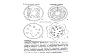
• 3. Occurrence of Cortical Bundles
• 4. Occurrence of Medullary Bundle
Common anomalous structures
that occur in stems
• 5. Occurrence of Internal Bundles
• 6. Occurrence of Internal Phloem or Intraxylary
(Perimedidlary, Medullary) Phloem
• 7. Absence of Vessels in Angiosperm
• 8. Occurrence of Separate Xylem and Phloem Bundles
1. Occurrence of Scattered Vascular Bundles
in Dicotyledonous Stem
• Normally the vascular bundles, in the dicotyledonous stem, are arranged
more or less in a ring.
• The followings are a few variations where the vascular bundles are more or
less irregularly scattered in the parenchymatous ground tissue of the stem.
• Examples: Papaver orientale and P. somniferum (Papaveraceae),
Bougainvillea (Nyctaginaceae), Piper betle and Peperomia langsdorfii
(Piperaceae), Nymphaea (Nymphaeaceae) etc.
2. Occurrence of Vascular Bundles Arranged in a
Ring in Monocotyledonous Stem
• In monocot stems the stele is atactostele, i.e. the vascular
bundles are scattered in the ground tissue.
• But the followings are the exceptions where the vascular
bundles are arranged more or less in one or two rings.
• Ex. Coix, Triticum, Oryza (Poaceae), Tamus communis
(Dioscoreaceae) etc.
3. Occurrence of Cortical Bundles
• In dicotyledonous stem the vascular bundles occur in the stele. But there are some
dicotyledonous stems where accessory vascular bundles are found in the cortex of
stem in addition to stelar bundles.
• In most species cortical bundles are leaf trace bundles.
• Nyctanthes arbortristis (Oleaceae), four inversely oriented cortical vascular
bundles occur with external xylem and internal phloem
• Bombax malabaricum (Bombacaceae), Calycanthus occidentalis, Chimonanthns
(Calycanthaceae) where four pericyclic bundles (leaf traces) occur with internal
phloem and external xylem
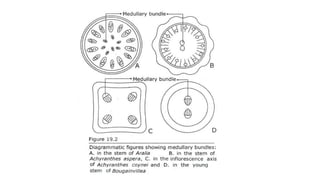
4. Occurrence of Medullary Bundle
• The supplementary bundle, which occurs in the pith of dicotyledonous
stem in addition to the normal ring of bundles, is referred to as
medullary bundles.
• Achyranthes aspera (Amaranthaceae) with two medullary bundles at
internode.
• Raphanus sativus (Cruciferae); Anthocleista (Potaliaceae);
Bougainvillea, Mirabilis longiflora: with four medullary bundles
5. Occurrence of Internal Bundles
• The supplementary bundles, which occur below the normal ring
of bundles and above the medullary bundles, are referred to as
internal bundle.
• These bundles are not so deeply situated in the pith to be
classified as medullary bundles.
• Ex. Rumex crispus, R. orientalis etc.
6. Occurrence of Internal Phloem or
Intraxylary (Perimedidlary, Medullary)
Phloem
• The phloem that occurs at the margin of pith in the form of a continuous
cylinder or strands is referred to as intraxylary phloem or internal phloem.
• It is also known as medullary phloem due to its occurrence at the margin
of pith.
• Thevetia, (Apocynaceae); Calotropis (Asclepiadaceae); Convolvulus,
Ipomoea, Evolvulus (Convolvulaceae); Barleria (Acanthaceae); Solanum,
Nicotiana (Solanaceae) etc.
7. Absence of Vessels in Angiosperm
• The xylem of angiosperms is characterized by the presence of
vessels. Exceptions are noted in families Tetracentraceae
(Tetracentron), Trochodendraceae (Trochodendron) and
Winteraceae (Drimys, Pseudoivintera, Bubbia, Zygogynum)
where vessels are absent.
9. Occurrence of Separate Xylem and
Phloem Bundles
• Usually the two vascular tissues xylem and phloem occur together
to form bundles.
• In abnormal cases bundles consisting of either xylem or phloem
strands only occur in addition to the normal bundles. In Cuscuta
fully developed phloem occurs interspersed between other
normal collateral bundles.
• Incomplete bundles consisting of phloem only are observed in
Rumex crispus, Ricinus communis, Xanthium strumarium,
Antigonon leptopus, Achxjranthes aspera, Mirabilisjalapa etc.,
Maheswari (1930).
• In Boerhaavia diffusa incomplete bundles are formed as a result
of centripetal differentiation of phloem from procambium cells
(Maheswari).
Anomalous secondary Growth in Nyctanthes
– Stem (Family – Oleaceae)
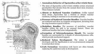
• Anomalous Behavior of Nyctanthes arbor-tristis Stem
• The stem of Nyctanthes arbor-tristis exhibits certain structural
peculiarities that deviate from the normal dicot stem structure.
These features include:
1. Absent or Reduced Vascular Cambium: Unlike typical
dicot stems, Nyctanthes has a limited or absent vascular
cambium, leading to minimal or no secondary growth.
2.Presence of Scattered Vascular Bundles: Vascular bundles
are scattered and show an irregular arrangement, deviating from
the ring formation typical of dicots.
3.Medullary Bundles: The presence of medullary vascular
bundles within the pith is an unusual feature. These are
additional bundles that arise independently.
4.Formation of Sclerenchymatous Sheath: The vascular
bundles are surrounded by a sheath of sclerenchymatous cells,
which provide mechanical support.
5.Reduced Xylem Development: The xylem is poorly
developed, and the lignification is not as pronounced compared
to normal dicot stems.
6.Cork Formation: Anomalous cork layers are often formed,
replacing the epidermis early in development.
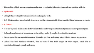
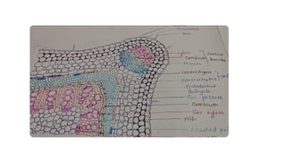
• The outline of T.S. appears quadrangular and reveals the following tissues from outside with-in-
• Epidermis:
• i. Single-layered epidermis consists of rectangular cells.
• ii. A thick uninterrupted cuticle is present on the epidermis. iii. Many multicellular hairs are present.
• 2. Cortex.
• It is few layered thick and is differentiated into outer region of collenchyma and inner parenchyma.
• Collenchyma is several layers deep in the ridges and a few cells deep in other regions.
• Parenchyma forms rest of the cortex. The cells are thin and many intercellular spaces are present.
• Cortex has four vascular bundles, one in each of the four bulges at four angles. Each one is
conjoint,collateral, exarch and open.
3. Endodermis. A distinct endodermis with caspaian strips is absent.
4. Pericycle.
• It is present just below the cortical region and separates cortical region
from the vascular tissues.
• It forms almost a complete ring of parenchymatous cells.
5. Vascular tissue system
• The structure shows secondary growth.
• The tissues occur in the following sequence-primary phloem, secondary phloem, cambium,
secondary xylem and primary xylem.
• The primary phloem occurs in small patches just below the pericycle.
• Secondary phloem is in the form of a complete ring. It consists of phloem parenchyma,
sieve tubes and
• companion cells.
• The cambium is single layered and is present between the secondary phloem and secondary
xylem.
• Secondary xylem occupies most of the region towards the centre of
the axis. It consists of tracheids, vessels and xylem parenchyma.
• A few vessels are very distinct. Annual rings are not very distinct.
• Primary xylem lies close to the pith. The protoxyleni is endarch.
Primary vascular bundles are conjoint, collateral, endarch and open.
• 6. Pith. A large parenchymatous pith is present in the centre
Abnormality:
• Cortical vascular bundles.
• The stem shows four vascular bundles in the
cortex, one each in four corners of the angular
stem.
• These bundles called as cortical bundles due to
their location in the cortex, are in fact leaf trace
bundles.
• Bundles arise at much lower a node.
Boerhaavia
• Anomalous Behavior of Boerhavia Stem
• The stem of Boerhavia exhibits certain anomalous secondary growth features that deviate from the typical dicot stem pattern.
These include:
1. Unusual Vascular Arrangement:
1. The vascular bundles are conjoint, collateral, and closed.
2. Unlike typical dicot stems, secondary growth occurs from successive cambial rings rather than a single continuous cambium.
2. Formation of Successive Cambial Rings:
1. After the primary cambium ceases its activity, new cambial rings are formed from the pericycle, producing additional vascular bundles in a
concentric manner.
3. Multiseriate Xylem Rays:
1. The xylem rays in the secondary xylem are unusually wide and multiseriate, which is not commonly observed in most dicots.
4. Development of Medullary Bundles:
1. In addition to the normal vascular bundles, accessory or medullary bundles are formed within the pith, an unusual feature.
5. Non-uniform Secondary Growth:
1. The secondary growth is irregular, resulting in an uneven distribution of tissues, contributing to the abnormal stem structure.
Vascular tissue system
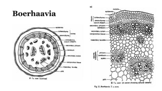
• 1. There' are many vascular bundles which are arranged in rings. A zone of secondary tissues
is also very dstinct.
• 2. The outermost ring has many bundles. Due to secondary growth, phloem occurs in the
form of crushed and obliterated patches.
• Abundant prosenchyma (conjunctive tissue) is present. Secondary phloem forms a complete
ring.
• Cambium that follows separates phloem and xylem. The primary xylem groups are situated
close to the pith. Protoxylem is endarch and the vascular bundles are conjoint, collateral,
endarch and open.
• 3. The innermost ring consists of two vascular bundles. Each bundle is
conjoint, collateral, endarch and open. The bundles lie close to the pith,
and are, therefore, known as medullary bundles. These bundles produce a
small amount of secondary phloem and secondary xylem in radial rows.
• 4. The middle ring consists of six or seven (upto fourteen) bundles. These
bundles are smaller than those of the inner ring. Each bundle is conjoint,
collateral, endarch and open.
• Pith. In the centre a small parenchymatous pith is persent.
Points of interest
• 1. Medullary bundles. The vascular bundles are arranged in three
rings. Out of these, two inner rings occur in the pith and are,
therefore, known as medullary bundles. The medullary bundles
possess fascicular (intrafascicular) cambium and produce a little
amount of secondary tissues.
2. Abnormal secondary growth.
• In Boerhaavia, vascular bundles of the outermost ring have fascicular cambium. Later, interfascicular cambium
also develops, thus forming a complete ring.
• The cambial ring produces secondary xylem and secondary phloem. A new ring (accessory cambium) appears
later in the region of the pericycle. This ring of cambium, also, as in earlier cases functions only for some time.
• Such many (upto twenty two) accessory cambia are produced successively, much farther away into the cortex
every time.
• This results in the formation of successive alternate zones of secondary xylem and secondary phloem.
• These rings or zones (of secondary tissues) are sometimes eccentrically developed. Cambia produce a very large
amount of prosenchyma into which xylem remains scattered and at times becomes indistinguishable from it.
Casuarina
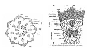
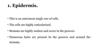
1. Epidermis.
• This is an outermost single row of cells.
• The cells are highly cuticularized.
• Stomata are highly sunken and occur in the grooves.
• Numerous hairs are present In the grooves and around the
stomata.
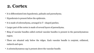
2. Cortex
• It is differentiated into hypodermis, palisade and parenchyma.
• Hypodermis is present below the epidermis.
• It is made of sclerenchyma, arranged in T - shaped patches.
• Larger part of the cortex is made of several layers of parenchyma.
• Ring of vascular bundles called cortical vascular bundles is present in the parenchymatous
region.
• These are situated only below the ridges. Each vascular bundle is conjoint, collateral,
endarch and open.
• A sclentnchymatous cap is present above the vascular bundle.
3. Endodermis.
• This is a single layer of cells which separates cortex from the
underlying vascular tissue.
Vascular tissue
• Vascular bundles are arranged in a ring.
• Each vascular bundle occurs below the groove.
• A sclerenchymatous patch, known as bundle cap is present just above the bundle.
• Each vascular bundle is conjoint, collateral, end arch and open.
• It shows a- small amount of secondary growth.
• A wide parenchymatous region is present between the two adjacent vascular bundles.
5. Pith.
1. A well-developed parenchymatous pith is present in the centre.
Xerophytic characters
• The anatomy shows following xerophytic characters.
• Presence of thick cuticle.
• Stomata sunken and covered with hairs.
• Sclerenchymatous hypodermis and bundle cap.
• Presence of palisade in the cortex.
• Well developed vascular tissue.
• The presence of well-developed cortex and the type of vascular bundles indicate
that the material is a dicot stem.
Anomalous secondary growth in
Dracaena stem
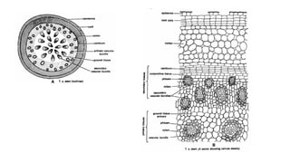
• Dracaena is a typical example of Anomalous secondary thickening in
monocots.
• Typically, secondary thickening is absent in monocots.
• Therefore, secondary thickening itself is an anomaly as Dracaena is a
monocot.
Dracaena young stem
• Young stem has typical monocot structure
• 1. Epidermis single layered.
• 2. Hypodermis is sclerenchymatous.
• 3. Numerous closed, collateral vascular bundles scattered in the
parenchymatous ground tissue.
• 4. Xylem is endarch.
Anomalous secondary thickening in Dracaena
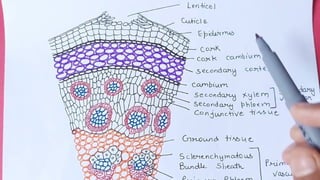
• In Dracaena, secondary growth is due to
• Extrastelar cambial ring in a monocot stem at the cortex.
• Abnormal activity of cambium
During Secondary thickening
• Formation of secondary meristem or secondary cambium occurs in the
inner region of parenchymatous cortex.
• The activity of cambium is abnormal.
• It produces secondary vascular bundles on its inner side only and
parenchymatous cells on the outer side.
• The secondary vascular bundles are amphivasal where phloem is
surrounded by xylem.
• Later cambium produces more parenchymatous cells to the inside that pushes
newly formed vascular bundles to the centre.
• The activity and position of vascular bundles keep on changing and vascular
bundles are arranged in concentric rings.
• The second ring of vascular bundles is alternating in position with the first ring.
• The vascular bundles in the last inner ring are embedded in a mass of lignified
conjunctive tissue.
• Cork cambium activity is normal and produces cork and secondary cortex at the
outer region.