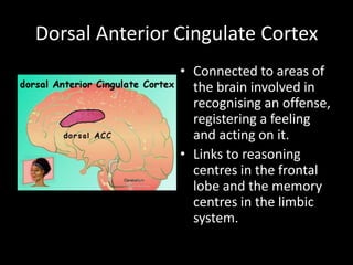
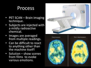
E
M
O
T
I
Anger
Dorsal Anterior Cingulate Cortex
                • Connected to areas of
                  the brain involved in
                  recognising an offense,
                  registering a feeling
                  and acting on it.
                • Links to reasoning
                  centres in the frontal
                  lobe and the memory
                  centres in the limbic
                  system.
Anger  Aggression
• Amygdala – fear
• Hypothalamus -
  rage
In sum of anger…
• Dorsal anterior cingulate cortex (daCC)
• Amygdala
• Hypothalamus
SADNESS
Dr Mark George
Process
• PET SCAN – Brain imaging
  technique.
• Subjects are injected with
  a mildly radioactive
  chemical.
• Images are averaged
  from multiple readings.
• Can be difficult to react
  to anything other than
  the machine itself!
• Solution – show scenes
  from films to evoke
  various emotions.
Findings
•   Dr. George conducted his studies with the
    same sex to avoid possible differences
    between sexes.
•   Findings:
•   Sad woman  increased activity in the
    structures of the limbic system near the face,
•   More activity in the left prefrontal cortex than
    in the right.
•   Activation of the amygdala
•   Brain areas involved in ordinary sadness
    almost completely shut down when a person
    is clinically depressed.
•   Sadness and depression seem to involve the
    same brain region, the left prefrontal cortex, in
    different ways.
•   It gets more active during ordinary sadness,
    but shuts down in people with clinical
    depression.
•   Suggests the left prefrontal cortex may burn
    itself out when sadness persists for several
    months.
In sum of sadness
•   Limbic system closer to the face
•   Amygdala
•   Left prefrontal cortex (sadness)
•   Less activity in prefrontal cortex (depression))
Happiness
   
Dr. George’s findings…
• Decrease of activity in the regions
  of the cerebral cortex that are
  committed to forethought and
  planning.
• These regions are in the temporal-
  parietal area of the cortex, located
  just over and a bit behind the
  ears, and the right prefrontal
  lobe, just behind the forehead.
• The areas become even less active
  when volunteers received
  injections of morphine or cocaine.
• Whilst the amygdala area activates
  during sadness, the structures
  change only slightly when a person
  is happy. The left amygdala seems
  to decrease activity, while the right
  amygdala increases activity.
In sum of happiness
• Temporal-parietal
• Right prefrontal lobe
• Amygdala
What about recognising
      emotions?
The Right Hemisphere Hypothesis
             (RHH)
V   S   H
a   p   y
l   e   p
e   c   o
n   i   t
c
e
    f
    i
        h
        e   V         RIGHT
                    HEMISPHERE
    c   s
–       i
        s   S   •   Negative emotions


            H
V   S   H
                        a   p   y
                        l   e   p
                        e   c   o


                                    V
                        n   i   t
       LEFT
                        c   f   h
    HEMISPHERE          e   i   e
                            c   s

                                    S
•   Positive emotions
                        –       i
                                s


                                    H
Evidence?
• Support for both…
    (Ley and Bryden, 1979; Reuter-Lorenz and Davidson, 1981; Natale et al., 1983; Reuter-Lorenz
    et al., 1983; McLaren and Bryson, 1987; Rodway et al., 2003)

• Brain damaged patients:
-   In some, lesions to the right hemisphere impair the perception of emotion,
    regardless of the valence. (Adolphs et al., 1996; Borod et al., 1998; Adolphs et al.,
    2000)
-   In others, it was found that damage to the left hemisphere impairs the perception
    of +ive emotions while damage to the right impair perception of -ive emotions.
    (Borod et al., 1986; Mandal et al., 1991)

• Chimeric studies on healthy participants
CHIMERIC FACES

• Facial expression stimuli artificially
designed to project a different emotion to
each half of the brain simultaneously
(Levy et al., 1972)


 •IDENTIFY THE
    EMOTION!
CHIMERIC FACES


Most people:


   But some others…
Fin…

01 emotional brain

  • 1.
  • 4.
  • 5.
    Dorsal Anterior CingulateCortex • Connected to areas of the brain involved in recognising an offense, registering a feeling and acting on it. • Links to reasoning centres in the frontal lobe and the memory centres in the limbic system.
  • 6.
    Anger  Aggression •Amygdala – fear • Hypothalamus - rage
  • 8.
    In sum ofanger… • Dorsal anterior cingulate cortex (daCC) • Amygdala • Hypothalamus
  • 9.
  • 10.
  • 11.
    Process • PET SCAN– Brain imaging technique. • Subjects are injected with a mildly radioactive chemical. • Images are averaged from multiple readings. • Can be difficult to react to anything other than the machine itself! • Solution – show scenes from films to evoke various emotions.
  • 12.
    Findings • Dr. George conducted his studies with the same sex to avoid possible differences between sexes. • Findings: • Sad woman  increased activity in the structures of the limbic system near the face, • More activity in the left prefrontal cortex than in the right. • Activation of the amygdala • Brain areas involved in ordinary sadness almost completely shut down when a person is clinically depressed. • Sadness and depression seem to involve the same brain region, the left prefrontal cortex, in different ways. • It gets more active during ordinary sadness, but shuts down in people with clinical depression. • Suggests the left prefrontal cortex may burn itself out when sadness persists for several months.
  • 13.
    In sum ofsadness • Limbic system closer to the face • Amygdala • Left prefrontal cortex (sadness) • Less activity in prefrontal cortex (depression))
  • 14.
    Happiness 
  • 15.
    Dr. George’s findings… •Decrease of activity in the regions of the cerebral cortex that are committed to forethought and planning. • These regions are in the temporal- parietal area of the cortex, located just over and a bit behind the ears, and the right prefrontal lobe, just behind the forehead. • The areas become even less active when volunteers received injections of morphine or cocaine. • Whilst the amygdala area activates during sadness, the structures change only slightly when a person is happy. The left amygdala seems to decrease activity, while the right amygdala increases activity.
  • 16.
    In sum ofhappiness • Temporal-parietal • Right prefrontal lobe • Amygdala
  • 17.
  • 18.
    The Right HemisphereHypothesis (RHH)
  • 19.
    V S H a p y l e p e c o n i t c e f i h e V RIGHT HEMISPHERE c s – i s S • Negative emotions H
  • 20.
    V S H a p y l e p e c o V n i t LEFT c f h HEMISPHERE e i e c s S • Positive emotions – i s H
  • 21.
    Evidence? • Support forboth… (Ley and Bryden, 1979; Reuter-Lorenz and Davidson, 1981; Natale et al., 1983; Reuter-Lorenz et al., 1983; McLaren and Bryson, 1987; Rodway et al., 2003) • Brain damaged patients: - In some, lesions to the right hemisphere impair the perception of emotion, regardless of the valence. (Adolphs et al., 1996; Borod et al., 1998; Adolphs et al., 2000) - In others, it was found that damage to the left hemisphere impairs the perception of +ive emotions while damage to the right impair perception of -ive emotions. (Borod et al., 1986; Mandal et al., 1991) • Chimeric studies on healthy participants
  • 22.
    CHIMERIC FACES • Facialexpression stimuli artificially designed to project a different emotion to each half of the brain simultaneously (Levy et al., 1972) •IDENTIFY THE EMOTION!
  • 24.
    CHIMERIC FACES Most people: But some others…
  • 25.