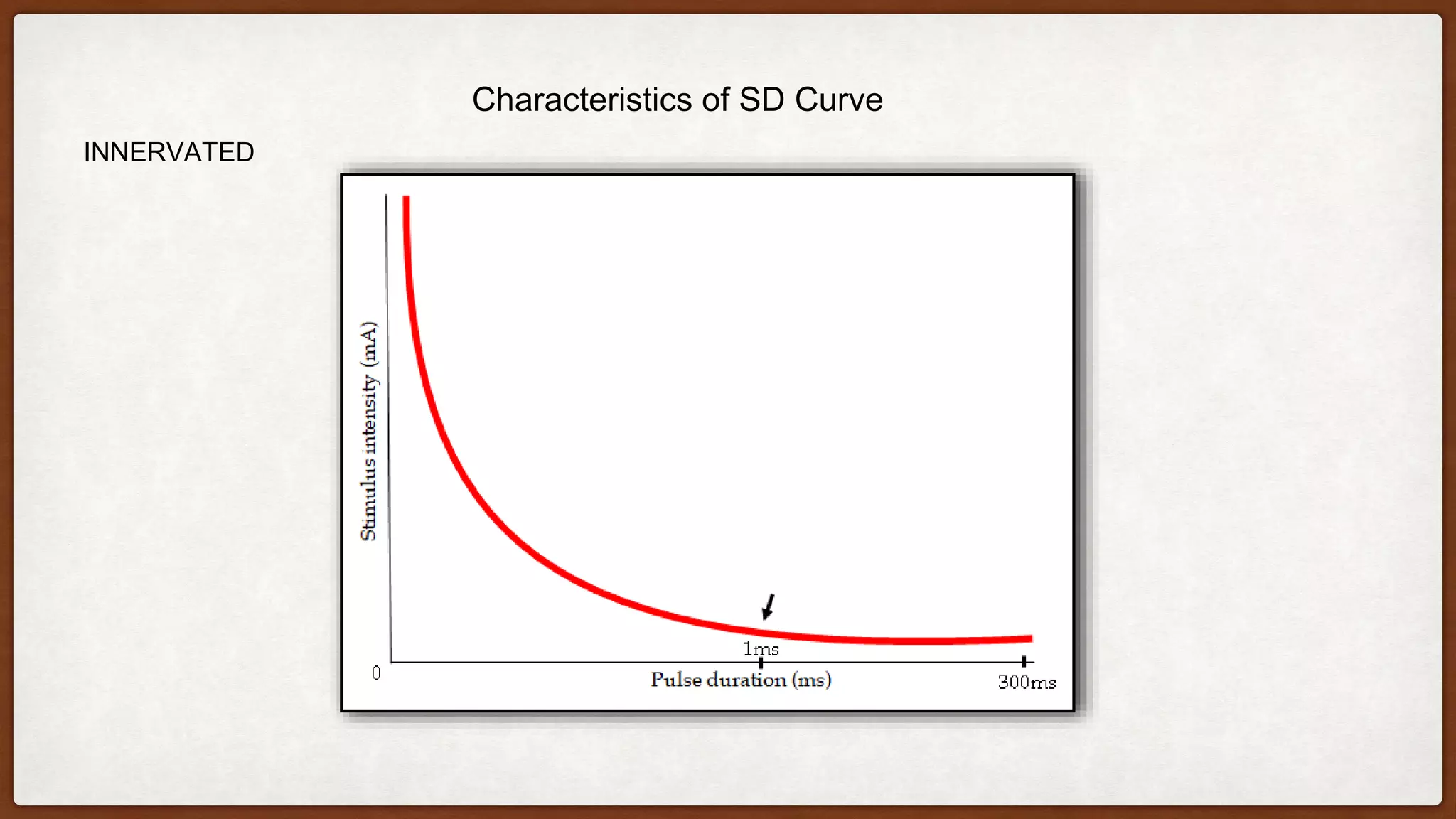
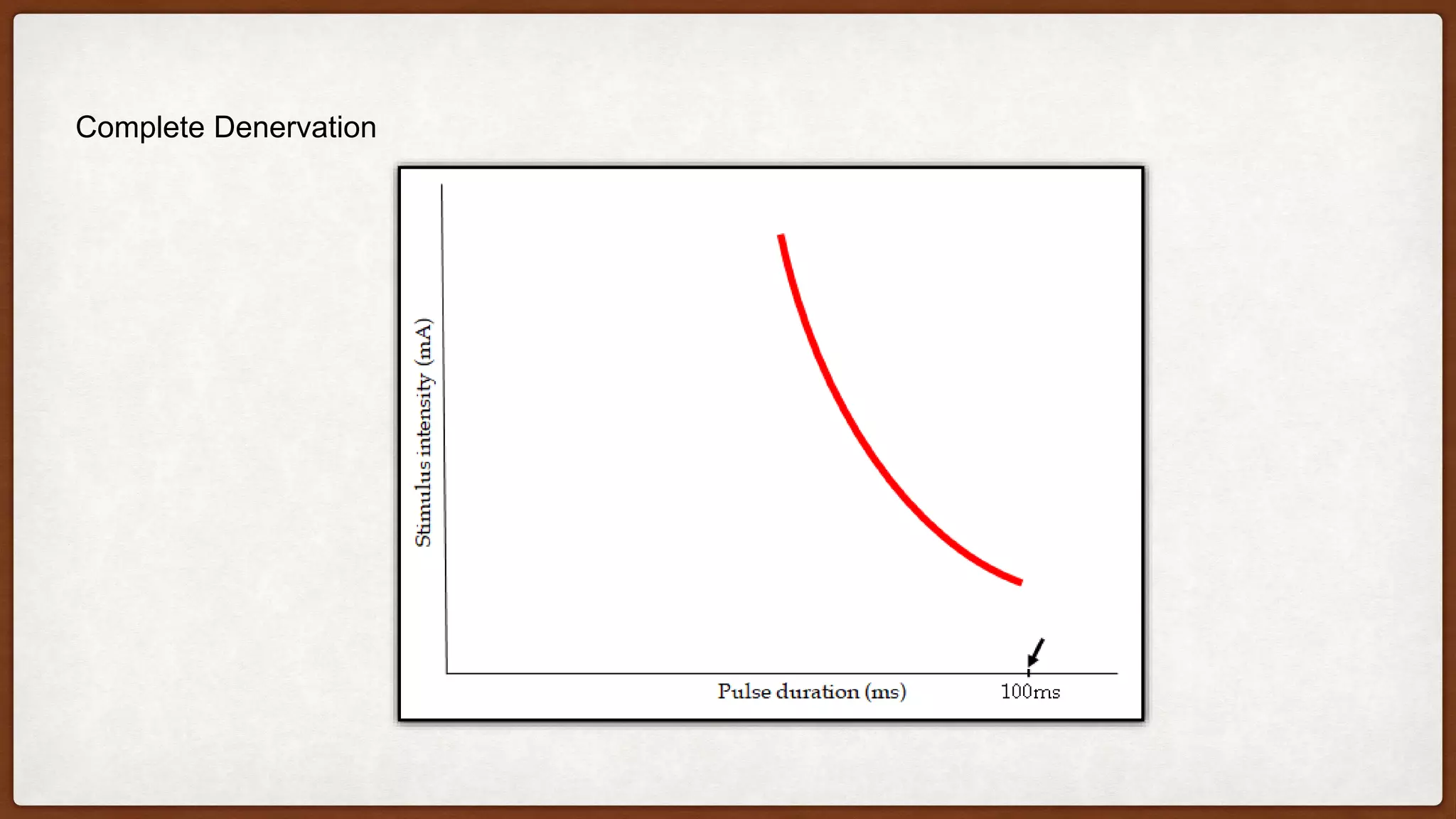
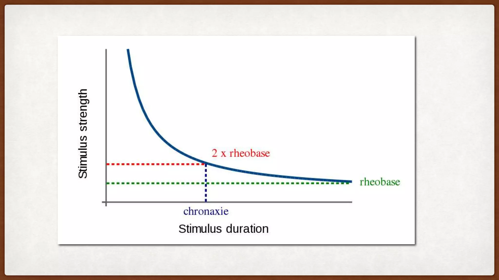
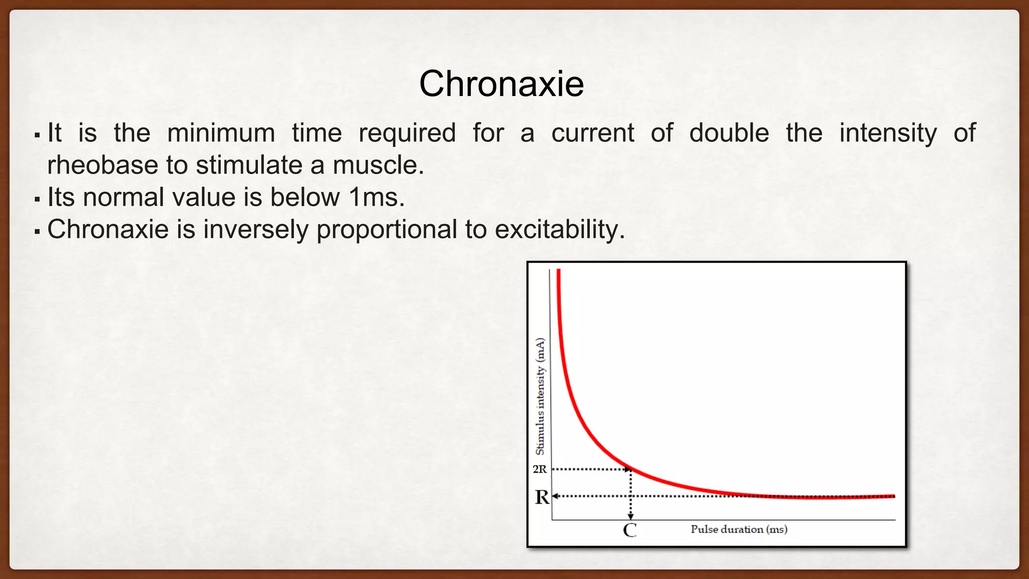
STRENGTH-DURATION
CURVE
(SD CURVE)
DR. PETETI SAIRAM,
MPT(NEURO)
INTRODUCTION
• The strength-duration curve is a graphical representation of the
relationship between the intensity of an electrical stimulus at the
motor point of a muscle and the length of time taken to elicit a
minimal contraction in that muscle.
• strength refers to the stimulus intensity on the vertical axis
• duration refers to the pulse duration on the horizontal axis.
▪ After 21st/22nd day, regeneration of nerve will start, generally
it take about 270 days to regenerate.
▪ SDC test can be done 10 – 14 days after the lesion has
occurred.
▪ The degeneration of nerve from the proximal to distal is called
Wallerian degeneration.
▪ SDC is used to identify denervation, partial innervation, and
compression.
▪ There are also other method for this purpose like EMG and
NCV.
Apparatus/ Material required:
▪ The apparatus used for plotting S-D Curve supplies rectangular impulses
of different duration.
▪ Impulse with duration of 0.01, 0.03, 0.1, 0.3, 10, 30, 100, 300 ms are
required.
▪ The stimulator may be of either the constant current or constant voltage
type.
▪ Moist saline pad.
▪ Electrodes.
▪ Leads.
▪ Bandages/ Micropore.
Methods of plotting SD Curve
▪ The patient must be warm, fully supported and in sufficient light.
▪ The Inactive electrode may be applied over some convenient area usually on the midline
of the body or the origin of the muscle group.
▪ Active electrode placed over motor point.
▪ Current is applied using the longest stimulation first and increased until a minimal
contraction is obtained.
▪ Intensity of current is noted and impulse is shortened. This procedure is repeated with
each stimulation in turn, the intensity of current being increased as required.
▪ A minimal contraction is used, as this make it easy to detect any change in strength, and
electrode should be placed in same point over the muscle throughout the test.
▪ The S-D Curve is plotted from the result of the test, and although it will be further to the
left with constant voltage than with the constant current stimulator.
Characteristics of SD Curve
INNERVATED
▪ This is also called a "nerve curve".
▪ All nerve fibers supplying the muscle are intact. The shape of the curve is a
continuous rectangular hyperbola.
▪ The same intensity is required to produce a response at longer durations.
The intensity increases steadily for shorter durations.
▪ The curve is usually seen rising at the 1ms mark.
Complete Denervation
• This is also called a "muscle curve".
• All nerve fibers supplying the muscle have degenerated.
• The curve is characteristically steep and shifted to the right.
• The intensity keeps increasing when lowering the duration below
100ms.
• Response not seen at very short durations.
Partial Denervation
• Some of the nerve fibers supplying the muscles have degenerated
while others are intact.
• A characteristic kink is present in the curve.
• The right side of the curve represents the denervated part of the
muscle while the left side represents the innervated fibers of the
muscle.
Rheobase
•It is the minimum intensity of current required to stimulate a muscle at infinite
duration.
•Its normal value ranges between 2 and 18 mA.
•The rheobase is greater for denervated muscles
•Mainly 100 to 300 ms duration are used to record rheobase.
•The pulse is always rectangular measured in miliamperes or volts.
•Normal values of rheobase are 2 to 18 mA or 5 to 35 volts.
•Normal value of rheobase of different muscle:
•Deltoid-14 volts,5mA.
•Triceps-18 volts,5mA.
•Abductor digiti minimi-30volts,8mA.
•Frontalis-14volts,4mA.
▪ Resistance of skin and subcutaneous tissue.
▪ Edema and inflamation.
▪ Ischemia and underlying pain.
▪ Temperature variation.
▪ Position of electrode.
▪ Amount of subcutaneous tissue.
▪ Degeneration.
▪ Deneravtion.
▪ Partial denervation generally produce no changes in rheobase.
Factors responsible for Rehobase
Chronaxie
▪ It is the minimum time required for a current of double the intensity of
rheobase to stimulate a muscle.
▪ Its normal value is below 1ms.
▪ Chronaxie is inversely proportional to excitability.
Factors responsible for Chronaxie
▪ Texture of skin.
▪ Ischemia.
▪ Edema.
▪ Fatigue.
▪ Position of stimulating electrode.
▪ Denervation.
▪ Partial denervation.
▪ Re-inervation.
▪ Nerve root lesion.
▪ Peripheral neuropathy.
▪ Myopathy (No significant change).
Advantages of SD Curve
▪ It is simple, reliable and cheaper.
▪ It is quick and easy to perform.
▪ Indicate proportion of denervation.
▪ Less time consuming.
▪ It requires minimal training.
Disadvantages of SD Curve
▪ In large muscles, only proportion of fibers may respond hence
picture is not clearly shown.
▪ It won’t point out the site of lesion
▪ It only provides qualitative data in relation to the degree of
denervation.
SD curve (Strength Duration Curve)

SD curve (Strength Duration Curve)

  • 1.
  • 2.
    INTRODUCTION • The strength-durationcurve is a graphical representation of the relationship between the intensity of an electrical stimulus at the motor point of a muscle and the length of time taken to elicit a minimal contraction in that muscle. • strength refers to the stimulus intensity on the vertical axis • duration refers to the pulse duration on the horizontal axis.
  • 3.
    ▪ After 21st/22ndday, regeneration of nerve will start, generally it take about 270 days to regenerate. ▪ SDC test can be done 10 – 14 days after the lesion has occurred. ▪ The degeneration of nerve from the proximal to distal is called Wallerian degeneration. ▪ SDC is used to identify denervation, partial innervation, and compression. ▪ There are also other method for this purpose like EMG and NCV.
  • 4.
    Apparatus/ Material required: ▪The apparatus used for plotting S-D Curve supplies rectangular impulses of different duration. ▪ Impulse with duration of 0.01, 0.03, 0.1, 0.3, 10, 30, 100, 300 ms are required. ▪ The stimulator may be of either the constant current or constant voltage type. ▪ Moist saline pad. ▪ Electrodes. ▪ Leads. ▪ Bandages/ Micropore.
  • 5.
    Methods of plottingSD Curve ▪ The patient must be warm, fully supported and in sufficient light. ▪ The Inactive electrode may be applied over some convenient area usually on the midline of the body or the origin of the muscle group. ▪ Active electrode placed over motor point. ▪ Current is applied using the longest stimulation first and increased until a minimal contraction is obtained. ▪ Intensity of current is noted and impulse is shortened. This procedure is repeated with each stimulation in turn, the intensity of current being increased as required. ▪ A minimal contraction is used, as this make it easy to detect any change in strength, and electrode should be placed in same point over the muscle throughout the test. ▪ The S-D Curve is plotted from the result of the test, and although it will be further to the left with constant voltage than with the constant current stimulator.
  • 6.
    Characteristics of SDCurve INNERVATED
  • 7.
    ▪ This isalso called a "nerve curve". ▪ All nerve fibers supplying the muscle are intact. The shape of the curve is a continuous rectangular hyperbola. ▪ The same intensity is required to produce a response at longer durations. The intensity increases steadily for shorter durations. ▪ The curve is usually seen rising at the 1ms mark.
  • 8.
  • 9.
    • This isalso called a "muscle curve". • All nerve fibers supplying the muscle have degenerated. • The curve is characteristically steep and shifted to the right. • The intensity keeps increasing when lowering the duration below 100ms. • Response not seen at very short durations.
  • 10.
  • 11.
    • Some ofthe nerve fibers supplying the muscles have degenerated while others are intact. • A characteristic kink is present in the curve. • The right side of the curve represents the denervated part of the muscle while the left side represents the innervated fibers of the muscle.
  • 12.
    Rheobase •It is theminimum intensity of current required to stimulate a muscle at infinite duration. •Its normal value ranges between 2 and 18 mA. •The rheobase is greater for denervated muscles •Mainly 100 to 300 ms duration are used to record rheobase. •The pulse is always rectangular measured in miliamperes or volts. •Normal values of rheobase are 2 to 18 mA or 5 to 35 volts. •Normal value of rheobase of different muscle: •Deltoid-14 volts,5mA. •Triceps-18 volts,5mA. •Abductor digiti minimi-30volts,8mA. •Frontalis-14volts,4mA.
  • 14.
    ▪ Resistance ofskin and subcutaneous tissue. ▪ Edema and inflamation. ▪ Ischemia and underlying pain. ▪ Temperature variation. ▪ Position of electrode. ▪ Amount of subcutaneous tissue. ▪ Degeneration. ▪ Deneravtion. ▪ Partial denervation generally produce no changes in rheobase. Factors responsible for Rehobase
  • 15.
    Chronaxie ▪ It isthe minimum time required for a current of double the intensity of rheobase to stimulate a muscle. ▪ Its normal value is below 1ms. ▪ Chronaxie is inversely proportional to excitability.
  • 16.
    Factors responsible forChronaxie ▪ Texture of skin. ▪ Ischemia. ▪ Edema. ▪ Fatigue. ▪ Position of stimulating electrode. ▪ Denervation. ▪ Partial denervation. ▪ Re-inervation. ▪ Nerve root lesion. ▪ Peripheral neuropathy. ▪ Myopathy (No significant change).
  • 17.
    Advantages of SDCurve ▪ It is simple, reliable and cheaper. ▪ It is quick and easy to perform. ▪ Indicate proportion of denervation. ▪ Less time consuming. ▪ It requires minimal training.
  • 18.
    Disadvantages of SDCurve ▪ In large muscles, only proportion of fibers may respond hence picture is not clearly shown. ▪ It won’t point out the site of lesion ▪ It only provides qualitative data in relation to the degree of denervation.