2. Introduction
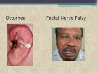
• Malignant otitis externa is an aggressive and
potentially life-threatening infection of the
softtissues of the external ear and surrounding
structures, quickly spreading to involve the
periosteum and bone of the skull base.
• Synonyms :
▫ Necrotizing Otitis Externa
▫ Skull Base osteomyelitis
3. • Malignant otitis externa is a misnomer as it is
not a neoplastic process.
• In 1968, Chandler described this otitis externa as
malignant because he observed an aggressive
clinical behavior, poor treatment outcome, and a
high mortality rate for the patients affected by
this disease.
4. Microbiology:
• Bacterial :
▫ Pseudomonas aeruginosa (95%)
▫ Staphylococcus aureus , S epidermidis
• Fungus
▫ Aspergillus Fumigatus, A. Flavus, A. Niger
5. Predisposing Factors
• Uncontrolled diabetes above 55 years of age.
• Immunocompromised - Acquired Immune
Deficiency Syndrome (AIDS) or malignancy and
are on immunosuppressive drugs like
azathioprine, methotrexate, cyclophosphamide
and cyclosporine.
• Malnutrition
• Anemia(Children)
• Organ Transplant Recipent
7. CLINICAL FEATURES
SYMPTOMS
• Pain-severe excruciating pain in external and
deep meatus (gets worse when moving head)
• Discharge:
Initial stage =Mucopurulent
Late stage=Purulent and blood stained
• Deafness-sensorineuronal hearing loss
• Facial nerve palsy-lower motor neuron
type(loss of taste sensation in anterior 2/3 of
the tongue on the affected side ,weakness in
facial muscle)
12. • External Auditory Canal(EAC) is narrow and
discharge present
• Sagging of the posterior meatal wall
• Tympanic membrane is normal in early cases
• May show bulge,congestion and granulation if
middle ear is involved
• Signs of multiple cranial nerve palsies in late
cases
13. STAGING AND CLASSIFICATION
• Stage 1:Clinical evidence of malignant otitis
externa with infection of soft tissue beyond the
external auditory canal,but negative Tc-99 bone
scan
• Stage 2:soft tissue infection beyond EAC with
positive Tc-99 bone scan
• Stage 3:As above,but with cranial nerve paralysis
3a -single
3b-multiple
• Stage 4:Meningitis,empyema,sinus thrombosis or
brain abscess
14. Diagnosis of malignant Otitis Externa
• There is no single pathognomonic criterion that
defines malignant otitis externa
• History: Age, Diabetic, may also give history of
trauma to ear by irrigation or cleaning
• Complete head and neck examination: Signs of
otitis externa with or without cranial nerve palsy
15. Investigations
• Ear swab for Culture & Sensitivity
▫ Positive for Pseudomonas
• Blood examination: Complete blood count
• ESR, CRP are raised
• Fasting Blood Sugar & HbA1C for the status of
the blood glucose level.
• RFT & LFT are not for diagnosis but are
important because the patient has to be treated
with antibiotics for longer period.
• Tissue biopsy to rule out malignancy.
16. Radiological Invstigations
▫ CT Scan/MRI with galium contrast will show
evidence of temporal bone involvement and extent
of the ear.
▫ TC 99 isotope bone scan – demonstrate increased
uptake in the region of skull base in active case.
17. Management
• Medical management:
▫ Glucose control
▫ Aural toilet
▫ Systemic anti-Pseudomonas antibiotics (treatment
of choice )
▫ Hyperbaric Oxygen Therapy
18. Medical Management
• Fluoroquinolones are active against P.
aeruginosa.
• For at least 6 to 8 weeks- oral and
intravenous ciprofloxacin
• Ceftazidime provide an alternative to
ciprofloxacin with or without Aminoglycoside
• Amphotericin B is the most commonly used
antifungal agent for fungal
19. Hyperbaric Oxygen (HBO):
• HBO increases the partial pressure of oxygen,
improving hypoxia and allowing greater
oxidative killing of bacteria.
• Used only as an adjunct to antimicrobial therapy
20. Surgical Management
• Local debridement of necrotic tissues and bone.
• Wide surgical excision of infected tissue and
bone may be required sometime.
21. Cranial Nerve Complications
• Cranial nerves can be affected by inflammation
along the skull base or by a neurotoxin produced
by Pseudomonas species.
• The facial nerve (VII) is affected most commonly,
usually at the stylomastoid foramen.
• As the disease progresses, cranial nerves IX, X,
and XI can be affected at the jugular foramen,
followed by XII at the hypoglossal canal.
• Cranial nerves V and VI can be affected if the
disease extends to the petrous apex.
22. Intracranial Complications
• Meningitis, brain abscess, and dural sinus
thrombosis.
• Sigmoid Sinus Thrombosis- if cranial
neuropathies related to jugular foramen is
present.
• Cavernous Sinus Thrombosis- if Cranial nerve V
& VI are involved.
Santorini's fissures: Vertical fissures in the anterior part of the cartilage of the external acoustic meatus (ear canal).
Tc 99= accumulates at sites of osteoblastic activity making it highly sensitivity for bony infection.
Gallium Scal 67ga= more specific than bone scanning since radioisotope is incorporated into granulocytes and bacteria.
used to follow response to treatment since uptake value return to normal with resolution of infection.
MOE contin. Deep boring extrosis pain.
Other ma throbing pain
Not limited to soft tissue > periosteum > bone so pain is deep boring pain as in osteomyelitis
Tregal tenderness +/-
Tympanic membrane normal
Granulation tissue +nt at junction of