
Recommended
Recommended
More Related Content
What's hot
What's hot (20)
Similar to 2003 pharmaceutical microbiology lab manual
Similar to 2003 pharmaceutical microbiology lab manual (20)
More from University of Zambia, School of Pharmacy, Lusaka, Zambia
More from University of Zambia, School of Pharmacy, Lusaka, Zambia (20)
Recently uploaded
Recently uploaded (20)
2003 pharmaceutical microbiology lab manual
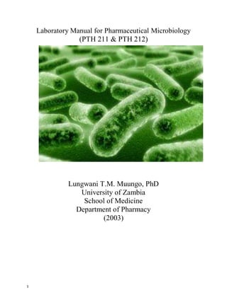
- 1. 1 Laboratory Manual for Pharmaceutical Microbiology (PTH 211 & PTH 212) Lungwani T.M. Muungo, PhD University of Zambia School of Medicine Department of Pharmacy (2003)
- 2. 2 Lab Manual Overall Objectives The objectives for each exercise of this Microbiology laboratory manual are simple: You are expected to know and understand all of the material for each exercise as well as any lab procedures used. In addition, you must be able to identify the different types of organisms covered in this manual (plus the characteristics of each organism) when viewed under the microscope or if a PowerPoint presentation, on the computer’s screen.. The development of good, safe microbiology techniques are essential and expected in this course. You are responsible for understanding and recognizing, therefore, all material and microbes covered in this manual. You are also responsible for correctly answering all exercise quiz questions that are found towards the end of each exercise before the next week’s exercise. All students are expected to have thoroughly studied the lab exercise before attending the lab that covers that particular exercise’s material. Your Name_________________________________________
- 3. 3 General Laboratory Safety: General Lab Safety 1. Do not eat, drink, store food or drink, bring food or drink or apply cosmetics while in the lab. (Water in bottles included.) 2. Always wear shoes at all times. 3. Never place any personal items on the floor. 4. Never place any personal items on the countertops. 5. If you have long hair, tie it back. 6. If you wear long sleeves, roll them back up your arm. 7. Disinfect your work area before lab starts and at lab’s end. 8. Disinfect your work area if you make a spill. 9. Wash your hands before and at the end of the lab period. 10. Wash your hands if they become contaminated with microbes. 11. Wash your hands before you leave the room for whatever reason. 12. Wash your hands when you remove your gloves. 13. For whatever reason, always scrub your hands for a minimum of 24 seconds. 14. Keep hands, pencils, pens etc. out of your mouth. 15. Keep your work area uncluttered. 16. Do not insert contact lenses in the lab. 17. Wear eye protection when heating chemicals of if you wear contact lenses. 18. Turn off Bunsen burners when not in use. They are a hazard and also a source of heat in the room. 19. No horseplay ever in any lab anywhere. 20. Do not experiment on your own. 21. Follow lab manual directions carefully and correctly. 22. Never pipette by mouth. 23. Call your instructor if there is microbial spillage. 24. Don’t touch broken glassware. Use a broom and dust pan to remove.
- 4. 4 25. Wear disposable gloves while staining or handling microbes. Student Conduct: 1. Again, do not eat, drink, store food or drink, bring food or drink or apply cosmetics while in the lab. (Water in bottles included.) 2. Come prepared for the day’s exercise. Study the exercise before you come to lab. 3. Don’t remove anything from the lab. 4. It is not permitted to apply cosmetics or insert contact lenses in lab. 5. Do not talk while the instructor is talking to the lab class. 6. Always bring your lab manual to lab. 7. Wash hands thoroughly after handling microbes and anytime you leave the lab. 8. At all times, practice safety—be mentally alert at all times!!! 9. When the instructor is talking, pay attention--you may learn something.
- 5. 5 Fire Safety: 1. Learn all exits from the lab the first day of lab. 2. Know the locations of Bunsen burners at all times. 3. Turn off Bunsen burners when not in use. 4. Tie long hair back; roll up long sleeves. 5. Find the fire extinguisher and fire blanket. 6. In case of fire, calmly leave the lab via exit doors. 7. In the case of fire in the laboratory, there are four exit doors through which you can exit the lab. Locate, during your first lab meeting, these four doors. All of these doors are located in the front of the lab (left and right sides). The other two doors up front lead into the chemistry lab and can be used for exit. 8. After leaving the building calmly, go immediately to the formation area described by your instructor on the first day of lab. Do not leave this area. 9. Your instructor will handle the fire emergency. 10.Locate all fire alarm boxes in the lab and the hallway during the first lab period and know their locations. 11.Advise the instructor immediately if a fire starts in lab. 12.Always be extra careful when using Bunsen burners.
- 6. 6 Contaminated Materials Disposal: 1. Place all plate cultures in an autoclavable bag for disposal when finished with them. 2. Place all microscope slides in the container marked “slides”. They will be immersed in a disinfectant solution in this container. 3. Remove all labels from plates or tubes before disposal. 4. Place all gloves in their respective autoclavable bag.
- 7. 7 Table of Contents Lab Name Page 1. The Microscope…………………………………………………………………………………………………… 6 2. Streak Plate Method…………………………………………………………………………………………… 15 3. Helminthes…………………………………………………………………………………………………………… 27 4. Simple Staining…………………………………………………………………………………………………… 56 5. Gram Staining…………………………………………………………………………………………………… 65 6. Winogradsky Columns………………………………………………………………………………………… 78 7. Wine Making………………………………………………………………………………………………………… 82 8. Soil Productivity…………………………………………………………………………………………………… 86 9. Serial Dilutions……………………………………………………………………………………………………… 90 10. Swabs: Environmental, throat, body, rectal………………………………………………………… 95 11. Bacterial DNA Extraction………………………………………………………………………………………… 101 12. Bacterial Transformation……………………………………………………………………………………… 110 13. Gel Electrophoresis……………………………………………………………………………………………… 115
- 9. 9 Materials 1. Microscope PowerPoint presentation on lab computer and on WebCT. 2. Binocular microscope. 3. The following prepared microscope slides. a. Computer Chip b. Human Helminth Eggs c. Drosophila Chromosome d. Cyanobacteria Water Bloom Objectives 1. Learn the location and function of the listed microscope’s parts. 2. Learn how to place a specimen onto a microscope and then properly use the microscope for specimen examination. 3. Learn how to properly use the different objective lenses to bring a specimen into perfect focus. 4. Learn how to properly clean the oil immersion lens. 5. Learn the specified metric measurements. 6. Be able to define resolution and N.A. . 7. Be able to compute the total magnification of a specimen. 8. To always follow proper hand washing and countertop disinfection procedures. 9. Learn all terms in bold print. INTRODUCTION The Microscope . You are expected to access WebCT and learn the Lab Exercise 1 material before you attend the first lab (using the microscope). The online exercise presentation has sound. Be sure to adjust your computer’s speakers using the second slide in the online exercise. The first slide has no sound but advises you to turn on your computer’s speakers. It is required that you go over this online material several times to ensure that you are well familiar with the microscope and its parts as well as any measurements and defined terms. You will be tested on these during the first lab quiz as well as the lab mid-term. The online material is also found on the hard drive of the lab’s computers but with no sound. Your lab microscope is known as a brightfield compound microscope since it contains more than one lens (thus compound and it has three separate lens systems) and the illumination source for your specimen is a light bulb in the base of the microscope. The lab’s microscopes are binocular which means there is not one but two eyepieces (or ocular lenses) through which you can view the specimen. The magnification of each ocular lens is 10X. The capital “X” after the number always indicates, in science, the magnifying power of a lens. Within one of the ocular lenses (or eyepieces) is a “pointer” that allows one to “point” to a particular area or object on a slide’s specimen that they are viewing. Then someone else can look into the eyepieces and see what they were referring to. One can adjust the distance between eyepieces (their intraocular distance) by gently pulling or pushing on them both (at the same time). At least one eyepiece has
- 10. 10 INTRODUCTION CONTINUED: a focus adjustment dial, the other may or may not. If not both, then you should first bring your specimen into focus using the eyepiece that does not have a focus dial. Then, you would next adjust the eyepiece that has the adjustment dial (thus bringing the specimen into perfect focus with it). If both have a dial, then choose one to initially focus the specimen; then adjust the other. A second group of lenses on your binocular microscope is the objective lenses. They are found on the rotating nosepiece and can be moved around in a circle. When moving objective lenses, one can tell when they are in their proper position for viewing a specimen because one can hear or feel them click into place. Try it a few times to make sure you understand and can do this. Each objective lens has its own name and magnification. In their order from the smallest in magnification and proceeding counterclockwise on the nosepiece to the highest in magnification would be the scanning lens (also called the low power lens), the medium power lens, the high dry (hi dry) lens or high power lens and then the most powerful one called the oil immersion lens. The oil immersion lens is the only lens you immerse in oil. Immersing the high dry lens in oil can and will ruin it. PLEASE DON’T DO THAT! PAY ATTENTION! The following are the magnifications for each of the objective lenses: Low Power lens - 4X Medium Power lens – 10X High Power lens - 40X Oil Immmersion lens – 100X How does one determine the total magnification of the specimen they are viewing? After all, one is looking through the eyepieces as well as one of the objective lenses. The circular area you see with your eyes when looking through the eyepieces is called your “microscopic field” or just your “field”. We call your microscope a “brightfield” microscope since the bulb makes your field bright and the specimen dark. The following formula allows you to determine the total magnification of the specimen you are viewing in your field: Total magnification = ocular lens magnification x objective lens magnification As an example, let’s say you’re looking at a specimen using the high dry lens. What is the total magnification of your specimen once in focus with this objective lens? Substituting in the formula: Total magnification = 10X x 40X = 400X Your specimen has been magnified 400 times. Be sure you can figure the total magnification of a specimen when using any of the four objective lenses.
- 11. 11 INTRODUCTION CONTINUED: Where is the third lens system in your microscope? It is in the condenser. The condenser lies beneath the stage. The condenser grabs as many light rays as possible coming from you illumination source (light bulb in our microscopes) and directs then straight upwards (in the form of a cone) into the specimen on your slide. Light rays coming from an illumination source such as a light bulb try to wander off in another direction besides “straight”. However, the condenser grabs as many of these wayward light rays as possible and directs them straight upwards in the form of a cone thus trying to assure that the specimen is uniformly illuminated. Once you have focused on a specimen, play with the condenser and see how it can “contrast” the specimen you’re looking at. It is an important structure on the microscope. I expect you to know what it does and how to use it properly. Before you start for the day, make sure the condenser is lowered slightly downwards from the stage (just a little bit) before you start the day’s exercise. Inside the condenser is an iris diaphragm. By moving the iris diaphragm lever on the outside of the condenser, one can affect the diameter and angle of the cone of light hitting the specimen and hence the illumination of the specimen. Be sure to move the iris diaphragm lever back and forth slowly to see what it does. When starting for the day, make sure the iris diaphragm is full open. What is the “resolution” of a microscope or its “resolving power”? One way to look at it is to say that it is the measurement of how far two points must be in order for the microscope to tell that they are separate points. The best compound microscopes have a resolution of down to about 0.2 micrometers. In other words, at the highest magnification you can get, you cannot see objects less than 0.2 microns apart. What is the “N. A.” or “Numerical aperture” of an objective lens? It is a measurement of a lens’s ability to capture light coming from the specimen. The higher the N. A. number, the greater the ability of that lens to capture light. Thus, if you use a lens with a higher N. A. than the preceding lens, you must add more light from your illumination source in order to properly illuminate your specimen. If you use a lens with a lower N. A. than the previous lens, then you need to lower your rheostat since you don’t need as much light in this new lens (as you did in the previous one). a. Which objective lens on your microscope has the least ability to capture light? b. Which objective lens has the greatest ability to capture light?
- 12. 12 INTRODUCTION CONTINUED: Measurements The metric system of measurements is important thorough out all of science. An ant is about 5 millimeters (5 mm) long. A dust mite is somewhere near 200 micrometers (200 microns or 200 m) long. The head of a pin is about 1 to 2 millimeters (1 – 2 mm) in diameter. An erythrocyte is approximately 8.5 to 11.5 microns (micrometers or m) in diameter. A leukocyte is about 10 m (micrometers) in diameter. Bacteria vary in length, depending on who they are, from 0.2 to 100 microns (micrometers) in length. A typical human hair is about 60,000 nanometers (60,000 nm) wide. DNA is about 2.5 nanometers (2.5 nm) wide. An atom of hydrogen is only 0.1 nanometers ( 1 angstrom ) in diameter. Did you know that there are nanobacteria? It is essential that you understand and can use these measurements in science, especially mm, nm and m. One meter is approximately 39.36 inches in length if you refer to the English measurement system. 1 centimeter = 1/100 meter (abbreviated: 1 cm) 1 millimeter = 1/1000 meter (abbreviated: 1 mm) 1 micrometer (1 micron or 1 m) = 1/1,000,000 meter 1 nanometer = 1/1,000,000,000 meter (abbreviated: 1 nm) 1 angstrom = 1/10 nanometer Measurement Practice Problems: 1. 1 meter = ? cm 2. 1 meter = ? mm 3. 1 meter = ? m 4. 1 meter = ? nm 5. ? micrometers ( ? m) = 1 mm 6. 20 mm = ? micrometers ( m) 7. 100 micrometers ( 100 m) = ? mm 8. 10 micrometers ( 10 m) = ? mm 9. 1 micrometer (1 m) = ? nm 10. 100 nm = ? micrometers (? m) 11. Which is the larger number? 1/1,000,000 or 1/1,000,000,000 12. 4 nm = ? angstroms 13. .5 nm = ? angstroms 14. 100 angstroms = ? nm 18. 1 micron = ? angstroms 15. 3 angstroms = ? nm 19. 1 nm = ? angstroms 16. 400 mm = ? cm 20. ? nm = 2 micrometer (2 m) 17. 3.5 cm = ? mm 21. ? angstroms = 1 micrometer
- 13. 13 Using the Microscope It is essential that you understand and use the microscope properly in lab. For an assignment, you are to study and learn the microscope’s WebCT presentation before your first lab meeting. Click on the following item in WebCT’s NAV bar or else the icon in “Course Home” ----- “Lab Exercise #1 Material”. Here, I will lecture to you online about the microscope as well as other exercise #1 items during this presentation. You will be responsible for learning and understanding all of the following items including their location on the microscope as well as their function or meaning (as appropriate): Ocular lens or eyepiece Base Magnification of each eyepiece Rheostat Interpupillary distance On/Off switch Eyepiece focus adjustment Illumination source Pointer What the “X” after a number means Arm Revolving nosepiece Objective lenses and their appropriate names The magnification of each objective lens Why the 40X lens can’t be placed in oil N. A. or numerical aperture N. A. of each objective lens The resolution (the limit of resolution) of a microscope Stage Stage Aperture Slide Holder Mechanical stage Mechanical stage control (adjustment) knobs Condenser Condenser control knob Iris diaphragm lever Iris diaphragm
- 14. 14 Setting Up and focusing on the specimen: Get Out the Microchip Slide. Use these steps as a guide: 1. Make sure the microscope is plugged firmly into an electrical outlet. 2. Make sure the microscope’s rheostat is on zero (0). 3. Make sure the condenser is lowered a little way from the stage. 4. Make sure the iris diaphragm is full open (use its lever). 5. Using the on/off switch, turn the microscope on. 6. Lower your stage all full down using coarse adjustment knob. 7. Firmly grasp your slide (labeled microchip) and place it gently into the slide holder. Please do not drop the slides------they cost money and are hard to replace. Center this slide over your stage aperture. 8. Ascertain that the medium power objective lens is properly positioned for use on the revolving nosepiece. 9. Look through your ocular lenses with both eyes. Make any adjustments to your interpupillary distance now. You are now looking at your microscopic field. Notice the pointer. 10. Note which eyepiece you cannot adjust with a focus dial (if there is only one). Use it first to bring your specimen into focus. Then adjust the other. 11. Using the coarse focus knob and while looking through your ocular lenses, slowly bring the specimen slide (stage) towards you or upwards. You cannot break the slide with this objective lens. It is true that some others state that for the initial focusing of your specimen, once all is ready, bring the stage all the way upwards and then slowly lower the stage away from you (while looking in both eyepieces). It doesn’t matter to me which way you do it. Just get the specimen in focus with medium power. 12. Once you have the specimen in focus, next, using the fine focus knob, “fine tune” your specimen’s focus. 13. From here on, when you go to higher magnifications, only use the fine focus knob. 14. Go to high power next and bring the chip into focus. 15. We will not use oil immersion today. 16. Next, your instructor will “walk” you through the other specimen slides. Please do not go ahead of the instructor.
- 15. 15 When finished for the day 1. First, make sure to turn your microscope’s rheostat all the way to zero (0). 2. If you have used oil immersion, thoroughly clean the oil immersion lens with lens paper and xylene. 3. Wrap the microscope’s electrical cord gently around the arm. 4. Carefully and properly, carry the microscope and return it to its storage area. 5. If we have used any live microorganisms in the lab, disinfect your countertop work area. Then wash your hands a minimum of 24 seconds. 6. Place all paper towels etc. into the waste can. Leave your work area clean. 7. Place all prepared slides back into their storage box. Do not break them! 8. Wash your hands a minimum of 24 seconds again before leaving the lab. Exercise Questions: 1. List 5 things that could affect the illumination of your specimen. 2. Where would you find the “pointer”? What does it allow you to do? 3. Which ocular lens should one use when first focusing on a specimen in our lab? 4. How do you properly carry a microscope? 5. What is the magnification of your ocular lens or eyepiece in lab? 6. What is the magnification of your high dry lens? What type of lens is it? 7. What is the magnification of your oil immersion lens? Your medium power lens? 8. Which lens does not require as much light going through it (in order to properly illuminate the specimen) when compared to all of the others? Which one requires the most light going through it in order to see the specimen? 9. What is the purpose of the revolving nosepiece? 10. Why do they call your microscope a brightfield, compound microscope? 11. Millie is looking at her slide specimen. It is magnified 400X. Which objective lens is she using? 12. Alamander is using the oil immersion lens. What will be the total magnification of his specimen? 13. On your microscope, what part is used to give you greater or lesser contrast of your specimen? 14. Where should your rheostat setting be if you’re using the oil immersion lens? 15. Where should the rheostat setting be if using the medium power lens? 16. Using mathematics (show your work here), would our microscopes resolve a specimen that is 1000 nm wide? 17. True or False: You can use the coarse focus knob on high power or oil immersion.
- 16. 16 Exercise Measurement Questions 1. This is a virus. Notice its length and width given in “ m’: .8 m x 0.015 m What would these dimensions be in nanometers? 2. This is a protein molecule called hemoglobin. Its length is: length = 15 nm What would be its length in m? 3. This is a bacterial cell. Its shape is called a “bacillus”. Its length and width are: 2.0 nm x 10 nm What would its length and width be in m? 4. This is a type of leukocyte called a neutrophil. Neutrophils average 10 – 14 m in diameter. How would you write this average diameter range in nm?
- 17. 17 Exercise No. 2 STREAK PLATE PROTOCOL History The modern streak plate procedure has evolved from attempts by Robert Koch and other early microbiologists to obtain pure bacterial cultures in order to study them, as detailed in an 1881 paper authored by Koch (5). Slices of sterilized potatoes became the first solid media employed on which to grow bacteria. This process was a procedure that worked only for a few organisms and only until the bacteria decomposed the potato surface. A search for other materials led to experimentation with the suitability of gelatin and agar-agar as solidifying agents. Gelatin was difficult to prepare and difficult to use at room temperature, let alone at the higher temperature of an incubator, and many bacteria digest the protein. Agar, because of its characteristics of melting only when boiled, rarely being digested by bacteria, and providing a substance in which other nutrients could be dissolved, proved to be a suitable material on which to grow bacteria. Agar was originally called agar-agar and is derived from seaweed. The agar that we use today is the same substance as agar-agar, but it has been processed by the manufacturer. Agar, as purchased 100 years ago, required filtering before it was clear enough to use in media (12). In the early eras of microbiology, making media was an extensive process of preparing the extracts of meat or other nutrient sources, as well as purifying and filtering the gelatin or agar. Before the invention of the autoclave, sterilizing the media properly was also time consuming. The 1939 edition of An introduction to Laboratory Technique in Bacteriology, an early microbiology lab manual, contains extensive instruction for students to prepare their own media from "scratch" (7) for use in the lab. Before R. J. Petri invented the petri dish, flat plates of glass covered by glass lids were most commonly used to culture organisms in gelatin. Even after agar was adopted and solid media were available, the streak plate was not commonly used. Historically, microbiologists most frequently used pour plates to isolate organisms for pure cultures. A pure culture was made from an isolated colony, represented only one species or strain, and traditionally arose through the growth of a single cell. Colonies are considered isolated if they are not touching any other colony. Isolated colonies were identified and transferred by streaking onto a new agar or gelatin plate using a sterile needle, a process called "picking colonies." More rarely, a researcher would try to isolate organisms directly on the surface of a gelatin or agar plate. A typical description of the streaking process was given by Huber Williams, revised by Meade Bolton in A Manual of Bacteriology published in 1908 (11). "...the isolation of bacteria may sometimes be effected by drawing a platinum wire containing material to be examined rapidly over the surface of a petri dish containing solid gelatin or agar; or over the surface of the slanted culture medium in a test tube; or by drawing it over the surface of the medium in one test tube, then without sterilizing, over the surface of another, perhaps over several in succession." Bacteriology textbooks and lab manuals from the early and mid 20th century did not mention the streak plate nor did they have our typical "isolation streak" exercise. For instance, isolation by streaking is absent from Buchanan and Buchanan, 1938 (2) and from Sherwood, Billings and Clawson's manual published in 1952 (10). During a literature search to pinpoint the first appearance of our modern streak plate, several papers published in the 1940s were found to mention streak plates. However, these did not describe the process or illustrate the results, and from the context, most probably referred to the process of picking colonies and creating a pure culture in fresh media. An early version of our modern isolation streak is found in Levine's An Introduction to Laboratory Technique in Bacteriology published in 1939 (7) and a similar version from 1954, in Salle's Laboratory Manual on Fundamental Principles of Bacteriology, 4th ed. (9). In that process, the student picked up organisms on a needle or loop and then either stabbed into the agar
- 18. 18 or spread the loopful of the culture at the upper end of the petri dish to thin it out. Then a series of strokes 1/4-inch apart was made over the rest of the plate. Dr. Salle noted that the first streaks would contain too many organisms but that the last streaks should give isolated colonies. He suggested that a second plate be inoculated without flaming the wire loop first, to give a better chance of obtaining isolated colonies. This process dilutes the bacteria as the plate is streaked, similar to the dilution observed in a modern streak plate. FIG. 1. An example of the one-directional streak pattern as described in the lab manuals by Levine and Salle (7, 9). The plate illustrated is a 100-mm petri dish.
- 19. 19 In 1958, in the first edition of Laboratory Exercises in Microbiology, Pelczar and Reid (8) presented a streak plate exercise. It utilized a 4-quadrant streak pattern, and the procedure described using both a loop and a needle in the streak and all streaks were in the same direction, rather than both back and forth. FIG. 2. A drawing representing the streak pattern recommended by Pelczar and Reid (8). All strokes of the loop or needle are done in a single forward direction, rather than in a back-and- forth pattern, as indicated by the arrowhead directions. The initial sector is at the top of the plate, followed clockwise by sectors 2, 3, and 4. The earliest appearance of the three sector streak pattern (called the T streak) commonly used today may be the 1961 photos published in Finegold and Sweeney (4). An illustration detailing how to perform this streak is in the 1968 edition of the Manual of BBL Products and Laboratory Procedures (1). In addition to the T streak, the BBL Manual illustrates two other streak patterns, neither of which is the simple monodirectional streak pattern used earlier in the century. Today, there are two most commonly used streak patterns, a three sector T streak and a four quadrant streak. Microbiology lab manuals since the 1970s have presented an isolation streak exercise. Lab manual editions published between 1970 and 2000 illustrated and described several streak pattern variations. However, today, almost all published microbiology lab manuals illustrate at least the T streak.
- 20. 20 FIG. 3. A three sector T streak of Serratia marcescens grown on trypticase soy agar. This illustrates a streak plate which has many isolated colonies. FIG. 4. This plate illustrates a streak plate which did not achieve isolation, and which would not be considered a good streak plate example. This photograph is by Dr. Min-Ken Liao, Furman University. Purpose The purpose of the streak plate is to obtain isolated colonies from an inoculum by creating areas of increasing dilution on a single plate. Isolated colonies represent a clone of cells, being derived from a single precursor cell. When culture media is inoculated using a single isolated colony, the resulting culture grows from that single clone. Historically, most microbiology research and microbial characterization has been done with pure cultures. Theory One bacterial cell will create a colony as it multiplies. The streak process is intended to create a region where the bacteria are so dilute that when each bacterium touches the surface of the agar, it is far enough away from other cells so that an isolated colony can develop. In this manner, spreading an inoculum with multiple organisms will result in isolation of the different organisms. PROTOCOL Mesophilic bacteria are generally streaked onto media solidified with 1.5% agar or agarose. Gelatin can be used if a high enough concentration of gelatin protein or a low enough incubation temperature is used. Thermophiles and hyperthermophiles can also be streaked onto growth media solidified with agar substitutes, such as Gelrite and guar gum.
- 21. 21 One-hundred-mm-diameter petri dishes are the most commonly used size of plate for streaking. The agar surface of the plate should be dry without visible moisture such as condensation drops. Traditionally, inoculated petri dishes are incubated with the agar side up to prevent condensed moisture from falling onto the agar surface, which would ruin the isolation by allowing bacteria to move across the moist surface creating areas of confluent growth instead. The inoculum for a streak plate could come from any type of source, for example clinical specimen, sedimented urine, environmental swab, broth, or solid culture. The two most common streak patterns are the three sector T streak and the four sector quadrant streak. In a streak plate, dilution is achieved by first spreading the specimen over the agar surface of one sector. If a cotton swab or disposable loop or needle was used to inoculate the first sector, it is now discarded into an appropriate container, while reusable loops, usually with nichrome or platinum wire (24 gauge), are flamed to incinerate any organisms on the loop. When cooled, the sterile loop is streaked through the initial sector and organisms are carried into the second sector where they are spread using a zig-zag movement. In a similar manner, the organisms present on the loop are incinerated after the second sector is streaked, and the third sector is streaked. For a four quadrant plate, the process is carried an extra step. Detailed procedure for a Three Sector Streak, the T Streak: Reference J. Lammert, Techniques in Microbiology, A Student Handbook (6) Materials: Specimen to be streaked; this protocol is written for a test tube culture Transfer loop (usually nichrome, a nickel-chromium alloy, or platinum; it may also be a single-use disposable plastic loop, which would be discarded between sectors rather than resterilized) Bunsen burner Sterile petri dish with appropriate bacterial media, such as trypticase soy agar Labeling pen Sterile cotton swabs (if necessary to remove condensation from the agar surface and from around the inner rim of the petri dish) A. Label a petri dish. Petri dishes are labeled on the bottom rather than on the lid. In order to preserve area to observe the plate after it has incubated, write close to the edge of the bottom of the plate. Labels usually include the organism name, type of agar, date, and the plater's name or initials. Using sterile cotton swabs, remove any visible water on the agar in the plate or around the inner rim of the petri plate. Observe the plate and mentally divide it into three sectors, a "T." The area above the "T" will become the first sector streaked. The plate will then be turned clockwise (if you are right handed) with the agar side up. The second sector will then be at the top for streaking and then the plate is turned again so that the third sector can be streaked. B. Sterilize the transfer loop before obtaining a specimen. In order to streak a specimen from a culture tube, metal transfer loops are first flamed so that the entire wire is red-hot. The incineration and flaming
- 22. 22 process is described below in the Tips section. When flaming, the wire loop is held in the light blue area of a bunsen burner just above the tip of inner flame of the flame until it is red-hot. If a hot incinerator is available, the loop may be sterilized by holding it inside the incinerator for 5 to 7 seconds. Once sterile, the loop is allowed to cool by holding it still. Do not wave it around to cool it or blow on it. When manipulating bacteria, transfer loops are usually held like a pencil. If plastic disposable loops are being utilized, they are removed from the packaging to avoid contamination and after being used, are discarded into an appropriate container. A new loop is recommended for each sector of an isolation streak plate. C. Open the culture and collect a sample of specimen using the sterile loop. Isolation can be obtained from any of a variety of specimens. This protocol describes the use of a mixed broth culture, where the culture contains several different bacterial species or strains. The specimen streaked on a plate could come in a variety of forms, such as solid samples, liquid samples, and cotton or foam swabs. Material containing possibly infectious agents should be handled appropriately in the lab (see biosafety references below), only by students with appropriate levels of skill and expertise. Remove the test tube cap. It is recommended that the cap be kept in your right hand (the hand holding the sterile loop). Curl the little finger of your right hand around the cap to hold it or hold it between the little finger and third finger from the back. See the illustration. Modern test tube caps extend over the top of the test tube, keeping the rim of the test tube sterile while the rim of the cap has not been exposed to the bacteria. The cap can also be placed on the disinfected table, if the test tube is held at an angle so that air contamination does not fall down into the tube and the test tube cap is set with the sterile rim on the table. Insert the loop into the culture tube and remove a loopful of broth. Replace the cap of the test tube and put it back into the test tube rack. D. Streak the plate. Inoculating the agar means that the lid will have to be opened. Minimize the amount of agar and the length of time the agar is exposed to the environment during the streak process. 1) Streak the first sector. Raise the petri dish lid to insert the loop. Touch the loop to the agar area on the opposite side of the dish, the first sector. Bacteria on the loop will be transferred to the agar. Spread the bacteria in the first sector of the petri dish by moving the loop in a back and forth manner across the dish, a zig-zag motion. Make the loop movements close together and cover the entire 21
- 23. 23 first region. The loop should glide over the surface of the agar; take care not to dig into the agar. 2) Between sectors. Remove the loop from the petri dish and obtain a sterile loop before continuing to the second sector. Either incinerate the material on the loop or obtain a sterile loop if using plastic disposable loops. The loop must be cool before streaking can continue. Metal loops can be touched to an uninoculated area of agar to test whether they are adequately cooled. If the loop is cool, there will be no sizzling or hissing and the agar will not be melted to form a brand. If a brand is formed, avoid that area when continuing with the streaking process. 3) Streak the second sector. Open the petri dish and insert the loop. Touch the cooled loop to the first sector once, invisibly drawing a few of the bacteria from the first sector into the second sector. The second sector is streaked less heavily than the first sector, again using a zig-zag motion. 4) Obtain a sterile loop for the third sector (see 2, above). 5) Streak the third sector. Open the petri dish and insert the loop. Touch the cooled loop (if the loop has been flamed) once into the second sector and draw bacteria from the second sector into the third sector. Streak the third sector with a zig-zag motion. This last sector has the widest gap between the rows of streaking, placing the bacteria a little further apart than in the previous two sectors. Watch closely to avoid touching the first sector as the streak is completed. 6) Final step. Flame the loop to incinerate any bacteria that are left on the loop. Allow the loop to cool before placing it near anything that is flammable. Invert the petri dish so that the agar side is up and incubate the plates. SAFETY The ASM advocates that students must successfully demonstrate ability to explain and practice safe laboratory techniques. For more information, visit the ASM Curriculum Recommendations: Introductory Course in Microbiology and read the section on laboratory safety. Three additional articles provide important information: Biosafety Levels-What We Need to Know About Them in Teaching Labs by 22
- 24. 24 Christina Thompson (2004) Update of Biosafety Level Designations by Erica Suchman (2004) Safety Recommendations from the Concurrent Sessions on Safety in the Microbiology Teaching Laboratory at the Undergraduate Microbiology Education Conference 2003 by Jackie Laxon (2003) COMMENTS AND TIPS A. Alternate streak patterns and different culture media A variety of alternate streak patterns exist. Some are used for specific inocula, such as a urine specimen. The patterns also differ in the number of sectors as well as in the number of times the loop is sterilized. The four quadrant streak pattern would be recommended for use when large amounts of bacteria are expected in the inoculum. The extra sector will provide additional dilution and increase the probability of isolated colonies on the plate. The four quadrant streak plate is described in a variety of references, e.g., in Cappuccino and Sherman's Microbiology, A Laboratory Manual, 8th ed. (3). Sometimes, cultures will be streaked on enrichment media or various selective and differential media. For instance, a culture which is expected to have a gram-negative pathogen will be streaked on a MacConkey agar plate, which inhibits the growth of gram-positive organisms. B. Incinerating material on transfer loops—flaming Reusable microbiological loops and needles are sterilized by flaming. A Bunsen burner is traditionally used for this process. Most microbiology manuals show the microbiologist positioned with his/her hand above the burner, with the loop placed into the flame. To avoid possible contact with the flame, the microbiologist might consider placing his/her hand below the flame with the loop/needle above the hand in the flame. The flame of the Bunsen burner should be adjusted to blue, with the darker blue cone of cooler air visible in the center of the flame. The loop or needle should be placed into the hotter part of the flame and kept there until it glows red. There is a possible aerosolization hazard if the loop or needle contains liquid or a bacterial clump. These loops and needles should be placed into the heat slowly so that the moisture evaporates rather than sputters. If an incinerator such as a Bacti-Cinerator is used to sterilize the loop, the loop is to remain inside the incinerator for 5 to 7 seconds. When warmed up (which will take 5 minutes), the temperature inside the incinerator is 815°C. 23
- 25. 25 The incinerator will take 5 to 10 minutes to warm up to working temperature. C. Several techniques decontaminate transfer loops between sectors of a streak plate: flame, dig into agar, flame once and rotate loop A variety of methods exist for removing organisms from the loop between sectors. Beginning students are generally taught to sterilize the loop between each sector by incinerating and then cooling the loop. Clinical microbiologists practice a variety of methods. Some flame once after the initial sector and then rotate the loop so that the next sectors can be streaked with an unused side of the loop. Other laboratorians (as clinical microbiologists name themselves) stab the loop several times into the agar to clear the loop between sectors. D. Isolated colony appearances Isolated colonies can be described using the traditional colony descriptions. The Colony Morphology Atlas-Protocol project provides information about bacterial colony appearance and characteristic photographs. The appearance of an organism can vary. For instance, a colony of an organism growing in a crowded sector of the plate will not grow as large as the identical organism growing in isolation. The media composition, pH, and moistness, as well as the length of time and temperature can all affect the organism's appearance. Colonies selected for subculturing should be colonies which are isolated, i.e., there is no other colony visibly touching the colony. Agar with a surface layer of water is not suitable for obtaining isolated colonies. Obvious water drops should be removed from the surface of the plate and from the rim of the plate by using sterile cotton swabs. Plates should be incubated agar side up, to avoid condensation that would drop onto the growing colonies on the agar surface. E. Flaming tube mouths Many protocols suggest flaming the tube mouth before and after removing organisms from a tube. Flaming was important when test tubes were capped with a cotton plug. Flaming would still be appropriate if a foam plug were being used. If a screw cap, KimKap, or similar test tube cap is used, the open end of the tube remains sterile since the cap normally covers that area. F. Rehearsing the streak procedure Some instructors have students practice the streaking procedure on a piece of paper. The process helps the student visualize the completed product and practice the fine muscle movements that are required in successful streaking for isolation. 24
- 26. 26 Students may also find that they can visualize the pattern better if they mark the back of the petri dish (for instance, a T streak divide the plate into three sectors). Before learning to streak, students should have had the opportunity to work with 1.5% agar media. Ideally they will have also previously had the opportunity to practice using a loop on a plate to determine the best angle of approach and the amount of force required to glide the loop over the surface of the agar without gouging the surface. G. Holding the plate while streaking If possible, adequate lighting should be available to help the microbiologist follow the tracings of the loop on the agar. For most labs, this means that the petri dish should be held in one's hand while being streaked in order to reflect the light properly. Additionally, the length of time the petri dish lid is removed should be minimized in order to limit contamination. There are several ways to hold the petri dish. Beginning students may find that they obtain the best results leaving the plate on the lab bench and lifting the lid to work. Other students may find that they can place the plate upside down on the workbench and lift the agar containing bottom, hold it to streak and then quickly replace it into the lid. Yet other students may have the manual dexterity to manipulate the entire dish in their hand, raising the lid with thumb and two fingers while balancing the plate in the rest of their hand. REFERENCES 1. BBL. 1973. BBL manual of products and laboratory procedures. Becton Dickson Microbiology Systems, Cockeysville, MD. 2. Buchanan, E. D., and R. E. Buchanan. 1938. Bacteriology for students in general and household science, 4th ed. Macmillan Company, New York, NY. 3. Cappuccino, J. G., and N. Sherman. 2008. Microbiology a laboratory manual, 8th ed. Pearson/Benjamin Cummings, San Francisco, CA. 4. Finegold, S. M., and E. E. Sweeney. 1960. New selective and differential medium for coagulase-positive staphylococci allowing rapid growth and strain differentiation. J. Bacteriol. 81(4):636–641. 6. Lammert, J. M. 2007. Techniques in microbiology. A student handbook. Pearson/Prentice Hall, Upper Saddle River, NJ. 7. Levine, M. 1939. An introduction to laboratory technique in bacteriology, revised ed. The Macmillan Company, New York, NY. 8. Pelczar, M. J., Jr., and R. D. Reid. 1958. Laboratory exercises in microbiology, p. 45– 47. McGraw-Hill Book Company, Inc., New York, NY. 9. Salle, A. J. 1954. Laboratory manual on fundamental principles of bacteriology, 4th ed., p. 39. McGraw-Hill Book Company, Inc., New York, NY. 25
- 27. 27 10. Sherwood, N. P., F. H. Billings, and B. J. Clawson. 1992. Laboratory exercises in bacteriology and diagnostic methods, 7th ed. The World Co., Lawrence, KS. 11. Williams, H. U. 1908. A manual of bacteriology, p. 100. Revised by B. M. Bolton. P. Blakiston's Son & Co., PA. 12. Williams, C. L., and H. P. Letton. 1916. A note on the preparation of agar agar culture media. J. Bacteriol. 1:547-548. http://jb.asm.org/cgi/reprint/1/5/547?maxtoshow=&HITS=10&hits=10&RES ULTFORMAT= &author1=williams&author2=letton&titleabstract=agar- agar&searchid=1&FIRSTINDEX=0&tdate=3/31/1931&resourcetype=HWCIT. 26
- 28. 28 Exercise No. 3: Endoparasites: Helminthes Materials 1. Helminthology PowerPoint presentation on lab computers’ hard drive. 2. The Helminthology PowerPoint presentation is also found online in WebCt under the “Image Database”. 3. Lab Manual (always bring your lab manual to lab) 4. Microbiology textbook by Talaro. Read the following pages: Objectives 1. Have the student learn and understand basic Helminthology/Parasitology terms. 2. Learn the basic facts, as presented in this exercise, for the following helminths: a. Ascaris lumbricoides b. Enterobius vermicularis c. Genus Ancyclostoma d. Genus Trichuris e. Trichinella spiralis 3. Be able to answer general questions about each picture on the Helminthology PowerPoint slides . 27
- 29. 29 INTRODUCTION Your travels throughout life will more than likely expose you to persons who have one or more helminth infestations. A general knowledge of some of the frequently encountered worms (helminths) is therefore necessary, not only for your personal and family’s health, but also for having the ability to intelligently discuss with your patients (or family members) the important details concerning the particular endoparasite they have been unfortunate enough to contract. People and patients ask questions. Hopefully, you will be able to answer most of their basic questions (as well as your own) if you study and learn the material presented on the following pages. The helminths we will study are endoparasites---they live inside the body. The lab’s “Helminthology” PowerPoint is also on WebCT under the “Image Database”. I. Terms You Need To Learn A. Parasitism A relationship (with two organisms living together) wherein one is harmed while the other benefits (benefits by extracting food from its living host). Examples of parasites: certain bacteria, fungi, protozoans, parasitic worms, etc. Viruses are considered nonliving parasites but do not obtain food – they only replicate in living cells and have no characteristics of life. B. Parasitology A branch of biology dealing with parasites. C. Helminthology The study of parasitic worms. D. Oviparous Parasite A Parasite that lays eggs. E. Viviparous Parasite A Parasite that gives birth to living young. 28
- 30. 30 I. Terms You Need To Learn Continued: F. Larva (plural: larvae) A microscopic, immature (baby) worm that hatched from an egg. Once in the body and depending on who the parasite is, larvae can be found in sputum, feces or tissue samples. G. Rhabditiform Larvae Larvae that must live part of their life cycle in the soil. Ancyclostoma has rhabditiform larvae. H. Adult Helminth The Helminth stage that can reproduce. I. Antihelminthic Agent A drug that kills the adult stage. Most available drugs (worm medicine).cannot kill immature, migrating larvae in the body The drugs can only kill the adult stage. J. Ectoparasites Parasites who live on superficial surfaces of the body. Examples are fleas, ticks, and lice. They are facultative parasites – they can leave their host without harm to themselves. K. Endoparasites Parasites who must live within the body. Examples are tapeworms, flukes, roundworms, hookworms, and heartworms. They are obligate parasites – they are unable to leave their host without harm to themselves. L. Host A parasite lives at the expense of another organism which is called its host. M. Definitive Host The host which eventually harbors the adult parasite. N. Intermediate Host The host which harbors the immature larval stage or stages only. Some parasites have only one intermediate host (such as a snail) whereas others can have two intermediate hosts (such as a snail and then a fish). A few have the same organism as both its inter- mediate host and definitive host (an example of this being 29
- 31. 31 Enterobius vermicularis nicknamed “roundworms”). We humans are both hosts for the helminths in this exercise. I. Terms You Need To Learn Continued: O. Aberrant Host A host that is accidental and not normally in the natural parasite cycle. Larvae do not continue normal cycle development in this type of host. Example: “Creeping Eruption” due to hookworm larvae. P. Monecious Both male and female reproductive organs are contained within one body. Q. Dioecious Separate sexes (male and female parasites who eventually mate). In this exercise, all of the helminthes are dioecious. R. Cycle The stages in the development of a parasite. Includes the required environment and hosts. S. Pathology The study of the macroscopic (autopsy) and microscopic (biopsy) damage done to the body by an invading parasite or disease process (ex. Disease process such as cancer). II. General Information A. Two helminths account for about 1 billion infestations yearly throughout the world: the genera Ascaris (nicknamed “roundworms”) and Enterobius (nicknamed “pinworms”). It is possible to be infested with both at the same time (as well as other helminths besides these). B. Some pathology of the helminths we will study: Depending on the helminth or parasitic infestation: 1. Adults cause damage to the intestinal mucosa (lining) thereby disrupting digestion and absorption. 2. All produce toxic waste products and other substances that damage the body (since you absorb them). 3. Migrating larvae produce bodily tissue damage throughout their migration phase. Both adults and migrating larvae cause damage. 4. If there are enough adults or migrating larvae, (the worm burden) they could and do cause death. 30
- 32. 32 5. Some can obstruct or perforate intestines. 6. One helminth we’ll study causes anemia. II. General Information Continued: C. Diagnosis (Dx) – How does one diagnose (Dx) that a patient has a helminth infestation? What do we look for? Generally, depending on the particular parasitic worm infecting a patient, the diagnosis depends on looking for and identifying: 1. Ova (eggs) – depending on the genus and species involved, one would begin by performing a lab procedure on the feces of a patient. Most helminthes that we will study live as adults in the intestinal tract. Thus, eggs from females will be deposited in one’s feces. Feces is the most common substance examined for ova in a lab. 2. Larvae (singular: larva)– immature forms that have hatched from ova. They are most often found in feces since many adult helminths target the digestive tract as their final home. 3. Adults – the full-grown parasite. One doesn’t find these often. D. Classification: Most important helminth pathogens are found in the: 1. Class Trematoda (flukes) 2. Class Cestoda (tapeworms) 3. Phylum Nematoda (roundworms)—we will study some of these. III. General Features of Nematodes: A. According to production: 1. Oviparous – egg-bearing and hatch larvae. 2. Viviparous – bear living young (filarial or microfilaria); these roundworms are called the filarial worms. We will not study them. B. According to pathology: 1. Intestinal – pathology occurs in the gut… destruction of lining affects digestion/ 31
- 33. 33 absorption. 2. Tissue – pathology occurs in the tissue III. General Features of Nematodes Continued: C. Groupings 1. Plant nematodes – at least 1,000 nematode species parasitize plants; estimated that they consume approximately 10% of all plant crops annually. None infest humans. They are economically important. 2. Human parasitic nematodes – nearly 50 species parasitize humans. Depending on the genus / species, they enter by food, contaminated water, simply swallowing contaminated soil or other object, or by skin penetration. Insects are vectors for some (the filarial worms). Poor hygiene often plays a tremendous role in obtaining nematode infestations (oral ovas ingestion). Children are notorious for lack of good hygiene. Remember: Feces, fingers, flies, fomites and filth. 3. There are several reasons why we can’t digest nematodes that exist in our digestive tracts. The first is that they have a tough outer layer called a cuticle which our digestive enzymes cannot penetrate. Also, many helminthes secrete enzymes that inactivate our digestive enzymes. 32
- 34. 34 EXERCISE #2 CONTINUED IV. THE PHYLUM NEMATODA All members of this phylum are generally referred to as the ”roundworms”. The members we will discuss are oviparous worms: “roundworms” (genus Ascaris), “pinworms” (genus Enterobius), “hookworms” (genus Ancyclostoma) and “whipworms” (genus Trichuria) In a different nematode grouping is the genus Trichinella (nickname “pork roundworm”) which is unusually considered to be both oviparous and viviparous. The quotation marks in this paragraph indicate nicknames or common names for these worms. Here are facts about the nematodes you need to know: Helminth #1: Ascaris lumbricoides (nickname: “roundworms”: Cycle: 33
- 35. 35 Ascaris lumbricoides Facts: 1. a roundworm (nematode) nicknamed (or common name) “roundworms”. 2. dioecious (separate sexes). 3. adult female – approximately 6 inches; male smaller. 4. definitive host also the intermediate host – autoinfection (autoinfestation) possible with your own eggs (the result of poor hygiene). We humans are considered both the intermediate and definitive host. 5. Ascaris infections often occur when children or adults eat dirt contaminated with ascarid eggs or by consuming food or drink contaminated with eggs (ova). Dogs and cats can transmit their roundworms to humans. (Get these animals checked for worms and dewormed if needed by 2 – 6 weeks of age---don’t wait!!). 6. Roundworm eggs can remain viable for months in the soil. They do not hatch in the soil. 7. Once the eggs are swallowed, they hatch in the intestinal tract. The larvae then penetrate the intestinal wall and over the next few weeks, migrate through the abdomen to the lungs. In the lungs the larvae molt (mature a little) and are eventually coughed up and swallowed. These larvae are microscopic but cause damage during their migration. I have seen death due to pneumonia when the worm burden was very, very large. Once these larvae have arrived again in the intestinal tract, they will mature into large adults who will mate. Females pass thousands of eggs in the person’s or animal’s feces each day. If you get these eggs on your hands or fomites (candy bars, cold drinks etc..) or swallow dirt (soil) containing the eggs, then infestation can occur. If a person has roundworms and does not wash their hands after using the bathroom, it is very possible for that person to have an autoinfestation with that worm. Poor hygiene, whether it be dirt or feces, is a common causative factor for obtaining this parasite. 34
- 36. 36 Ascaris lumbricoides Facts Continued: 8. Remember, the migrating roundworm larvae cause tissue damage as well as intestinal adults. If there are enough larvae and damage, death can result. We have no drugs to kill larval stages. 9. Adults damage the intestinal wall thus disrupting digestion and absorption. They can perforate the intestinal wall yielding peritonitis. Sufficient numbers of adults leads to malnutrition, diarrhea, fluid electrolyte loss and death. They can partially obstruct or totally obstruct the intestines which in either case is an emergency situation. Other facts about Ascaris Lumbricoides Distribution: Although the highest frequency of ascariasis is found in the tropical areas, it is also common in many temperate regions of the world. Pathology: The maturing and adult worms live in the small intestine deriving their nourishment from semi-digested food. The detrimental effect on the host’s nutrition is approximately proportional to the number of worms. The survival time of mature A. lumbricoides in the human intestine is relatively short, generally not exceeding a year. Epidemiology: Ascariasis is a disease due to fecal contamination of the soil. In most hyper- endemic areas infected small children in and around the home provide the major source for the infection by their promiscuous defecation. Infective-stage eggs remain viable for weeks or months; only desiccation, freezing, heat, and direct sunlight are detrimental to them. 35
- 37. 37 Other facts about Ascaris Lumbricoides Continued: Life Cycle: The adult Ascaris is the largest roundworm parasitizing the human intestinal tract. When ingested, the fertile eggs hatch in the duodenum and the emerging robust larvae penetrate into the nearby intestinal wall, enter the mesenteric venules and via the liver eventually make their way into the alveoli of the lungs. The period from exposure to maturity requires 8 to 12 weeks. They are eventually coughed up, swallowed, and mature in the intestinal tract. Therapeusis: There is no conclusive evidence that any antihelminthic presently employed in intestinal ascariasis is lethal to the larval worms migrating through the lungs. Obstructive jaundice in a child with intestinal ascariasis indicates the need for surgical intervention to remove the worm from the common bile duct. Prognosis is usually excellent following appropriate treatment, but at times it is grave when there is massive larval invasion of the lungs, in case of hypersensitization, or when surgical complications develop. Some larvae can wander and cause blindness in an eye. Adults have been known to come out of the nose of the individual. Prophylaxis: The problem of control is concerned directly with home and community sanitation. 36
- 38. 38 Helminth #2: Enterobius Vermicularis– nickname: pinworms Cycle: a. Facts: 1. as adults are approximately ¼ inch long 2. dioecious; spread via fomites or autoinfestation. Dust particles can spread eggs/larvae. 3. mate in lower colon causing colitis. 4. can cause appendicitis. The appendix is part of the immune system. 5. can enter female vaginal tract and migrate up to fallopian tubes causing PID. 6. nocturnal – female migrates at night to lay eggs outside anal opening. Eggs mature in a few hours. 7. major symptoms: vaginal and/or anal pruritis 37
- 39. 39 Detailed facts for Enterobius Vermicularis – The pinworm, seatworm, or threadworm causing enterobiasis or oxyuriasis. Distribution: More common in persons living in cool or temperate zones than in strictly tropical areas. Although it is almost exclusively a parasite of man, it has been found, on a few occasions, in the chimpanzee. It is not found in dogs or cats. Pathology: First recognizable symptom is pruritus as the worms emerge from the rectum and crawl over the perianal and perineal skin. As worms in various stages of development frequently are seen in the appendix, and occasionally are found deep in the inflamed mucosa of the colon, pinworms often are suspected of causing appendicitis. At times worms enter the female genital tract and become encapsulated within the uterus or fallopian tubes dragging bacteria with them and causing PID. Epidemiology: Pinworm infestation is more prevalent in large family groups, in schools and mental institutions than it is in the population at large. Cool, moist atmosphere is optimal for survival of the eggs; dry heat and good ventilation produce rapid death of the enclosed larvae. Life Cycle: The eggs discharged on the skin are essentially mature and within a few hours contain a fully developed infective-stage larva. They are swallowed either on fomites (even dust particles in the air) or by autoinfestation (anus- to-mouth). Chlorine in swimming pools does not kill them. In all cases one becomes both a definitive and intermediate host. On reaching maturity in the cecal area of the intestine, the worms mate and complete the life cycle in 15 to 28 days. Therapeusis: Two groups of drugs are useful in infections of this nature – the piperazine compounds and cyanine dyes. 38
- 40. 40 Detailed facts for Enterobius Vermicularis Continued: Prophylaxis: The infection can be controlled by personal and group hygiene and mass chemotherapy. Infection in a family group can be reduced by developing habits of personal hygiene in the children; by providing small children with closed sleeping garments and by keeping the fingernails short. Eggs of pinworms are not killed by chlorination of water in swimming and wading pools. Dust particles can spread. Dx: Scotch tape analysis. Your instructor will explain this. =============================================================== Helminth #3: The genera Ancyclostoma and Necator: Nicknamed “Hookworms”: A. A few examples of hookworm genera and species: 1. Ancyclostoma duodenale – “old world hookworm” in man 2. Ancyclostoma caninum – a dog hookworm 3. Ancyclostoma braziliense – a dog hookworm 4. Necator Americana – “New world hookworm” in man 39
- 41. 41 The genera Ancyclostoma and Necator: Nicknamed “Hookworms” Continued: Discussion 1. Especially prevalent in the South. 2. Symptoms: There are two possibilities that can occur when hookworm larvae penetrate the skin. What will occur depends on the genus/species we are referring to: a. Skin penetration and eventual migration through the body until the larvae reach the intestinal tract where they mature into adults who mate. Skin penetration sites can cause eruptions and itching; however, the larvae will begin a migration to the GI tract. b. Skin penetration which will not usually migrate beyond the skin and subcutaneous tissues. This is due to the fact that we are not a true host for this particular hookworm (aberrant host). This is usually referred to as the creeping eruption (or cutaneous larval migrans). This occurs because we are not the normal host of these particular genera and species. The immune system and the inflammatory response usually keep these larvae from migrating further into the body. Thiobendazole and ethylene chloride are treatments for this. Pathology 1. If we are a true definitive host, the penetrating larvae migrate from the skin and eventually reach the intestinal tract where they mature into male and female adults. In the intestinal tract adults feed on blood and other tissues. They attach to the wall of the gut with sharp ridges or teeth. When they are finished with that site of attachment, they move over to a new site and reattach (called grazing). The old site continues 40
- 42. 42 to hemorrhage since the hookworms secrete an anticoagulant. One adult hookworm can cause a The genera Ancyclostoma and Necator: Nicknamed “Hookworms” Continued: Pathology Continued: blood loss of up to .4cc of blood per day. They can live for several years. Disruption of nutrition can also occur – deficiencies of iron and protein results in weight loss. Anemia often occurs with these parasites and is the number one cause of death and pathology. In humans and animals death from anemia and malnutrition can occur. These worms have teeth. 2. Digested blood in the stool is called melena. Black, tarry stools often indicate digested blood. Frank blood (red in color) in the feces can also occur. Weight loss, anemia, low albumin, diarrhea, and abdominal pains are a few of the symptoms that can be seen. Life Cycle 1. Eggs from the adult female pass in the feces and contaminate the soil. 2. Eggs hatch in moist soil releasing free-living larvae that feed on bacteria and organic debris (rhabditiform larvae). 3. The rhabditiform larvae grow, molt (mature some), and become Infective larvae that will penetrate the skin and cause ground itch Or creeping eruption. Sometimes the larvae bring bacteria into the skin with them creating an infection as well. When it does, it is referred to as “ground itch”. 4. In aberrant hosts creeping eruption occurs when the infective larvae in the soil penetrate the skin. 5. In a normal definitive host, after skin penetration, blood vessels carry the larvae to the heart and lungs. They penetrate the lung tissue and eventually some are coughed up and swallowed. Upon reaching the intestinal tract, they will mature into adults who mate. 6. Migrating larvae do cause damage to the host. Of course, 41
- 43. 43 the adults cause damage as previously described. Enough of either can cause death. Other facts for Hookworms: Ancyclostoma duodenale – The “Old World Hookworm” Distribution: Europe, Asia, Africa, North and South America. Pathology: Man is probably the only normal host of A. duodenale. Papules and vesicles accompanied by intense itching and burning, edema, and erythema, and known as “ground itch” or “dew itch”, mark the site of entry of the larvae. Epidemiology: Local hemorrhages, a pneumonitis, or bronchitis may be produced by larvae reaching the lungs. In chronic infections cardiac symptoms may be evident, and infected children may show retardation. Life Cycle: Embryonation to the first larval stage takes place in 24 to 48 hours on moist, sandy loam in about 25 degrees C. Free-living larval stages on the soil are similar to those of Necator americanus. Therapeusis: There is no consistently successful treatment for hookworm infection. Ancyclostoma duodenale – The “Old World Hookworm” continued: Prophylaxis: Two major lines of attack are indicated, namely (1) antihelmenthic treatment of all infected individuals to reduce to a minimum the sources of soil infestation, and (2) selective treatment of individuals showing evidence of hookworm disease. Equally important is the sanitary disposal or sterilization of human feces to prevent infestation of the soil (and of animals also). 42
- 44. 44 Other facts for Hookworms Continued: Ancyclostoma caninum – The Common Hookworm of Dogs and Cats Distribution: Common in temperate climates. Pathology: It has been reported on four occasions as an incidental intestinal parasite of man. Occasionally, creeping eruption of the human skin is caused by A. caninum. Epidemiology: In light infections, the blood loss can be completely compensated and in moderately heavy infections compensated by an adequate, well-balanced diet containing iron, other minerals, quality animal proteins and vitamin A. In severe hookworm disease even with a highly fortified diet, the hematopoietic mechanism is unable to produce new supplies of normal red blood cells as rapidly as they are lost. Moreover, underlying protein deficiency in the diet, even with adequately absorbed iron intake, may contribute measurably to the anemia of hookworm patients, a majority of whom subsist essentially on carbohydrates. In hookworm areas exposure begins fairly early in childhood and is repeated again and again throughout life. Life Cycle: The life cycle is similar to that of A. duodenale but prenatal infection is common in the dog. Therapeusis: There is no consistently successful treatment for hookworm infection. To date, best results have been obtained with tetrachlorethylene. In animals, pryantel pamoate and others are efficient. Ancyclostoma duodenale – The “Old World Hookworm” continued: Prophylaxis: Sanitary improvements, health education, nutrition and improved agricultural practices. 43
- 45. 45 Other facts for Hookworms Continued: Ancyclostoma braziliense – Dog Hookworm Distribution: This species was first described from the intestines of cats and dogs in Brazil and it is now found in many warm areas of the world. Pathology: Clinical interest in A. braziliense is concerned primarily with human skin exposure to larvae of this species derived from feline and canine hosts, causing a dermatitis referred to as “creeping eruption”. Epidemiology: Human intestinal infection with A. braziliense does not occur or is relatively uncommon. Life Cycle: Similar to A. duodenale. Therapeusis: Same drugs as others for hookworms. Prophylaxis: Sanitary improvements, health education, and removal of infected animals. Necator americanus – “New World” hookworm, causing human hookworm infection of warm climates. Distribution: Southern United States, Mexico, Central America, the West Indies and South America east of the Andes. This hookworm was introduced into the Western hemisphere with the importation of African slaves. Pathology: Intense itching and burning, edema and erythema mark the site of entry into the skin of the filariform larvae. 44
- 46. 46 Other facts for Hookworms Continued: Epidemiology: Identification of eggs in fecals is definitive, but larvae from older stool samples do hatch from the eggs and must then be distinguished from larvae of Stroniloides and Trichostrongylus. N. americanus is attached to the upper levels of the small intestine onto the mucosa from mid duodenum through the jejunum. Heavy infections may extend far down into the ileum and occasionally into the cecum. Life Cycle: When the host’s feces are deposited on moist sandy loam, in a warm, shaded location, hatching takes place in 24 to 48 hours. Optimal conditions include good aeration of the top soil, which must be moist but not saturated with water, and be protected from the direct sun and at 31 to 34.5 degrees C. On contact with exposed human skin, the hatched larvae penetrate under epidermal scales or into hair follicles. The most common area for invasion is the tender skin between the toes. From here they migrate through the tissues eventually reaching the lungs. From here they are eventually swallowed and mature in the intestinal tract. Therapeusis: Iron must be administered to replace that lost through intestinal hemorrhage caused by the grazing worms. Occasionally, in humans, whole blood transfusion may be needed. Liver is not indicated for the patient with hookworm disease. Prophylaxis: Similar to A. duodenale. 45
- 47. 47 Helminth #4: The Genus Trichuris – Nickname “Whipworms” Life Cycle: Similar to hookworms. Pathology: Similar to hookworms: severe anemia, mucosal damage with diarrhea and hemorrhage. Severe infection can lead to rectal prolapse (since adults inhabit the colon) and intussusception of the intestines. These are due to severe smooth muscle spasms in the intestinal wall caused by these worms. Examples: Trichuris vulpis - dogs Trichuris trichiura - humans =========================================================== 46
- 48. 48 Helminth #5: A Nematode in a class all by itself Trichinella spiralis – The “pork roundworm” also called Trichinella. Discussion: 1. One ingests encysted larvae by eating undercooked pork. 2. Most Americans have a few worms in their body if they have eaten pork meat. Incidence on the rise in the U.S. 3. The usual infection is by ingestion of encysted larvae in undercooked pork meat; venison, other game meat (bear). Horse meat has also rendered infections. 4. Once in our intestines, cysts release larvae that mature into male and female adults in about 2 days. The males 47
- 49. 49 die after mating. Before the females also die in the intestinal tract, they produce eggs which hatch inside of them (oviparous) and then living larvae are released (viviparous) who then penetrate the intestinal wall. The larvae migrate through blood and lymph vessels to the liver, heart, lungs, and skeletal muscles as well as other tissues. Upon reaching the skeletal muscles, especially tongue, eye, and chewing muscles, they form a cyst. Cysts last for years (10-12). We do not term the baby worms “filarial” since no vector is involved. Trichinella Discussion Continued: 5. The cycle is the same in pigs. a. Pig eats undercooked garbage containing infested pork (slop). b. Cycle same as in humans – larvae penetrate intestinal wall and migrate to target tissues where they encyst. 6. This parasite causes damage as adults. Females penetrate the intestinal wall to some degree releasing toxins which mimic food poisoning. Wandering larvae damage blood vessels and tissues. Death can result from kidney or heart failure, respiratory disorders, and reactions to toxins. Encysted larvae cause muscle pain. Most damage occurs if larvae burrow into brain or heart muscle. 7. Just because one has Trichinosis doesn’t mean they will die. It depends on the worm burden. COOK MEAT THOROUGHLY. 8. The cycle suggests that Trichinella is both oviparous and viviparous, unusual to say the least. (Note: Trichinella is a roundworm or nematode that is not a filarial worm.) Facts for Trichinella spiralis – Trichina worm, causing trichinosis. Distribution: Germany, Poland, Spain, Hungary, and the lower Danube countries. It has come to be recognized as a widely disseminated, clinically important disease in the United States and parts of Latin America, with epidemic outbreaks in small or moderate-size groups of the population. Pathology: Only larvae reaching the striated muscle survive; 48
- 50. 50 they cause a myositis and eventually calcification of the cysts. The diaphragm, tongue, pectoral and intercostals muscles are especially involved. Sudden onset of edema of the upper eyelids is one of the earliest signs, appearing about the 11 th day of infection. If myocardial failure develops, it occurs between the 4 th and 8 th week. By about the 3 rd month, most symptoms disappear; vague muscular pains and fatigue may persist for months. Trichinella spiralis – The “pork roundworm” Trichinella facts continued: Epidemiology: Infection with the roundworm T. spiralis results from ingesting raw or inadequately cooked or processed pork or pork products containing encysted larvae. Life Cycle: See previous discussion. Thereapeusis: There is no specific therapy for trichinosis. Hence, palliative and supportive treatment must be relied upon to carry the patient through the critical phase. In patients critically ill due to allergic reactions to, or intoxication with substances derived from the worms, administration of ACTH may ameliorate the crisis. Prognosis: In heavy infections this is poor to grave; in lighter infections it is fair to good. In severe epidemics 0.5 to 30% of the patients succumb, with an average of about 3%. Prophylaxis: It has been demonstrated that refrigeration at minus 18 degrees C. for 24 hours provides essentially safe pork. However, one can’t buy a freezer going that low nor do grocery companies have freezers going that low. Thorough cooking of pork at 55 degrees C. also will kill the larvae. There is a possibility that irradiation may eventually be used to kill Trichinella cysts in carcasses. ============================================= When Finished with Lab: Properly shut down your computer. Make sure the CPU is OFF and the monitor is OFF. Lab Exercise Questions: 49
- 51. 51 You are to correctly answer the exercise questions That begin on the next page, You are responsible for having completed then before the next lab exercise. MICROBIOLOGY LAB QUIZ FOR EXERCISE #2 1. In the Lab what do laboratory technologists look for in order to diagnose a helminth infestation in a patient? a. b. c. 2. In a Lab what do lab technologists look in in order to diagnose a helminth infestation in a patient? a. b. c. 3. Helminths that produce eggs are termed __________. 4. Helminths that give birth to living young are termed _________. 5. Antihelminthic drugs usually kill which form or stage of the parasite? 6. What is a host? 7. What is a definitive host? 8. What is an intermediate host? 9. Why can’t we digest parasitic helminths? a. b. 10. What is an aberrant host? 11. What are larvae? 50
- 52. 52 12. What two genera of helminths account for the most infestations throughout the world? What are their nicknames or common names? a. b. 13. Define pathology. Lab Exercise Questions Continued: 14. List four (4) examples of pathology (that we discussed) which parasitic helminths could cause to the body. a. b. c. d. 15. How and from where does one obtain an Ascaris infection? 16. How long can Ascaris eggs remain viable in the soil? 17. What happens after a person swallows an Ascaris egg? (Be specific, descriptive and complete). 18. How do maturing Ascaris larvae in the lungs get to the intestinal tract where they will mature into mating adults? 19. What drugs can be used against migrating larvae such as Ascaris larvae? 20. List the common name for Enterobius vermicularis. 21. List the ONLY two definitive hosts for Enterobius. a. b. 22. List the ONLY two intermediate hosts for Enterobius. a. b. 51
- 53. 53 23. Where do the adult Enterobius males and females mate? 24. List two different areas inside the female body where one could find Enterobius. They must be from different systems. (By the way, they don’t live in the stomach, esophagus, or small intestine and anal opening isn’t an answer.) a. b. Lab Exercise Questions Continued: 25. List two different anatomical areas inside a male where Enterobius could be found. One belongs to the immune system, the other to the digestive system. a. b. 26. What do the female pinworms do when they come out of the anal opening usually at night? (Note: The anal opening is, for our purposes, to be considered outside the body and not inside). 27. List two possible, different, system signs (symptoms) of a pinworm infestation in a female. a. b. 28. What are the usual signs (symptoms) in a male? 29. List the two MAJOR (general) ways or means one obtains a pinworm infestation. a. b. 30. Can dust particles in the air carry pinworm eggs? 31. Can swimming and wading pools be infested with pinworm eggs? 32. What is the scotch tape analysis and what are we looking for? 33. Discuss the procedure for a scotch tape analysis. 34. What is the MAJOR method by which hookworms enter the body? 52
- 54. 54 35. In hookworm infestations, where we are a true definitive host, what is the major consequence or pathology to us? 36. In hookworm infestations, where we are not a true definitive host, what is the disease it causes in us? 37. A hookworm larvae, migrating under the skin, and for which we are not a true definitive host, cause the condition known as the _________ __________. Lab Exercise Questions Continued: 38. Do hookworms have rhabditiform larvae? 39. In general, for hookworms in which we are a true definitive host, where in the body do the adults reside and mate? 40. What damage do the hookworm adults cause and list two pathological results to the body of this damage. 41. Are hookworm females oviparous, viviparous, monoecious or dioecious? 42. Give the common name or nickname for the following genera: a. Ascaris = b. Ancyclostoma = c. Trichuris = d. Necator = e. Enterobius = f. Trichinella = 43. What is a rhabditiform larva? 44. Which roundworm has rhabditiform larvae? 45. How do hookworms attach to the intestinal wall? 46. What is grazing? 47. What happens to the old “site” in grazing and why? 48. How much blood loss could possibly result from 10 hookworm adults in one day’s time? 49. What is melena? 53
- 55. 55 50. What does melena look like? 51. Which helminth causes melena? 52. What do rhabditiform larvae feed upon? 53. One hookworms larvae enter the body and reach the intestinal tract, they undergo a body migration phase. How do hookworm larvae eventually wind up again in the intestinal tract? Lab Exercise Questions Continued: 54. How does mankind obtain Trichinosis? 55. How do pigs obtain Trichinosis? 56. How does the cycle differ between pigs and humans? 57. What happens to the encysted larvae in the undercooked meat that you just ate? 58. What happens, and where does it happen, to the male adult Trichinella worm? 59. What happens, and where does it happen, to the female adult Trichinella worm? 60. What does the female do, before she dies, to continue the cycle in Trichinella? 61. Where are the eggs hatched in the Trichinella cycle? 62. What happens to the female’s larvae after birth? 63. List four (4) different meats from which one can obtain Trichinosis. a. b. c. d. 64. How many Americans, who have eaten pork, have Trichinosis? 65. Where, in general, do the larvae from the female Trichinella worm like to go in the body? 54
- 56. 56 66. How does one try to avoid obtaining Trichinosis? 67. Does freezing meat kill the Trichinella larvae? What about microwaving? 68. Do migrating larvae, in all the roundworms we’ve studied, cause damage to the host? 69. Do Ascaris worms have rhabditiform larvae? Lab Exercise Questions Continued: 70. Do the adult worms, in all the roundworms we’ve studied, cause damage to the host? 71. Is it possible to die from the damage caused by migrating larvae in the roundworms we’ve studied? 72. In life, what two possible outcomes could happen to Trichinella encysted larvae? 73. Trichuris has a life cycle similar to which type of roundworm? 74. Where do Trichuris adults live and mate? 75. What can Trichuris adults cause to happen to the smooth muscle sheets in the intestinal wall? 76. What could be two results of this problem in question #75? ============================================================ 55
- 57. 57 EXERCISE No. 4: THE SIMPLE STAIN PROCEDURE Materials 1. Microscope 2. Immersion oil, lens paper, xylene, Chinese marker 3. Microscope slides 4. New Methylene Blue stain 5. Microbiology loop and Bunsen burner 6. Water in glass beakers 7. Soap/paper towels for washing hands (At sink in first row) 8. Lysol disinfectant and paper towels for disinfecting your counter top 9. Bacterial plate cultures of Escherichia coli and Bacillus cereus or other species (I may substitute, at times, Bacillus megaterium for Bacillus cereus.) 10. Patience, organization and time management 11. Disposable, sterile nitrile gloves and protective eyewear. 12. Slide disposal beaker and glove disposal bag in the back of the room Objectives 1. To understand why we stain specimens. 2. To describe what the ideal stain is. 3. To learn how to properly place bacteria onto a glass slide (and keep them there). 4. To learn how to accurately stain the bacteria. 5. To define aseptic technique. 6. To describe at least 5 aseptic techniques we will have employed in lab during this exercise. 7. To practice lab safety at all times. 8. To be able to give two reasons why we heat-fix a slide. 9. To properly light and use Bunsen burners. 10. To properly “flame” the microbiology loop. 11. To properly disinfect your work area before you start the exercise and at its conclusion. 12. To learn how and when to properly wash your hands. 13. To learn how to properly dispose of used specimen slides and used gloves. EXERCISE #4 CONTINUED: 56
- 58. 58 DISCUSSION A rod-shaped or cylindrical bacterium is termed a bacillus (plural = bacilli pronounced bas-il’e). Notice the little “b”. If I use the word Bacillus (which has a capitalized “B”), then I am now referring to the genus Bacillus whose shape is a bacillus. A lower case “b” refers to a shape. Bacilli are found everywhere in nature – soil, water, intestines, mouth, on skin, etc. and many are essential due to their role in ecology – decomposers and nitrifyers. Some species can cause disease – Clostridium tetani (tetanus), Bacillus anthracis (anthrax), Escherichia coli strain 0157 (not all strains out there of Escherichia coli are harmful; many Escherichia coli strains are beneficial – you have huge numbers of them in your gut). The H7: 0157 strain secretes a toxin that can destroy your kidneys. Dairy cattle were first detected as having this strain. Cook raw hamburger meat or beef extremely well to avoid getting this strain of Escherichia coli. Some bacilli (notably the genera Clostridium & Bacillus) are capable of producing a survival structure called an endospore. Many genera of bacilli do not. Be sure to identify endospores in your stain today if you can see any. Their thick, calcium/dipicolinic acid cell wall and very low metabolism allow them to endure a long time as a survival structure. Upon reactivation in better environmental conditions, each one germinates into a vegetative cell that can reproduce by binary fission. Endospores themselves do not reproduce. They are simply a survival structure. This exercise is designed to allow you to observe stained bacilli and observe/determine their arrangement (be sure you understand the difference between shape and arrangement) as well as possibly find endospores. Observe the slides closely for the presence of endospores. Bacillus cereus and Bacillus megaterium produce endospores; they are soil microbes. Escherichia, a fecal bacterium, does not. Shape and arrangement are always given in the singular. Do not abbreviate in my course. Arrangement is simply how bacteria are placed together in nature or “arranged”. If they are always singular or separated from one another, then their shape is a bacillus, their arrangement is a single bacillus. If two bacilli are arranged together in pairs, then their shape is a bacillus and their arrangement is a diplobacillus. If three or more are attached to one another, then their shape is a bacillus, their arrangement is a streptobacillus. There is one organism that belongs to the “bacillus” family that has different shapes: Corynebacterium diptheriae which causes diphtheria. We call its shape pleomorphic since it has different shapes. Corynebacterium has no arrangement. A curved or comma-shaped bacillus is described as being a “vibrio” in shape. 57
- 59. 59 Asceptic Techniques NOW IS THE TIME TO BEGIN PRACTICING ASEPTIC TECHNIQUES. These are techniques that you employ to prevent contaminating the pure culture (contains only one genus and species), you and your lab environment with the organisms you are working with. Some examples of aseptic techniques we will use in lab are: 1. Flaming your loop 2. Disinfecting your countertop 3. Washing your hands 4. Getting in and out of the culture quickly and only opening its lid 45 degrees (or using vertical retrieval). 5. Heat-fixing the slide Procedures and Tips for Exercise #4: 1. Each student will make a bacterial, stained smear from each culture plate: one containing Escherichia coli, the other containing Bacillus cereus (or other species of Bacillus). 2. Preparing the slide for the Simple Stain: a. In order to stain a slide containing organisms, you can, if you want to, first draw a half dollar-sized circle, using a Chinese marker, on the BOTTOM of your slide. Always label the top of the slide with the bacterium’s initials. You will eventually place your bacteria onto the top of your slide, working in the circle (work area) you have drawn with the Chinese marker. b. All lab work is always performed while sitting down. Also, wear nitrile gloves when working with the bacteria. c. When first using your microbiology loop, thoroughly flame it before entering the culture plate. Then, after allowing it to cool, (approximately 10 seconds), gently touch the surface of the plate culture and obtain a very small amount of the bacteria within your loop. CAUTION: Do not get a lot of the organisms onto your microbiology loop. If you do 58
- 60. 60 Procedures and Tips for Exercise #4 Continued: c. so, your smear will be so thick with organisms that you will not be able to see individual cells under the microscope nor determine their shape and arrangement. Instead, you will have a dark, massive, dark purple mess. The loop can contain billions of bacteria. You want to see individual cells intensely stained and separated (the ideal smear). d. Heat-fixing the slide: Heat-fixing a slide accomplishes two important tasks: 1) It causes the organisms to adhere to the glass surface. If you didn’t do this, then they would “wash off” during the staining procedure. 2) It usually kills them. WARNING!! DO NOT slowly pass the slide through the flame when heat- fixing. If you do, you will “cook” the organisms. They will not stain properly if cooked and you will not be able to determine their shape or arrangement. This is a common, MAJOR, mistake students make. You have fried them. e. Using the microscope: Start off on medium power and eventually work your way up to oil immersion. You EVENTUALLY observe ALL bacteria under oil immersion in order to make your decision about their shape and arrangement. No cover slip is needed. Do not place the high dry lens into oil. Your instructor will discuss how to use the oil immersion lens. Remember, it has already been shown to you on WebCT’s Lab Exercise 1 presentation—so, there’s no excuse. To usually determine arrangement on a slide, scan the entire slide and look for the largest, orderly arrangement when under oil immersion. Make no decisions concerning the bacteria until under oil immersion. 59
- 61. 61 The Simple Stain Procedure Now you are ready to stain your bacteria. New Methylene Blue stains the cell wall of bacteria. It does not enter the cell. Follow these steps carefully. All work is done sitting down. Watch out for Bunsen burners!!!!! Pay attention to what you are doing: THE SIMPLE STAIN PROCEDURE STEPS 1. Label the top of your slide with the bacterium’s initials. If you want to draw a circular work area on the bottom of the slide then do so. Do not stain the bottom of the slide-----only the top of the slide with the bacteria gets stained. 2. Using a dropper, place a small drop of water towards the opposite end of the slide (middle is ok). A microbiology loop can also be used instead of the dropper method. If you use the loop, then I advise you to use at least two loops of water. 2. Using your microbiology loop, obtain some bacteria from the surface of the pure culture plate. Remember aseptic technique here. Also, have you flamed your loop before entering the plate? Place the bacteria on your loop into the drop(s) of water on your slide and then, using a rapid stirring motion, spread them outwards into a thin film of bacteria over your slide’s surface (spread them out to the size of between a quarter and a half dollar). 4. Let this film air dry. 5. Once air-dried, heat-fix the film by quickly passing the slide through the Bunsen flame (upper part of flame). 6. Place several drops of new Methylene Blue stain onto the slide. You don’t use many drops of stain; only enough to cover most of the bacterial smear. You can gently rock the slide to help cover most of the bacteria. 7. Let stand for several minutes and then gently rise off the stain by dipping the slide in beaker water. 8. Gently blot the slide dry using bilbous paper (not lens paper!!!). 9. Once the slide is dry, eventually work your microscope up to oil immersion for your final decision on shape and arrangement. 60
- 62. 62 When Finished With The Lab: A. When you are finished for the Lab period, place your slides in the glass disposal beaker in the back of the room. Remove your gloves and place them in the glove disposal container (bag). B. Clean your microscope oil immersion lens and high power lens using xylene and lens paper before you leave the lab. Then place your microscope back in its original location. C. THE LAST THING YOU MUST ACCOMPLISH IS TO THOROUGHLY DISINFECT YOUR COUNTERTOP WITH LYSOL AND THEN THOROUGHLY WASH YOUR HANDS BEFORE LEAVING THE LAB. Shapes and Arrangements: Do not abbreviate in my course. For credit, All shapes and arrangements are to be given in the singular and properly spelled. No credit for abbreviations. The following shapes and arrangements are listed under the “bacillus” family of bacteria: 1. Shape: Bacillus Arrangements: a. single rod b. chain of rods. c. paired rods
- 63. 63 61
- 64. 64 Shapes and Arrangements Continued: 2. Shape: vibrio Arrangement: single vibrio 3. Shape: pleomorphic Arrangement: None Exercise Questions 1. What is a bacillus? 2. What is meant when we write: Bacillus? 3. List three (3) genera of bacilli that can cause disease. (note: genera is the plural of genus) a. b. c. 4. Which genus has some strains that are not harmful to us (or to animals), lives in our colon but has some strains that cause disease? 5. What strain in question #4 above causes severe and sometimes fatal disease in humans? 6. List two (2) genera of bacilli that produce endospores. a. b. 7. Do all bacilli produce endospores? 8. Which bacillus is a major inhabitant of the flora of your gut? 62
- 65. 65 Exercise Questions Continued: 9. What is the purpose of an endospore? 10. Cell-wise, where does an endospore form? 11. List two (2) reasons why an endospore can survive for a long time. 12. Is an endospore considered to be alive? 13. What happens to an endospore when it “comes back to life”? 14. What is the a) shape and b) arrangement of the following bacilli? a. a) b) b. a) b) c. a) b) d. a) b) e. a) b) 15 Define aseptic technique. 16. List five things you do in Lab to practice aseptic technique. a. b. c. d. e. 17. What two things does “heat-fixing” bacteria accomplish? a. b. 63
- 66. 66 Exercise Questions Continued: 18. What is the major problem that could occur when students heat-fix a slide? 19. What happens if you get too many organisms on your microbiology loop and then make your smear? 20. Describe the ideal smear. 21. New Methylene Blue is used for what type of staining procedure? 22. Why do we stain cells or specimens? 23. List two specific roles that bacilli perform in nature (that we discussed). 24. Name the genus and species we mentioned that is pleomorphic. 64
- 67. 67 EXERCISE No. 5: Gram Stain (A Differential staining Procedure) Materials 1. Microscope 2. Immersion oil, lens paper, xylene, Chinese marker 3. Microscope slides, bilbous paper 4. Crystal Violet, Gram Iodine, Acetone Alcohol, Safrinin 5. Microbiology loop and Bunsen burners 6. Glass beaker for slide disposal (in back of room) 7. Soap/paper towels for washing hands (at 1 st row’s sink) 8. Lysol Disinfectant and paper towels for disinfecting your counter top 9. Bacterial plate culture of Micrococcus luteus, 10. Water in beakers 11. Patience, organization and time management 12. Disposable nitrile gloves and disposal bag in back of room 13. Protective eyewear Objectives 1. To be able to identify different coccal arrangements under the microscope. 2. To become more proficient at using oil immersion and staining. 3. To learn the name and roll of each chemical used in the gram stain procedure and how each affects gram positive and gram negative bacteria being stained. 4. To list the basic components of a gram positive cell wall and a gram negative cell wall. 5. To be able to describe the final color of Gm+ and Gm- bacteria and why they ended up these colors. 6. To be able to list a genus that doesn’t gram stain, why it doesn’t do so and what staining procedure is used for them. 7. To be able to list 4 things a gram stain could tell you about the bacteria being stained. 8. To learn to perform the gram stain procedure. 65
- 68. 68 Discussion The purpose of this lab exercise is to familiarize you with the shape and arrangement of cocci and how to perform the gram stain procedure. You will be performing the gram stain on Micrococcus luteus. Like the bacilli, the cocci are ubiquitous in nature. Cocci are spherical bacteria. Depending on the genus and species, they may be arranged singularly (single coccus), in pairs (diplococcus), in chains of three or more (streptococcus), in square groups of four (tetrad or gaffkya such as seen in the genera Micrococcus and Gaffkya), cuboidal packets of 8 or sarcina (as seen in the genus Sarcina or Sporosarcina) or in grape-like clusters (staphylococcus). Sporosarcina is the only genus of coccus to produce endospores. Many cocci are free-living saprophytes in nature. Some are pathogens in man and animals with examples being Staphylococcus aureus (abscess formation – boils, etc.), Streptococcus pyogenes (tonsillitis, the flesh-eating strep), and Neisseria gonorrheae (causes gonorrhea---gonorrhea is a gonococcus). Cocci usually have no flagella. A pure culture is growth, on a culture medium or agar medium, of only one genus and species of organism. No other genus and species of organism is present. If another is present, then it is a “mixed” culture. In laboratories, the clinician needs pure cultures to work with. Since some infections are often mixed, techniques, such as the streak plate technique, are employed in the beginning to separate the different bacteria when placing them onto the plate. Then, each isolate can be worked with separately and each genus and species eventually identified. Small, individual (isolated) growth areas on a culture medium are referred to as colonies. The entire plate and all of its growth is termed a culture. Some of the colonies may “run” together and make a large, confluent growth area. In the laboratories they are interested in INDIVIDUAL, ISOLATED PURE colonies; not those that are confluent or running together (This is because you may have a “mixed” culture and confluent growth areas may contain more than one genus and species). Bacteria of different genera and species usually have different appearing colonies on the isolation plate. Sometimes the color of each colony is different, or its texture (softness, roughness), size, etc. is different. Sometimes it is difficult to tell that there are different genera and species present. Laboratory medical technologists are well trained in microbiology and can easily tell different appearing colonies from one another on a plate. Pus, sputum, urine, tissues, etc. can contain more than one bacterial genus and species. Therefore, what you isolate on your culture plate may be a “mixed” culture. However, it may contain only one genus and species. Occasionally, you isolate nothing and have to 66