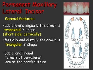
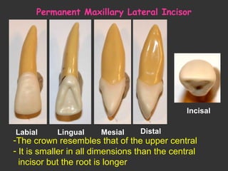
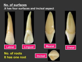
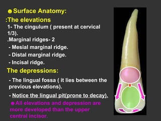
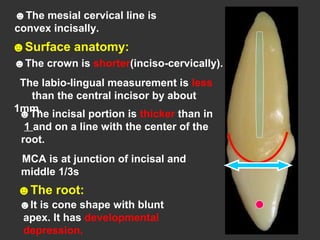
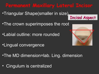
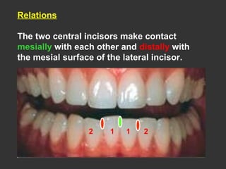
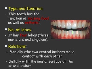
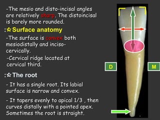
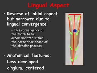
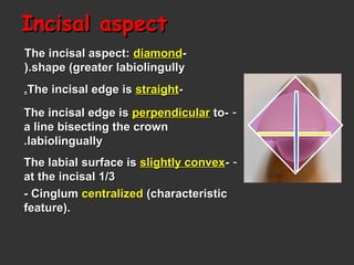
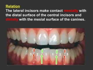
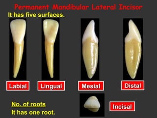
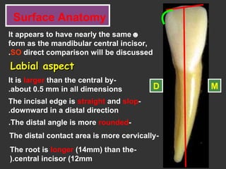
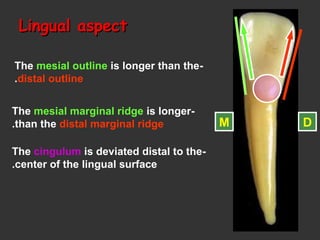
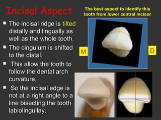
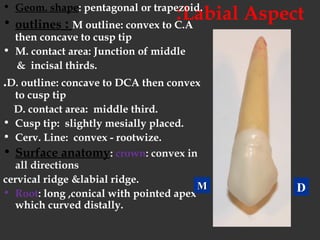
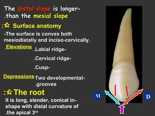
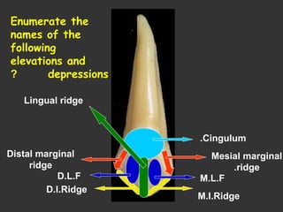
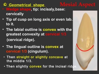
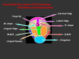
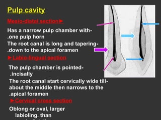
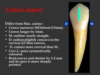
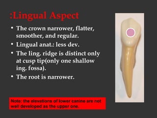
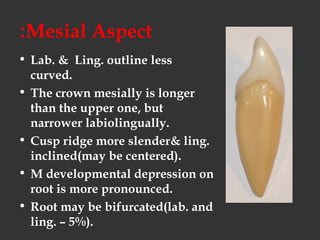
Surface AnatomySurface Anatomy
of Permanentof Permanent
TeethTeeth
By: Dr. 3mmar
Dr: MahMouDDr: MahMouD 3MMar3MMar abD El-haMiEDabD El-haMiED
MobileMobile: 0106 39 700 30: 0106 39 700 30
EmailEmail::m.3mmar9428m.3mmar9428@yahoo.com@yahoo.com
Addres:Addres: knowledge village centerknowledge village center..
Hihan st, mansoura opposite GalaaHihan st, mansoura opposite Galaa gategate
Permanent teeth divided intoPermanent teeth divided into:-:-
a) anterior teetha) anterior teeth::
11..incisorsincisors ((centralcentral && laterallateral((
22..caninecanine..
b)posterior teethb)posterior teeth::
11..pre-molarspre-molars
22..MolarsMolars
Surface Anatomy of
Permanent Teeth
C
I
I
C
P
P
M
M
I, for incisor
C, for canine
P, for premolar
M, for molar
Anterior teeth
And
Canines
Incisors
They are divided into:
Incisors
There are four maxillary incisors
and four mandibular incisors.
♥ Two central incisors
contact with each other in the
midline (mesially) and with
the lateral incisors distally.
♥ Two lateral incisors contact
with the central incisor mesially
and with the canine distally.
General Feature
1.Eight in number.
2.The crowns are trapezoidal in shape (labially
and lingually(.
3.The crowns are triangular in shape (proximally(.
4.The labial and lingual crest of curvatures are at
the cervical third of the crown.
5.Presence of mamelons on the incisal ridge.
6.Each incisor has 4 lobes (3 labial and 1 lingual(..
7.Have single root.
8.Function : incise and cut food material, esthetic,
and speaking.
For easy teeth description
We have to speak about
• Geometric outline of the crown.
• Outlines of the crown and root.
• Surface anatomy of the crown and root.
• Number of lobes (four lobes 3 labially and 1
lingually).
• Pulp cavity.
• Function (mastication (incising), esthetic, speaking).
• chronology
•Enamel organ appearance.
•Beginning of calcification.
•Crown completed.
•Eruption.
•Root completed.
Chronology:
Permanent
Maxillary Central Incisor
All teeth have 5 aspects
Facial Lingual Mesial Distal
Incisal
1
No. of rootsNo. of roots
It has one rootIt has one root..
Chronology of Permanent Maxillary
Central Incisor
Appearance of dental organ  5 m I. U.
Beginning of calcification-------- 3-4 months
Crown completion--------------Crown completion-------------- 4-5 years4-5 years
Eruption ---------------------Eruption --------------------- 7- 8years7- 8years
Root completion----------------Root completion---------------- 10 years10 years
Surface Anatomy
Labial aspect
☻Geometrical outline of the
crown: Trapezoid
-The short side cervically
-The long side incisally
☻The outline:
-Mesial outline is slightly convex.
-Distal outline is more convex.
-Incisal outline is straight and
perpendicular to the long axis of
the tooth(mamelons in newly
erupted tooth(.
-The cervical line is convex
root-wards.
D M
-The mesio-incisal angle is nearly
sharp.
The disto-incisal angle is rounded.
M.I
angle
D.I
angle
☻Surface anatomy:
1.crown:The surface is smoothly
convex, it is flattened incisally. Two
shallow vertical developmental
grooves divide the labial surface
into 3 portions of lobes
Cervical ridge located at
cervical third.
2.Roots: single root has
cone shape with blunt apex
Lingual AspectLingual Aspect1.Geometric shape of crown:
2.Outline form:
• The reverse of the labial aspect
3.Surface anatomy:
• Elevations:
• Cingulum → in cervical 1/3
• Mesial & distal marginal ridges
• Incisal ridge
• Depressions:
• Lingual fossa
• Root shape:
• Root apex: the reverse of the labial
Lingual aspect
-It has the same geometrical outline
and outline as the labial surface.
M D
- The mesial and distal sides
of the crown and root
converge lingually ( the lingual
surface is narrower than the
labial surface(.
- This convergence to
accommodate with the
horse shoe shaped of
the alveolar process.
The facial surface is
larger than the lingual
surface.
☻Surface anatomy:
The elevations:
1-The cingulum ( present
at cervical 1/3.
2-Marginal ridges.(well
developed MMR
and DMR(
3- Incisal ridge.
The depressions:
- The lingual fossa ( it lies
between the previous
elevations(.
Mesial surface Distal surface
Permanent Maxillary Central Incisor
Proximal Aspects
Mesial aspect
☻Geometrical outline of the crown:
☻Triangular in shape; the base
cervically and the apex incisally.
☻The incisal ridge is on a line
that bisects the center of the root.
☻The outline:
☻The labial outline is convex at
cervical 1/3 ( cervical ridge(.
-Then become flat to the incisal ridge.
☻The lingual outline is convex at
cervical 1/3 (cingulum(.
- Then concave at the lingual fossa.
- Then slightly convex for the incisal
ridge.
☻The mesial cervical line is
convex incisally. Its curvature is
greater than any other teeth in the
mouth.
☻Surface anatomy:
☻The mesial surface is convex
with the maximum convexity at the
the incisal 1/3 ( the mesial contact
area(
☻The root:
☻It is cone shape with blunt
apex.
DistalDistal
AspectAspect
Permanent Maxillary Central Incisor
1.Geometric shape of crown:
2.Outline form:
• The reverse of the mesial
3.Anatomical features:
• Contact area level:
more cervical, broader
4.Cervical line curvature:
concave rootwise
• Root shape: conical
• Root apex: blunt,centralized
Distal aspect
☻Similar to the mesial aspect but
differ in.
-The crown appear thicker at the
incisal 1/3.
-The cervical line curvature is less
than mesial ( by 1 mm(.
-The contact area located at
the junction of incisal and
middle 1/3s.(the distal contact
area(
Incisal aspect
☻Triangular in shape.
-The base is placed labially and the
apex lingually.
-M-D>lab-ling.
☻The labial surface is
broad and flat. The cervical
portion of the crown is
convex ( cervical ridge(.
☻The lingual outline tapers
lingually to the cingulum (lingual
convergence(.
-The cingulum is shifted distally.
-The crown superimposes the root.
D M
►Mesio-distal section
-The pulp chamber is wide
conforming the shape of the
crown.
-It presents three pulp horns
corresponding to the three
mamelons.
-The root canal tapers towards
the apex.
►Labio-lingual section
-The pulp chamber
pointed incisally, then
follows the increase in
the crown dimension
cervically.
-The root canal tapers gradually
as it traverse the root ending in a
constriction at the apex (the
apical foramen(.
☻Pulp cavity
Triangular
Trapezoidal
Pulp Cavity
Pulp is circular
Pulp Cavity
PermanentPermanent
MaxillaryMaxillary
LateralLateral IncisorIncisor
By: dr. 3mmar
Permanent MaxillaryPermanent Maxillary
Lateral IncisorLateral Incisor
General features:
•Labially and lingually the crown is
trapezoid in shape
(short side: cervically)
•Mesially and distally the crown is
triangular in shape
•Labial and lingual
“crests of curvature”
are at the cervical third
Malformations Of The Upper Permanent
Lateral Incisor
Peg-shaped
lateral
incisor.
Missing lateral
incisor.
13
Chronology of Permanent Maxillary
Lateral Incisor
Appearance of dental organ  5 m. I. U.
Beginning of calcification----- 1 year
Crown completion--------------Crown completion-------------- 4-5 years4-5 years
Eruption -----------------Eruption ----------------- 8-98-9 yearsyears
Root completion----------------Root completion---------------- 11 years11 years
Permanent Maxillary Lateral Incisor
Labial Lingual Mesial Distal
Incisal
-The crown resembles that of the upper central
- It is smaller in all dimensions than the central
incisor but the root is longer

Permanent Maxillary Lateral Incisor
Variations from Normal:
1.1.Congenitally missingCongenitally missing
2.2.Peg lateralPeg lateral
3.3.Lingual pit & grooveLingual pit & groove
4.4.Lingual tubercleLingual tubercle
No. of surfaces
It has four surfaces and incisal aspect.
Labial Lingual Mesial
Distal
Incisal
No. of roots
It has one root
1. Geometric shape of crown:
2. Outline form:
• Mesial outline: straight
crest of curvature at the junction of middle
and incisal 1/3s (contact area).
• Distal outline: convex
crest of curvature at the center of middle
1/3.
• - DI angle: more rounded than MI angle
3. Anatomical features:
• Crown: elevations & depressions(less
prominent) & surfce more convex
• Root : cone shape with sharp apex,curved
distally
Permanent Maxillary Lateral Incisor
Labial AspectLabial Aspect
DM
 More convex
Lingual AspectLingual Aspect1. Geometric shape of crown:
2. Outline form:
• Is similar to that of upper central
3.Anatomical features:
• Elevations: (more prominent)
• Cingulum → in cervical 1/3
• Mesial & distal marginal ridges
• Incisal ridge
• Depressions:
• Lingual fossa: deeper & circumscribed
• A palatal pit is enclosed by cingulum
• Root shape: cone shape
• Root apex: pointed
☻Surface Anatomy:
The elevations:
1- The cingulum ( present at cervical
1/3).
2-Marginal ridges.
- Mesial marginal ridge.
- Distal marginal ridge.
- Incisal ridge.
The depressions:
- The lingual fossa ( it lies between the
previous elevations).
☻All elevations and depression are
more developed than the upper
central incisor.
- Notice the lingual pit(prone to decay).
MesialMesial
AspectAspect
1.Geometric shape of crown:
2.Outline form:
• labial outline:……….
→ crest of curvature at…….
• lingual outline: ………
→ crest of curvature at…….
3.Anatomical features:
• Mesial marginal ridge
• Contact area level
4.Cervical line curvature:
concave rootwise
• Root shape: conical
Permanent Maxillary Lateral Incisor
☻The mesial cervical line is
convex incisally.
☻Surface anatomy:
☻The incisal portion is thicker than in
1 and on a line with the center of the
root.
MCA is at junction of incisal and
middle 1/3s
☻The root:
☻It is cone shape with blunt
apex. It has developmental
depression.
☻The crown is shorter(inciso-cervically).
The labio-lingual measurement is less
than the central incisor by about
1mm.
DistalDistal
AspectAspect
Permanent Maxillary Lateral Incisor
1.Geometric shape of crown:
2.Outline form:
• The reverse of the mesial
3.Anatomical features:
• Contact area level:
• more cervical, broader
4.Cervical line curvature:
• concave rootwise(less than mesial)
• Root shape: conical, more convex
• Root apex: sharp,centralized
• Developmental depression on root
Incisal AspectIncisal Aspect
•Triangular Shape(smaller in size)
•The crown superimposes the root
•Labial outline: more rounded
•Lingual convergence
•The MD dimension=lab. Ling. dimension
• Cingulum is centralized
Permanent Maxillary Lateral Incisor
• Is similar but smaller than that of central
incisor.
• It consists of a single root canal and a pulp
chamber.
☻Pulp cavity
Permanent
Mandibular Incisors
By: Dr. 3mmar
Permanent Mandibular
Incisors
They are fourThey are four
in number.in number.
They haveThey have smallersmaller
mesio-distal dimensionmesio-distal dimension
than any of the otherthan any of the other
teeth.teeth.
The contact areas:The contact areas:
near the incisal edge.near the incisal edge.
1 12 2
Relations
The two central incisors make contact
mesially with each other and distally with
the mesial surface of the lateral incisor.
11 22
Labial
surface:
lingually
inclined
The crownThe crown
and root areand root are
narrownarrow
mesiodistallymesiodistally
but are widebut are wide
labiolinguallylabiolingually
The smallest tooth in
the permanent dentition
Permanent
Mandibular Central Incisor
Chronology:
• Appearance of dental organ 5 m.i.u
• First evident of calcification 3-4 m
• Enamel completed 4-5 y
• Eruption 6-7 y
• Root completed 9 y
 Type and functionType and function::
- This tooth has the- This tooth has the
function offunction of incising foodincising food
as well asas well as estheticesthetic..
 No. of lobesNo. of lobes::
- It has- It has fourfour lobes (threelobes (three
mamelons and cingulum).mamelons and cingulum).
 RelationsRelations::
- Mesially :the two central incisors make- Mesially :the two central incisors make
contact with each othercontact with each other
- Distally with the mesial surface of the- Distally with the mesial surface of the
lateral incisor.lateral incisor.
It has five surfaces.
Labial Lingual Mesial Distal
Incisal
Permanent Mandibular Central Incisor
Labial Aspect
• Geometrical shape of crown :
Trapezoid
• Mesial & Distal outlines:
straight, then taper evenly to
to the narrow cervix.
• Incisal margin:
-The short side cervically.-The short side cervically.
-The long side incisally.-The long side incisally.
- It is straight and perpendicular to- It is straight and perpendicular to
the long axis of the tooth(mamelons).the long axis of the tooth(mamelons).
The cervical line :The cervical line :
convex root-wards.convex root-wards.
-The mesio and disto-incisal angles-The mesio and disto-incisal angles
are relativelyare relatively sharpsharp. The distoincisal. The distoincisal
is barely more rounded.is barely more rounded.
Surface anatomy:
-The surface is-The surface is convexconvex bothboth
mesiodistally and inciso-mesiodistally and inciso-
cervically.cervically.
-Cervical ridge located at-Cervical ridge located at
cervical third.cervical third.
The root:
- It has a single root. Its labial- It has a single root. Its labial
surface is narrow and convex.surface is narrow and convex.
- It tapers evenly to apical 1/3 , then- It tapers evenly to apical 1/3 , then
curves distally with a pointed apex.curves distally with a pointed apex.
Sometimes the root is straight.Sometimes the root is straight.
D M
Lingual Aspect
• Reverse of labial aspect
but narrower due to
lingual convergence
• Anatomical features:
Less developed
cinglum, centered
- This convergence of- This convergence of
the tooth to bethe tooth to be
accommodated withinaccommodated within
the horse shoe shape ofthe horse shoe shape of
the alveolar process.the alveolar process.
☻Surface anatomy:
The elevations:
1- The cingulum ( present at cervical
1/3).
2-Marginal ridges.
- Mesial marginal ridge.
- Distal marginal ridge.
3- Incisal ridge.
The depressions:
☻All elevations and depression are
poorly developed than the upper incisor.
- The lingual fossa ( it lies between
the previous elevations).
The Mesial AspectThe Mesial Aspect
☻Geometric outline of the crown:
☻Triangular in shape; the base
cervically and the apex incisally.
☻☻The incisal ridge isThe incisal ridge is lingual tolingual to a linea line
that bisects the centre of the root.that bisects the centre of the root.
☻The outline:
☻The labial outline is convex at
cervical 1/3 ( cervical ridge).
☻The lingual outline is convex at
cervical 1/3 (cingulum).
-Then become straight to the incisal
ridge.
- Then straight to slightly concave
at the lingual fossa.
- Then slightly convex for the incisal ridge.
The Mesial AspectThe Mesial Aspect
☻The root:
☻Is flat with longitudinal
devlopmental depression.
☻The root tapers apically
(pointed apex).
The Distal AspectThe Distal Aspect
☻Similar to the mesial aspect but
differs in.
- The cervical line curvature is less
than mesial ( by 1 mm).
-The distal contact area at the same
level as the mesial surface.
-The distal developmental depression
on the root is deeper than mesial and
may have developmental groove.
Incisal aspectIncisal aspect
--The incisal aspect:The incisal aspect: diamonddiamond
shape (greater labiolingullyshape (greater labiolingully).).
--The incisal edge isThe incisal edge is straightstraight..
---The incisal edge isThe incisal edge is perpendicularperpendicular toto
a line bisecting the crowna line bisecting the crown
labiolinguallylabiolingually..
---The labial surface isThe labial surface is slightly convexslightly convex
at the incisal 1/3at the incisal 1/3
- Cinglum- Cinglum centralizedcentralized (characteristic(characteristic
feature).feature).
Pulp cavity
☻☻Pulp cavityPulp cavity
►Mesio-distal section
►Labio-lingual section
- The outline of the pulp
cavity conform to the
crown and root outline.
- The pulp chamber has 2
or 3 pulp horns. The root
canal tapers to the apical
foramen.
►Cervical cross section
- Oval and usually constricted
mesio-distally and wide labio-
lingually.
Oval
MandibularMandibular
LateralLateral
IncisorIncisor
By: Dr. 3mmarA
Chronology
Appearance of the dental organ 5 m.i.u
First evidence of calcification 3-4 months.
Enamel completed 4-5 years
Eruption 7-8 years
Root completed 10 years
Type and function
This tooth has the function of incising food as well as
esthetic.
No. of lobes
It has four lobes (three mamelons and cingulum)
Relation
The lateral incisors make contact mesially with
the distal surface of the central incisors and
distally with the mesial surface of the canines.
11 223 3
It has five surfaces.
No. of roots
It has one root.
LabialLabial LingualLingual MesialMesial DistalDistal
IncisalIncisal
Permanent Mandibular Lateral Incisor
Surface Anatomy
☻It appears to have nearly the same
form as the mandibular central incisor,
SO direct comparison will be discussed.
Labial aspectLabial aspect
-It is larger than the central by
about 0.5 mm in all dimensions.
-The incisal edge is straight and slop
downward in a distal direction.
MD
-The distal angle is more rounded.
-The root is longer (14mm) than the
central incisor (12mm).
-The distal contact area is more cervically
Lingual aspectLingual aspect
-The mesial outline is longer than the
distal outline.
-The mesial marginal ridge is longer
than the distal marginal ridge.
-The cingulum is deviated distal to the
center of the lingual surface.
M D
Mesial aspect Distal aspect
-The distal surface is
shorter inciso-cervically.
-Both cervical lines are
less than those of the
central incisor.
-The distal contact area is
more cervical than the
mesial one.
-Root depressions are
seen on both the mesial
and distal surfaces.
Incisal Aspect
 The incisal ridge isThe incisal ridge is tiltedtilted
distally and lingually asdistally and lingually as
well as the whole tooth.well as the whole tooth.
 The cingulum is shiftedThe cingulum is shifted
to the distal.to the distal.
 This allow the tooth toThis allow the tooth to
follow the dental archfollow the dental arch
curvature.curvature.
 So the incisal edge isSo the incisal edge is
not at a right angle to anot at a right angle to a
line bisecting the toothline bisecting the tooth
labiolingullay.labiolingullay.
M D
The best aspect to identify this
tooth from lower central incisor
Incisal aspectIncisal aspect
- The crown appear
to be twisted on
root.
- The distal lobe is
larger than the
mesial lobe.
- This form fits the narrow U –shaped
mandibular arch.
- The incisal edge
inclines disto- lingually.
- The cingulum is
deviated distally.
Identify the aspect and tell THREE differences between
the lateral and central incisor.
2-Incisal edge.
3-The incisal angles.
1-The size.
☻Labial aspect.
Answer
Pulp cavity
Very similar to that of lower central incisor.
Hony please,
just calm down.
Let me explain.
The Permanent
Canines
*corner stone of the mouth*
carnivores
By: Dr. 3mmar
The Permanent Canines
General features:
• 2 Max. & 2 Mand.
• Longest tooth in the dental arch.
• Convex on all surfaces(self-cleansing quality).
• Highly developed middle lobe(lab. ridge)
• Lab.&ling. : pentagonal or trapezoidal.
• M&D: triangular.
• Functions: cutting & tearing of food.
-There are two maxillary and two mandibular canines.
-They are called corner stones of the mouth.
-It has long root for good anchorage in the bone.
-The middle labial lobe is well developed forming cusp
and labial ridge.
-The bone ridge over the labial surface of the root is called
canine eminence (supports facial expression).
The Permanent
Maxillary Canine
Chronology:
• Appearance of dental organ 6 m.i.u
• First evident of calcification 4 - 5 m
• Enamel completed 6 - 7 y
• Eruption 11-12 y
• Root completed 14-15 y
No. of surfacesNo. of surfaces
It has five surfacesIt has five surfaces..
LabialLabial LingualLingual MesialMesial DistalDistal
IncisalIncisal
No. of rootsNo. of roots
It has one rootIt has one root..
Labial Aspect:• Geom. shape: pentagonal or trapezoid.
• outlines : M outline: convex to C.A
then concave to cusp tip
• M. contact area: Junction of middle
& incisal thirds.
.D. outline: concave to DCA then convex
to cusp tip
D. contact area: middle third.
• Cusp tip: slightly mesially placed.
• Cerv. Line: convex - rootwize.
• Surface anatomy: crown: convex in
all directions
cervical ridge &labial ridge.
• Root: long ,conical with pointed apex
which curved distally.
DMM
--TheThe distal slopedistal slope is longeris longer
than thethan the mesial slopemesial slope..
 Surface anatomy:
-The surface is convex both-The surface is convex both
mesiodistally and inciso-cervically.mesiodistally and inciso-cervically.
Elevations
--Cervical ridgeCervical ridge..
--Labial ridgeLabial ridge..
Depressions --Two developmentalTwo developmental
groovesgrooves..
The root:
-It is long, slender, conical in
shape with distal curvature of
the apical 3rd
.
--CuspCusp..
DMM
Lingual Aspect• Crown narrower
Surface anatomy:
The elevations: (well developed(
-The cingulum:large – pointed like a cusp
-The marginal ridges (mesial and distal(.
-The lingual ridge.
-Mesio- and disto-incisal ridges.
The depressions:
-Two lingual fossae.
Geom. Outline: petagonal or
trapezoidal
The root: narrower lingually than
Labially(lingual convergence(.
Cingulum.
Mesial marginal
ridge.
Distal marginal
ridge
M.L.FD.L.F
M.I.Ridge
D.I.Ridge
Lingual ridge
Enumerate the
names of the
following
elevations and
depressions?
Mesial Aspect
• Wedge shape, tip: incisaly,base:
cervically
• Tip of cusp on long axis or even lab.
to it.
• The labial outline is convex with the
greatest convexity at cervical 1/3
(cervical ridge(.
• The lingual outline is convex at
cervical 1/3 (cingulum(.
 Geometrical shapeGeometrical shape
• Then straight or slightly concave at
the middle 1/3.
• Then slightly convex for the incisal ridge.
 M. contact area:M. contact area: at theat the
junction of incisal & middle 1/3.junction of incisal & middle 1/3.
 The cervical line:The cervical line: curvescurves
towards the cusp.towards the cusp.
 Root:Root: is conical & blunt apexis conical & blunt apex
may be curved lab.may be curved lab.
 Broad with a shallowBroad with a shallow
developmental depression.developmental depression.
Distal Aspect
• Similar to M aspect
except:
• Cerv. L: less curved.
• DMR: heavier & more
irregular.
• Contact area: more
cervically& surface
above it more concave.
• Root dev. depression:
more prominent(help to
anchor in the alveoli
and prevents rotation(.
• Lab. ling. > M D
• Cusp tip directed
towards lab. & mesial
• M cusp slop < D cusp
slop
• The labial surface is
convex
 All elevations andAll elevations and
depressions can be seendepressions can be seen
from this aspect.from this aspect.
Incisal Aspect
Diamond in shape
Enumerate the names of the following
elevations and depressions?
Cervical ridgr.
Labial ridge.
D. slope.
D.M.R
Cusp tip.
M. slope.
Lingual ridge
M.M.R
Lingual fossae.
Cingulum.
Pulp cavityPulp cavity
►Mesio-distal section
►Labio-lingual section
-Has a narrow pulp chamber with
one pulp horn.
-The root canal is long and tapering
down to the apical foramen.
-The pulp chamber is pointed
incisally.
-The root canal start cervically wide till
about the middle then narrows to the
apical foramen.
Oblong or oval, larger
labioling. than
►Cervical cross section
The Mandibular
Canine
By: Dr. 3mmar
The Mandibular canine
Chronology:
• Appearance of dental organ 6m.i.u
• First evident of calcification 4-5m
• Enamel completed 6-7y
• Eruption 9-10 y
• Root completed 12-13y
The Mandibular Canine
LabialLabial LingualLingual MesialMesial DistalDistal IncisalIncisal
No. of roots
It has one root
Labial aspect:
Differ from Max. canine :
• Crown narrower MD(about 0.5mm).
• Crown longer by 1mm.
• M. outline: nearly straight.
• D. outline:slightly concave in the
cervical 1/3 then convex,
• D. contact more cervical than M.
• Cerv.L: more symmetrically
cotoured.
• Root:convex and shorter by 1-2 mm
and its apex is more sharply
pointed.
D M
Lingual Aspect:
• The crown narrower, flatter,
smoother, and regular.
• Lingual anat.: less dev.
• The ling. ridge is distinct only
at cusp tip(only one shallow
ing. fossa).
• The root is narrower.
Note: the elevations of lower canine are not
well developed as the upper one.
Mesial Aspect:
• Lab. & Ling. outline less
curved.
• The crown mesially is longer
than the upper one, but
narrower labiolingually.
• Cusp ridge more slender& ling.
inclined(may be centered).
• M developmental depression on
root is more pronounced.
• Root may be bifurcated(lab. and
ling. – 5%).
Distal Aspect:
• DMR is more pronounced.
• Cerv. L. is less curved.
• D contact area more cerv.
• Dev. dep. on the root is
more deep than that on the
mesial.
Incisal Aspect:
• Mesial surface less curved
• Cusp tip is located mesioling.
• The D. cusp slop: slightly ling.
(distolingual twist to match the
dental arch)
• Cinglum shifted distally.
DM
ConvexFlat or concave
(Often similar to that of upper canine(
Incisal aspect.
Outline and surface anatomy
The thickness is greater than the width.The thickness is greater than the width.
The labial surface appeared convexThe labial surface appeared convex
and even more than the incisorsand even more than the incisors
The cingulum forms a shorter arcThe cingulum forms a shorter arc
than the one labiallythan the one labially..
The elevations and depressionsThe elevations and depressions
in the labial and lingual surfacesin the labial and lingual surfaces
appeared in this aspectappeared in this aspect
Mesial half resemmble incisorsMesial half resemmble incisors
& distal half ressemble& distal half ressemble
premolarspremolars
The pulp cavity:
• Similar to max. canine
but less large.
• May have 1 or 2 root
canals (lab. &ling.) that
joined at apex or
remain separate if root
bifurcated
Thank you
Dr: 3mmar
Any Question?

2 anterior teeth

  • 2.
    Surface AnatomySurface Anatomy ofPermanentof Permanent TeethTeeth By: Dr. 3mmar
  • 3.
    Dr: MahMouDDr: MahMouD3MMar3MMar abD El-haMiEDabD El-haMiED MobileMobile: 0106 39 700 30: 0106 39 700 30 EmailEmail::m.3mmar9428m.3mmar9428@yahoo.com@yahoo.com Addres:Addres: knowledge village centerknowledge village center.. Hihan st, mansoura opposite GalaaHihan st, mansoura opposite Galaa gategate
  • 4.
    Permanent teeth dividedintoPermanent teeth divided into:-:- a) anterior teetha) anterior teeth:: 11..incisorsincisors ((centralcentral && laterallateral(( 22..caninecanine.. b)posterior teethb)posterior teeth:: 11..pre-molarspre-molars 22..MolarsMolars
  • 5.
    Surface Anatomy of PermanentTeeth C I I C P P M M I, for incisor C, for canine P, for premolar M, for molar
  • 6.
  • 7.
    Incisors There are fourmaxillary incisors and four mandibular incisors. ♥ Two central incisors contact with each other in the midline (mesially) and with the lateral incisors distally. ♥ Two lateral incisors contact with the central incisor mesially and with the canine distally.
  • 8.
    General Feature 1.Eight innumber. 2.The crowns are trapezoidal in shape (labially and lingually(. 3.The crowns are triangular in shape (proximally(. 4.The labial and lingual crest of curvatures are at the cervical third of the crown.
  • 9.
    5.Presence of mamelonson the incisal ridge. 6.Each incisor has 4 lobes (3 labial and 1 lingual(.. 7.Have single root. 8.Function : incise and cut food material, esthetic, and speaking.
  • 10.
    For easy teethdescription We have to speak about • Geometric outline of the crown. • Outlines of the crown and root. • Surface anatomy of the crown and root. • Number of lobes (four lobes 3 labially and 1 lingually). • Pulp cavity. • Function (mastication (incising), esthetic, speaking). • chronology
  • 11.
    •Enamel organ appearance. •Beginningof calcification. •Crown completed. •Eruption. •Root completed. Chronology:
  • 12.
  • 13.
    All teeth have5 aspects Facial Lingual Mesial Distal Incisal 1 No. of rootsNo. of roots It has one rootIt has one root..
  • 14.
    Chronology of PermanentMaxillary Central Incisor Appearance of dental organ  5 m I. U. Beginning of calcification-------- 3-4 months Crown completion--------------Crown completion-------------- 4-5 years4-5 years Eruption ---------------------Eruption --------------------- 7- 8years7- 8years Root completion----------------Root completion---------------- 10 years10 years
  • 15.
    Surface Anatomy Labial aspect ☻Geometricaloutline of the crown: Trapezoid -The short side cervically -The long side incisally ☻The outline: -Mesial outline is slightly convex. -Distal outline is more convex. -Incisal outline is straight and perpendicular to the long axis of the tooth(mamelons in newly erupted tooth(. -The cervical line is convex root-wards. D M
  • 16.
    -The mesio-incisal angleis nearly sharp. The disto-incisal angle is rounded. M.I angle D.I angle ☻Surface anatomy: 1.crown:The surface is smoothly convex, it is flattened incisally. Two shallow vertical developmental grooves divide the labial surface into 3 portions of lobes Cervical ridge located at cervical third. 2.Roots: single root has cone shape with blunt apex
  • 17.
    Lingual AspectLingual Aspect1.Geometricshape of crown: 2.Outline form: • The reverse of the labial aspect 3.Surface anatomy: • Elevations: • Cingulum → in cervical 1/3 • Mesial & distal marginal ridges • Incisal ridge • Depressions: • Lingual fossa • Root shape: • Root apex: the reverse of the labial
  • 18.
    Lingual aspect -It hasthe same geometrical outline and outline as the labial surface. M D - The mesial and distal sides of the crown and root converge lingually ( the lingual surface is narrower than the labial surface(. - This convergence to accommodate with the horse shoe shaped of the alveolar process. The facial surface is larger than the lingual surface.
  • 19.
    ☻Surface anatomy: The elevations: 1-Thecingulum ( present at cervical 1/3. 2-Marginal ridges.(well developed MMR and DMR( 3- Incisal ridge. The depressions: - The lingual fossa ( it lies between the previous elevations(.
  • 20.
    Mesial surface Distalsurface Permanent Maxillary Central Incisor Proximal Aspects
  • 21.
    Mesial aspect ☻Geometrical outlineof the crown: ☻Triangular in shape; the base cervically and the apex incisally. ☻The incisal ridge is on a line that bisects the center of the root. ☻The outline: ☻The labial outline is convex at cervical 1/3 ( cervical ridge(. -Then become flat to the incisal ridge. ☻The lingual outline is convex at cervical 1/3 (cingulum(. - Then concave at the lingual fossa. - Then slightly convex for the incisal ridge.
  • 22.
    ☻The mesial cervicalline is convex incisally. Its curvature is greater than any other teeth in the mouth. ☻Surface anatomy: ☻The mesial surface is convex with the maximum convexity at the the incisal 1/3 ( the mesial contact area( ☻The root: ☻It is cone shape with blunt apex.
  • 23.
    DistalDistal AspectAspect Permanent Maxillary CentralIncisor 1.Geometric shape of crown: 2.Outline form: • The reverse of the mesial 3.Anatomical features: • Contact area level: more cervical, broader 4.Cervical line curvature: concave rootwise • Root shape: conical • Root apex: blunt,centralized
  • 24.
    Distal aspect ☻Similar tothe mesial aspect but differ in. -The crown appear thicker at the incisal 1/3. -The cervical line curvature is less than mesial ( by 1 mm(. -The contact area located at the junction of incisal and middle 1/3s.(the distal contact area(
  • 25.
    Incisal aspect ☻Triangular inshape. -The base is placed labially and the apex lingually. -M-D>lab-ling. ☻The labial surface is broad and flat. The cervical portion of the crown is convex ( cervical ridge(. ☻The lingual outline tapers lingually to the cingulum (lingual convergence(. -The cingulum is shifted distally. -The crown superimposes the root. D M
  • 26.
    ►Mesio-distal section -The pulpchamber is wide conforming the shape of the crown. -It presents three pulp horns corresponding to the three mamelons. -The root canal tapers towards the apex. ►Labio-lingual section -The pulp chamber pointed incisally, then follows the increase in the crown dimension cervically. -The root canal tapers gradually as it traverse the root ending in a constriction at the apex (the apical foramen(. ☻Pulp cavity Triangular Trapezoidal
  • 27.
  • 28.
  • 29.
  • 30.
    Permanent MaxillaryPermanent Maxillary LateralIncisorLateral Incisor General features: •Labially and lingually the crown is trapezoid in shape (short side: cervically) •Mesially and distally the crown is triangular in shape •Labial and lingual “crests of curvature” are at the cervical third
  • 31.
    Malformations Of TheUpper Permanent Lateral Incisor Peg-shaped lateral incisor. Missing lateral incisor. 13
  • 32.
    Chronology of PermanentMaxillary Lateral Incisor Appearance of dental organ  5 m. I. U. Beginning of calcification----- 1 year Crown completion--------------Crown completion-------------- 4-5 years4-5 years Eruption -----------------Eruption ----------------- 8-98-9 yearsyears Root completion----------------Root completion---------------- 11 years11 years
  • 33.
    Permanent Maxillary LateralIncisor Labial Lingual Mesial Distal Incisal -The crown resembles that of the upper central - It is smaller in all dimensions than the central incisor but the root is longer
  • 34.
     Permanent Maxillary LateralIncisor Variations from Normal: 1.1.Congenitally missingCongenitally missing 2.2.Peg lateralPeg lateral 3.3.Lingual pit & grooveLingual pit & groove 4.4.Lingual tubercleLingual tubercle
  • 35.
    No. of surfaces Ithas four surfaces and incisal aspect. Labial Lingual Mesial Distal Incisal No. of roots It has one root
  • 36.
    1. Geometric shapeof crown: 2. Outline form: • Mesial outline: straight crest of curvature at the junction of middle and incisal 1/3s (contact area). • Distal outline: convex crest of curvature at the center of middle 1/3. • - DI angle: more rounded than MI angle 3. Anatomical features: • Crown: elevations & depressions(less prominent) & surfce more convex • Root : cone shape with sharp apex,curved distally Permanent Maxillary Lateral Incisor Labial AspectLabial Aspect DM  More convex
  • 37.
    Lingual AspectLingual Aspect1.Geometric shape of crown: 2. Outline form: • Is similar to that of upper central 3.Anatomical features: • Elevations: (more prominent) • Cingulum → in cervical 1/3 • Mesial & distal marginal ridges • Incisal ridge • Depressions: • Lingual fossa: deeper & circumscribed • A palatal pit is enclosed by cingulum • Root shape: cone shape • Root apex: pointed
  • 38.
    ☻Surface Anatomy: The elevations: 1-The cingulum ( present at cervical 1/3). 2-Marginal ridges. - Mesial marginal ridge. - Distal marginal ridge. - Incisal ridge. The depressions: - The lingual fossa ( it lies between the previous elevations). ☻All elevations and depression are more developed than the upper central incisor. - Notice the lingual pit(prone to decay).
  • 39.
    MesialMesial AspectAspect 1.Geometric shape ofcrown: 2.Outline form: • labial outline:………. → crest of curvature at……. • lingual outline: ……… → crest of curvature at……. 3.Anatomical features: • Mesial marginal ridge • Contact area level 4.Cervical line curvature: concave rootwise • Root shape: conical Permanent Maxillary Lateral Incisor
  • 40.
    ☻The mesial cervicalline is convex incisally. ☻Surface anatomy: ☻The incisal portion is thicker than in 1 and on a line with the center of the root. MCA is at junction of incisal and middle 1/3s ☻The root: ☻It is cone shape with blunt apex. It has developmental depression. ☻The crown is shorter(inciso-cervically). The labio-lingual measurement is less than the central incisor by about 1mm.
  • 41.
    DistalDistal AspectAspect Permanent Maxillary LateralIncisor 1.Geometric shape of crown: 2.Outline form: • The reverse of the mesial 3.Anatomical features: • Contact area level: • more cervical, broader 4.Cervical line curvature: • concave rootwise(less than mesial) • Root shape: conical, more convex • Root apex: sharp,centralized • Developmental depression on root
  • 42.
    Incisal AspectIncisal Aspect •TriangularShape(smaller in size) •The crown superimposes the root •Labial outline: more rounded •Lingual convergence •The MD dimension=lab. Ling. dimension • Cingulum is centralized Permanent Maxillary Lateral Incisor
  • 43.
    • Is similarbut smaller than that of central incisor. • It consists of a single root canal and a pulp chamber. ☻Pulp cavity
  • 45.
  • 46.
    Permanent Mandibular Incisors They arefourThey are four in number.in number. They haveThey have smallersmaller mesio-distal dimensionmesio-distal dimension than any of the otherthan any of the other teeth.teeth. The contact areas:The contact areas: near the incisal edge.near the incisal edge. 1 12 2
  • 47.
    Relations The two centralincisors make contact mesially with each other and distally with the mesial surface of the lateral incisor. 11 22
  • 48.
    Labial surface: lingually inclined The crownThe crown androot areand root are narrownarrow mesiodistallymesiodistally but are widebut are wide labiolinguallylabiolingually The smallest tooth in the permanent dentition
  • 49.
    Permanent Mandibular Central Incisor Chronology: •Appearance of dental organ 5 m.i.u • First evident of calcification 3-4 m • Enamel completed 4-5 y • Eruption 6-7 y • Root completed 9 y
  • 50.
     Type andfunctionType and function:: - This tooth has the- This tooth has the function offunction of incising foodincising food as well asas well as estheticesthetic..  No. of lobesNo. of lobes:: - It has- It has fourfour lobes (threelobes (three mamelons and cingulum).mamelons and cingulum).  RelationsRelations:: - Mesially :the two central incisors make- Mesially :the two central incisors make contact with each othercontact with each other - Distally with the mesial surface of the- Distally with the mesial surface of the lateral incisor.lateral incisor.
  • 51.
    It has fivesurfaces. Labial Lingual Mesial Distal Incisal Permanent Mandibular Central Incisor
  • 52.
    Labial Aspect • Geometricalshape of crown : Trapezoid • Mesial & Distal outlines: straight, then taper evenly to to the narrow cervix. • Incisal margin: -The short side cervically.-The short side cervically. -The long side incisally.-The long side incisally. - It is straight and perpendicular to- It is straight and perpendicular to the long axis of the tooth(mamelons).the long axis of the tooth(mamelons). The cervical line :The cervical line : convex root-wards.convex root-wards.
  • 53.
    -The mesio anddisto-incisal angles-The mesio and disto-incisal angles are relativelyare relatively sharpsharp. The distoincisal. The distoincisal is barely more rounded.is barely more rounded. Surface anatomy: -The surface is-The surface is convexconvex bothboth mesiodistally and inciso-mesiodistally and inciso- cervically.cervically. -Cervical ridge located at-Cervical ridge located at cervical third.cervical third. The root: - It has a single root. Its labial- It has a single root. Its labial surface is narrow and convex.surface is narrow and convex. - It tapers evenly to apical 1/3 , then- It tapers evenly to apical 1/3 , then curves distally with a pointed apex.curves distally with a pointed apex. Sometimes the root is straight.Sometimes the root is straight. D M
  • 54.
    Lingual Aspect • Reverseof labial aspect but narrower due to lingual convergence • Anatomical features: Less developed cinglum, centered - This convergence of- This convergence of the tooth to bethe tooth to be accommodated withinaccommodated within the horse shoe shape ofthe horse shoe shape of the alveolar process.the alveolar process.
  • 55.
    ☻Surface anatomy: The elevations: 1-The cingulum ( present at cervical 1/3). 2-Marginal ridges. - Mesial marginal ridge. - Distal marginal ridge. 3- Incisal ridge. The depressions: ☻All elevations and depression are poorly developed than the upper incisor. - The lingual fossa ( it lies between the previous elevations).
  • 56.
    The Mesial AspectTheMesial Aspect ☻Geometric outline of the crown: ☻Triangular in shape; the base cervically and the apex incisally. ☻☻The incisal ridge isThe incisal ridge is lingual tolingual to a linea line that bisects the centre of the root.that bisects the centre of the root. ☻The outline: ☻The labial outline is convex at cervical 1/3 ( cervical ridge). ☻The lingual outline is convex at cervical 1/3 (cingulum). -Then become straight to the incisal ridge. - Then straight to slightly concave at the lingual fossa. - Then slightly convex for the incisal ridge.
  • 57.
    The Mesial AspectTheMesial Aspect ☻The root: ☻Is flat with longitudinal devlopmental depression. ☻The root tapers apically (pointed apex).
  • 58.
    The Distal AspectTheDistal Aspect ☻Similar to the mesial aspect but differs in. - The cervical line curvature is less than mesial ( by 1 mm). -The distal contact area at the same level as the mesial surface. -The distal developmental depression on the root is deeper than mesial and may have developmental groove.
  • 59.
    Incisal aspectIncisal aspect --Theincisal aspect:The incisal aspect: diamonddiamond shape (greater labiolingullyshape (greater labiolingully).). --The incisal edge isThe incisal edge is straightstraight.. ---The incisal edge isThe incisal edge is perpendicularperpendicular toto a line bisecting the crowna line bisecting the crown labiolinguallylabiolingually.. ---The labial surface isThe labial surface is slightly convexslightly convex at the incisal 1/3at the incisal 1/3 - Cinglum- Cinglum centralizedcentralized (characteristic(characteristic feature).feature).
  • 60.
  • 61.
    ☻☻Pulp cavityPulp cavity ►Mesio-distalsection ►Labio-lingual section - The outline of the pulp cavity conform to the crown and root outline. - The pulp chamber has 2 or 3 pulp horns. The root canal tapers to the apical foramen. ►Cervical cross section - Oval and usually constricted mesio-distally and wide labio- lingually. Oval
  • 62.
  • 63.
    Chronology Appearance of thedental organ 5 m.i.u First evidence of calcification 3-4 months. Enamel completed 4-5 years Eruption 7-8 years Root completed 10 years Type and function This tooth has the function of incising food as well as esthetic. No. of lobes It has four lobes (three mamelons and cingulum)
  • 64.
    Relation The lateral incisorsmake contact mesially with the distal surface of the central incisors and distally with the mesial surface of the canines. 11 223 3
  • 65.
    It has fivesurfaces. No. of roots It has one root. LabialLabial LingualLingual MesialMesial DistalDistal IncisalIncisal Permanent Mandibular Lateral Incisor
  • 66.
    Surface Anatomy ☻It appearsto have nearly the same form as the mandibular central incisor, SO direct comparison will be discussed. Labial aspectLabial aspect -It is larger than the central by about 0.5 mm in all dimensions. -The incisal edge is straight and slop downward in a distal direction. MD -The distal angle is more rounded. -The root is longer (14mm) than the central incisor (12mm). -The distal contact area is more cervically
  • 67.
    Lingual aspectLingual aspect -Themesial outline is longer than the distal outline. -The mesial marginal ridge is longer than the distal marginal ridge. -The cingulum is deviated distal to the center of the lingual surface. M D
  • 68.
    Mesial aspect Distalaspect -The distal surface is shorter inciso-cervically. -Both cervical lines are less than those of the central incisor. -The distal contact area is more cervical than the mesial one. -Root depressions are seen on both the mesial and distal surfaces.
  • 69.
    Incisal Aspect  Theincisal ridge isThe incisal ridge is tiltedtilted distally and lingually asdistally and lingually as well as the whole tooth.well as the whole tooth.  The cingulum is shiftedThe cingulum is shifted to the distal.to the distal.  This allow the tooth toThis allow the tooth to follow the dental archfollow the dental arch curvature.curvature.  So the incisal edge isSo the incisal edge is not at a right angle to anot at a right angle to a line bisecting the toothline bisecting the tooth labiolingullay.labiolingullay. M D The best aspect to identify this tooth from lower central incisor
  • 70.
    Incisal aspectIncisal aspect -The crown appear to be twisted on root. - The distal lobe is larger than the mesial lobe. - This form fits the narrow U –shaped mandibular arch. - The incisal edge inclines disto- lingually. - The cingulum is deviated distally.
  • 71.
    Identify the aspectand tell THREE differences between the lateral and central incisor. 2-Incisal edge. 3-The incisal angles. 1-The size. ☻Labial aspect. Answer
  • 72.
    Pulp cavity Very similarto that of lower central incisor.
  • 74.
    Hony please, just calmdown. Let me explain.
  • 76.
    The Permanent Canines *corner stoneof the mouth* carnivores By: Dr. 3mmar
  • 77.
    The Permanent Canines Generalfeatures: • 2 Max. & 2 Mand. • Longest tooth in the dental arch. • Convex on all surfaces(self-cleansing quality). • Highly developed middle lobe(lab. ridge) • Lab.&ling. : pentagonal or trapezoidal. • M&D: triangular. • Functions: cutting & tearing of food.
  • 78.
    -There are twomaxillary and two mandibular canines. -They are called corner stones of the mouth. -It has long root for good anchorage in the bone. -The middle labial lobe is well developed forming cusp and labial ridge. -The bone ridge over the labial surface of the root is called canine eminence (supports facial expression).
  • 79.
    The Permanent Maxillary Canine Chronology: •Appearance of dental organ 6 m.i.u • First evident of calcification 4 - 5 m • Enamel completed 6 - 7 y • Eruption 11-12 y • Root completed 14-15 y
  • 80.
    No. of surfacesNo.of surfaces It has five surfacesIt has five surfaces.. LabialLabial LingualLingual MesialMesial DistalDistal IncisalIncisal No. of rootsNo. of roots It has one rootIt has one root..
  • 81.
    Labial Aspect:• Geom.shape: pentagonal or trapezoid. • outlines : M outline: convex to C.A then concave to cusp tip • M. contact area: Junction of middle & incisal thirds. .D. outline: concave to DCA then convex to cusp tip D. contact area: middle third. • Cusp tip: slightly mesially placed. • Cerv. Line: convex - rootwize. • Surface anatomy: crown: convex in all directions cervical ridge &labial ridge. • Root: long ,conical with pointed apex which curved distally. DMM
  • 82.
    --TheThe distal slopedistalslope is longeris longer than thethan the mesial slopemesial slope..  Surface anatomy: -The surface is convex both-The surface is convex both mesiodistally and inciso-cervically.mesiodistally and inciso-cervically. Elevations --Cervical ridgeCervical ridge.. --Labial ridgeLabial ridge.. Depressions --Two developmentalTwo developmental groovesgrooves.. The root: -It is long, slender, conical in shape with distal curvature of the apical 3rd . --CuspCusp.. DMM
  • 83.
    Lingual Aspect• Crownnarrower Surface anatomy: The elevations: (well developed( -The cingulum:large – pointed like a cusp -The marginal ridges (mesial and distal(. -The lingual ridge. -Mesio- and disto-incisal ridges. The depressions: -Two lingual fossae. Geom. Outline: petagonal or trapezoidal The root: narrower lingually than Labially(lingual convergence(.
  • 84.
    Cingulum. Mesial marginal ridge. Distal marginal ridge M.L.FD.L.F M.I.Ridge D.I.Ridge Lingualridge Enumerate the names of the following elevations and depressions?
  • 85.
    Mesial Aspect • Wedgeshape, tip: incisaly,base: cervically • Tip of cusp on long axis or even lab. to it. • The labial outline is convex with the greatest convexity at cervical 1/3 (cervical ridge(. • The lingual outline is convex at cervical 1/3 (cingulum(.  Geometrical shapeGeometrical shape • Then straight or slightly concave at the middle 1/3. • Then slightly convex for the incisal ridge.
  • 86.
     M. contactarea:M. contact area: at theat the junction of incisal & middle 1/3.junction of incisal & middle 1/3.  The cervical line:The cervical line: curvescurves towards the cusp.towards the cusp.  Root:Root: is conical & blunt apexis conical & blunt apex may be curved lab.may be curved lab.  Broad with a shallowBroad with a shallow developmental depression.developmental depression.
  • 87.
    Distal Aspect • Similarto M aspect except: • Cerv. L: less curved. • DMR: heavier & more irregular. • Contact area: more cervically& surface above it more concave. • Root dev. depression: more prominent(help to anchor in the alveoli and prevents rotation(.
  • 88.
    • Lab. ling.> M D • Cusp tip directed towards lab. & mesial • M cusp slop < D cusp slop • The labial surface is convex  All elevations andAll elevations and depressions can be seendepressions can be seen from this aspect.from this aspect. Incisal Aspect Diamond in shape
  • 89.
    Enumerate the namesof the following elevations and depressions? Cervical ridgr. Labial ridge. D. slope. D.M.R Cusp tip. M. slope. Lingual ridge M.M.R Lingual fossae. Cingulum.
  • 90.
    Pulp cavityPulp cavity ►Mesio-distalsection ►Labio-lingual section -Has a narrow pulp chamber with one pulp horn. -The root canal is long and tapering down to the apical foramen. -The pulp chamber is pointed incisally. -The root canal start cervically wide till about the middle then narrows to the apical foramen. Oblong or oval, larger labioling. than ►Cervical cross section
  • 91.
  • 92.
    The Mandibular canine Chronology: •Appearance of dental organ 6m.i.u • First evident of calcification 4-5m • Enamel completed 6-7y • Eruption 9-10 y • Root completed 12-13y
  • 93.
    The Mandibular Canine LabialLabialLingualLingual MesialMesial DistalDistal IncisalIncisal No. of roots It has one root
  • 94.
    Labial aspect: Differ fromMax. canine : • Crown narrower MD(about 0.5mm). • Crown longer by 1mm. • M. outline: nearly straight. • D. outline:slightly concave in the cervical 1/3 then convex, • D. contact more cervical than M. • Cerv.L: more symmetrically cotoured. • Root:convex and shorter by 1-2 mm and its apex is more sharply pointed. D M
  • 95.
    Lingual Aspect: • Thecrown narrower, flatter, smoother, and regular. • Lingual anat.: less dev. • The ling. ridge is distinct only at cusp tip(only one shallow ing. fossa). • The root is narrower. Note: the elevations of lower canine are not well developed as the upper one.
  • 96.
    Mesial Aspect: • Lab.& Ling. outline less curved. • The crown mesially is longer than the upper one, but narrower labiolingually. • Cusp ridge more slender& ling. inclined(may be centered). • M developmental depression on root is more pronounced. • Root may be bifurcated(lab. and ling. – 5%).
  • 97.
    Distal Aspect: • DMRis more pronounced. • Cerv. L. is less curved. • D contact area more cerv. • Dev. dep. on the root is more deep than that on the mesial.
  • 98.
    Incisal Aspect: • Mesialsurface less curved • Cusp tip is located mesioling. • The D. cusp slop: slightly ling. (distolingual twist to match the dental arch) • Cinglum shifted distally. DM ConvexFlat or concave (Often similar to that of upper canine(
  • 99.
    Incisal aspect. Outline andsurface anatomy The thickness is greater than the width.The thickness is greater than the width. The labial surface appeared convexThe labial surface appeared convex and even more than the incisorsand even more than the incisors The cingulum forms a shorter arcThe cingulum forms a shorter arc than the one labiallythan the one labially.. The elevations and depressionsThe elevations and depressions in the labial and lingual surfacesin the labial and lingual surfaces appeared in this aspectappeared in this aspect Mesial half resemmble incisorsMesial half resemmble incisors & distal half ressemble& distal half ressemble premolarspremolars
  • 100.
    The pulp cavity: •Similar to max. canine but less large. • May have 1 or 2 root canals (lab. &ling.) that joined at apex or remain separate if root bifurcated
  • 101.