SlideShare a Scribd company logo
1 of 51
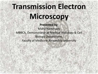
Transmission Electron
Microscopy
Presented by:
Maha Hammady
MBBCh, Demonstrator at Medical Histology & Cell
Biology Department,
Faculty of Medicine Alexandria university
Maha Hammady 4-1-2021
Transmission electron microscopy (TEM) is a significant tool in demonstrating
the ultrastructure of cells and tissues both in normal and disease states. In
particular, TEM can be crucial in the diagnosis of various renal pathologies,
the recognition of subcellular structural defects or the deposition of
extracellular material
Maha Hammady 4-1-2021
Tissue preparation for transmission
electron microscopy
-to obtain a high-quality image
and optimize the resolution of
the instrument, it is necessary to
section the tissue to a thickness
of around 80 nm.
-Sectioning at this level requires
tissues to be embedded in a rigid
material which can withstand
both the vacuum in the
microscope column and the heat
generated as the electron beam
passes through the section. The
most suitable embedding
material is the resins
Maha Hammady 4-1-2021
1-Specimen handling
-it is crucial that samples are fixed as soon as possible after the biopsy is
taken.
-The standard approach is to immerse the specimen in fixative immediately
on collection.
-Once in fixative, the specimen is cut into smaller samples using a scalpel or
razor blade. The final tissue blocks are in the form of small cubes
(approximately 1 mm3).
Maha Hammady 4-1-2021
1-Specimen handling
-Manual tissue processing is best performed by keeping the tissue sample in
the same vial throughout, and using a fine pipette to change solutions.
Maha Hammady 4-1-2021
2-Fixation
-The fixatives used in TEM generally comprise a fixing agent in buffer (to
maintain pH). The standard protocol involves primary fixation with an
aldehyde, usually glutaraldehyde, to stabilize proteins, followed by secondary
fixation in osmium tetroxide to retain lipids this is termed (double fixation).
-4F1G Fixative is the most commonly used primary fixative, it is formed of (4%
Formaldehyde & 1% Glutaraldehyde phosphate buffered saline, pH 7.4). using
this aldehyde mixture overcome the disadvantages of glutaraldehyde (a slow
penetration rate) and formaldehyde (less stable fixation) when applied
individually.
-The use of cold primary fixative (4°C) helps to minimize postmortem
changes.
Maha Hammady 4-1-2021
2-Fixation
Crosslinking is the process of chemically joining two or more molecules by a covalent
bond. ... Attachment between groups on two different proteins results in
intermolecular crosslinks that stabilize a protein-protein interaction.
Maha Hammady 4-1-2021
2-Fixation
-Volume of fixative should be at least 10 times
the volume of the tissue and it is also vital to
ensure that the tissue remains completely
submerged in the fixative.
-The time required for optimal fixation depends
on a range of factors. These include the type of
tissue, the size of the sample. In most
circumstances immersion of 0.5–1.0 mm3 blocks
of tissue for 2–6 hours is sufficient.
-Specimens fixed in aldehyde solutions should be
washed thoroughly in buffer before post-fixation
in osmium tetroxide to prevent interaction
between the fixatives which can cause
precipitation of reduced osmium.
Maha Hammady 4-1-2021
-The use of osmium tetroxide fixation to preserve
lipid is fundamental to TEM .it is usually used as a
secondary fixative at a concentration of 1 or 2%,
the penetration rate of osmium tetroxide is also
higher in stabilized tissue. So, immersion for 60–90
minutes is sufficient for most specimens. Osmium
tetroxide is generally used at room temperature.
-Osmium tetroxide is usually supplied in crystalline
form, sealed in glass ampoules. Extreme care
should be exercised when preparing this material,
gloves and eye protection should always be worn. It
is essential to only handle osmium tetroxide in a
fume-hood, as the vapor will also fix other tissues,
including the eyes and nasal tissues of the handler.
2-Fixation
Maha Hammady 4-1-2021
-The most sensitive cellular indicators of autolytic/degenerative change are
mitochondria and endoplasmic reticulum, both of which may show signs of
swelling (a reflection of osmotic imbalance) only a few minutes after the cells
are separated from a blood supply
2-Fixation
Maha Hammady 4-1-2021
3-Dehydration
-epoxy resins are immiscible with water and specimens must be dehydrated
prior to resin infiltration.
-Dehydration is performed by passing the specimen through increasing
concentration of an organic solvent to prevent the damage which would
occur with extreme changes in solvent concentration.
-The most frequently used dehydrants are acetone.
Maha Hammady 4-1-2021
4-Embedding
-Embedding mixture is prepared by mixing of Epoxy resin (araldite),
plasticizer, hardener and accelerator in a flask. The mixture is kept in oven at
60 oC for 10 minutes to eliminate air bubbles.
Maha Hammady 4-1-2021
4-Embedding
-After dehydration the tissue is infiltrated
with liquid resin mixture. The resin is
introduced gradually, beginning with a
50:50 mix of transition solvent (propylene
oxide) and resin followed by a 25:75 mix,
then finally pure resin.
- An hour in each of the preliminary
infiltration steps is usually adequate,
although some recommend leaving
samples in pure resin for 24 hours.
-Gentle agitation using a low-speed,
angled rotator during these steps will assist
resin infiltration.
Maha Hammady 4-1-2021
-Once infiltrated, tissue samples are placed in an appropriate capsule or mold
(various shapes and sizes are available) which is filled with resin. The resin is
polymerized using heat. Soft polyethylene capsules (resistant to 75°C) are
recommended for general embedding.
4-Embedding
Embedding mold Embedding capsule
Maha Hammady 4-1-2021
During polymerization, epoxy resins form cross-links, creating a three-
dimensional polymer of great mechanical strength. As well as their properties
of uniform polymerization and low shrinkage (usually less than 2%), epoxy
resins also preserve tissue ultrastructure, are stable in the electron beam,
section easily and are readily available.
4-Embedding
Maha Hammady 4-1-2021
5- Trimming
Once polymerized, blocks must be cleared of excess resin (trimmed) to
expose the tissue for sectioning. The final trimmed area should resemble a
flat-topped pyramid with a square or trapezium-shaped face
Maha Hammady 4-1-2021
6- Sectioning
Ultra-microtomes diamond knives are used to get an ultra-thin section
(approximately 60 nm). thin sections are floated out for collection as they are
cut
Maha Hammady 4-1-2021
7- Mounting
-The ultra-thin sections are mounted on copper grids (200 square mesh) 3.5
mm in diameter
Maha Hammady 4-1-2021
8-Staining
-Grids are placed on filter-paper in petri-dishes and allowed to dry.
- After drying, sections are stained with urinyl acetate for 10 minutes, and
then they are rinsed with distilled water before being stained by lead citrate
for another 10 minutes.
-This staining step increases contrast pf the resulting images
Maha Hammady 4-1-2021
9- Examination and photography
-Sections are examined and photographed by TEM
Maha Hammady 4-1-2021
The Mitochondria
Maha Hammady 4-1-2021
• Mitochondria are the cellular organelles
responsible for generating energy derived
from the breakdown of lipids and
carbohydrates, which is converted to ATP by
the process of oxidative phosphorylation.
• Mitochondria were among the first subcellular
organelles examined by electron microscopy.
In the early 1950s
The Mitochondria
Maha Hammady 4-1-2021
Ultra-structure of mitochondria:
• Mitochondria are surrounded by a double-
membrane system, consisting of inner and outer
mitochondrial membranes separated by an
intermembrane space .The inner membrane
forms numerous folds (cristae), which extend into
the interior (or matrix) of the organelle. Each of
these components plays a distinct functional role,
with the matrix and inner membrane
representing the major working compartments of
mitochondria
Maha Hammady 4-1-2021
Ultra-structure of mitochondria:
Maha Hammady 4-1-2021
Shape and organization of cristae
On the basis of their appearance in may be
broadly classified as either lamellar, tubular:
1-The lamellar form is the commonest ,cristae
have are parallel to one another, and frequently
they are orientated perpendicular to the long
axis of the mitochondrion.
2-Tubular cristae may be circular in cross-
section and they have uniform internal diameter
Maha Hammady 4-1-2021
Shape and organization of cristae
Drawings to compare the appearance of sections through lamelliform
and tubular cristae.
Maha Hammady 4-1-2021
Shape and organization of cristae
Electron micrograph of section of rat brown adipocyte tissue,
showing the regular arrangement of the cristae the majority of
which extend the full width of the mitochondrion
Maha Hammady 4-1-2021
Shape and organization of cristae
Electron micrograph of section of human uterine mucosa epithelial cell in
the middle of the menstrual cycle, showing mitochondrion with parallel
arrays of cristae characteristically in two groups, the intervening matrix
contains DNA.
Maha Hammady 4-1-2021
Shape and organization of cristae
Concentrically arranged cristae have been observed in cardiac
muscle in invertebrate ,mammalian spermatids ,fascicular zone
of the mouse adrenals and some skeletal muscles .In some of the
latter the cristae membranes show a wavy outline with abrupt,
sharp angles.
Electron micrographs of
mitochondria in the
peripheral cytoplasm of
red fibers in rat soft
palate muscle.
Maha Hammady 4-1-2021
Shape and organization of cristae
Tubular cristae (arrowheads) are present in many cell types, being a common form in
steroid-producing cells. Monkey Leydig cell
Maha Hammady 4-1-2021
Shape and organization of cristae
• The prismatic tubular cristae in the hamster
brain mitochondria form a hexagonal lattice
Electron micrograph of mitochondrion in a
section of an astrocyte from hamster
brain.
Maha Hammady 4-1-2021
Shape and organization of cristae
• tubulovesicular
Maha Hammady 4-1-2021
Distribution:
• Ordinarily mitochondria are evenly distributed
in the cytoplasm. They may, however, be
localized in certain regions
Maha Hammady 4-1-2021
Distribution:
1-proximal convoluted tubules:
they are found in the basal region.
Maha Hammady 4-1-2021
Distribution:
2-distal convoluted tubules:
they are found
in the basal region.
Maha Hammady 4-1-2021
Distribution:
3-In cardiac muscle:
1-Perinuclear mitochondria are present at nuclear
poles, they are spherical.
2-interfibrillar mitochondria are elongated and they
present between the myofibrils forming
longitudinal rows. IFM occupy the entire space
between Z-lines.
3-Subsarcolemmal mitochondria present beneath
sarcolemma, they are variable in their shapes, they
can show oval, spherical, polygonal, or horse-shoe
patterns.
Maha Hammady 4-1-2021
Distribution:
3-In cardiac muscle:
Maha Hammady 4-1-2021
Distribution:
3-In cardiac muscle:
Maha Hammady 4-1-2021
Distribution:
3-In cardiac muscle:
Maha Hammady 4-1-2021
Distribution:
4-In skeletal muscles :
Maha Hammady 4-1-2021
Distribution:
4-In skeletal muscles :
Electron microscopy imaging of
muscle fibers from a patient with
mitochondrial myopathy. from a
patient carrying a deletion
associated with Kearns-Sayre
syndrome. Note the dark, granular
structures indicated with the white
arrows. These inclusions are
aggregates of diseased
mitochondria and are a
characteristic signature of
mitochondrial dysfunction .
Maha Hammady 4-1-2021
Distribution:
5-In sperms :
the mitochondria lie in the middle piece of the
sperm forming the mitochondrial sheath.
Maha Hammady 4-1-2021
Distribution:
6-in close association with lipid droplet:
One of the frequently observed associations is
between mitochondria and lipid droplets
Rat hepatocyte mitochondria, showing close
association with a lipid droplet
Maha Hammady 4-1-2021
Distribution:
7-proximity to sites of
energy utilization:
in ciliated cells for energy
needed for ciliary motion
Human bronchial mucosa, showing
mitochondrial aggregates in the
apical cytoplasm adjacent to the
ciliary border.
X8000
Maha Hammady 4-1-2021
Shape:
• The shape is variable but is characteristic for a
cell or tissue type
• this too is dependable upon environment or
physiological conditions
Maha Hammady 4-1-2021
Size:
• The size of mitochondria also varies. In majority of the cells,
width is relatively constant, about 0.5µ, but the length varies
depends on the functional stage of the cell.
• Megamitochondria: Regardless o f internal configuration,
mitochondria rarely are larger than 1 µm
• in diameter or length. been reported to occur in human
tumors. In the pleomorphic adenoma of the submandibular
gland
Maha Hammady 4-1-2021
Size:
• Megamitochondria: Regardless o f internal configuration, mitochondria
rarely are larger than 1 µ m in diameter or length. been reported to occur
in human tumors. Some o f these megamitochondria contained expanded
cristae.
pleomorphic adenoma of the human submandibular gland: A huge mitochondrion with
arabesque cristae next to the nucleus. Bar :2 µ m
Maha Hammady 4-1-2021
Number:
• The mitochondria content of a cell is difficult to determine, but in
general, it varies with the cell type and functional stage. Thus cells
with high metabolic activity have a high number of mitochondria,
while those with low metabolic activity have a lower number.
• It is estimated that in liver mitochondria constitute 30 to 35 % of
the total content of the cell (normal liver cell contains about 100 to
1600 mitochondria)
• in kidney, 20 % of the total content of the cell
• In lymphoid tissue the value is much lower.
• but this number diminishes during regeneration and also in
cancerous tissue. This last observation may be related to decreased
oxidation that accompanies the increase to anaerobic glycolysis in
cancer.
Maha Hammady 4-1-2021
summary
• Mitochondria are the cellular organelles
responsible for generating energy
• Mitochondria are surrounded by a double-
membrane system, consisting of inner and outer
mitochondrial membranes separated by an
intermembrane space .The inner membrane
forms numerous folds (cristae), which extend into
the interior (or matrix) of the organelle.
• cristae are classified as either lamellar, tubular.
• Mitochondria in general vary in their shape, size,
number and distribution among cells.
Maha Hammady 4-1-2021

More Related Content

What's hot

Transmission Electron Microscope
Transmission Electron MicroscopeTransmission Electron Microscope
Transmission Electron Microscope20was
 
Transmission electron microscope
Transmission electron microscopeTransmission electron microscope
Transmission electron microscopeSenthil Arasan
 
Confocal microscope presentation pt
Confocal microscope presentation  ptConfocal microscope presentation  pt
Confocal microscope presentation ptShrigouri4605
 
Confocal laser scanning microscopy (clsm)
Confocal laser scanning microscopy (clsm)Confocal laser scanning microscopy (clsm)
Confocal laser scanning microscopy (clsm)Nethravathi Siri
 
3D tumor spheroid models for in vitro therapeutic screening: a systematic app...
3D tumor spheroid models for in vitro therapeutic screening: a systematic app...3D tumor spheroid models for in vitro therapeutic screening: a systematic app...
3D tumor spheroid models for in vitro therapeutic screening: a systematic app...Arun kumar
 
Optical microscopy
Optical microscopyOptical microscopy
Optical microscopyULVAN OZAD
 
Electron microscopy
Electron microscopyElectron microscopy
Electron microscopyGanga Huvin
 
Introduction to Atomic Force Microscopy
Introduction to Atomic Force MicroscopyIntroduction to Atomic Force Microscopy
Introduction to Atomic Force MicroscopyMd Ataul Mamun
 
Confocal laser scanning microscopy
Confocal laser scanning microscopyConfocal laser scanning microscopy
Confocal laser scanning microscopyBaskar Selvam
 
Applications of atomic force microscope(by kanupriya)
Applications of atomic force microscope(by kanupriya)Applications of atomic force microscope(by kanupriya)
Applications of atomic force microscope(by kanupriya)KANUPRIYASINGH19
 
Phase contrast microscope
Phase contrast microscopePhase contrast microscope
Phase contrast microscopeSalman Khan
 
Electron microscope - SEM and TEM
Electron microscope - SEM and TEMElectron microscope - SEM and TEM
Electron microscope - SEM and TEMgaurav raja
 
Confocal microscopy Likhith K
Confocal microscopy Likhith KConfocal microscopy Likhith K
Confocal microscopy Likhith KLIKHITHK1
 
Cryo-Electron Microscopy
Cryo-Electron MicroscopyCryo-Electron Microscopy
Cryo-Electron MicroscopyRabia Aziz
 

What's hot (20)

Scanning electron microscope
Scanning electron microscope  Scanning electron microscope
Scanning electron microscope
 
Transmission Electron Microscope
Transmission Electron MicroscopeTransmission Electron Microscope
Transmission Electron Microscope
 
Transmission electron microscope
Transmission electron microscopeTransmission electron microscope
Transmission electron microscope
 
Confocal microscope presentation pt
Confocal microscope presentation  ptConfocal microscope presentation  pt
Confocal microscope presentation pt
 
Confocal laser scanning microscopy (clsm)
Confocal laser scanning microscopy (clsm)Confocal laser scanning microscopy (clsm)
Confocal laser scanning microscopy (clsm)
 
3D tumor spheroid models for in vitro therapeutic screening: a systematic app...
3D tumor spheroid models for in vitro therapeutic screening: a systematic app...3D tumor spheroid models for in vitro therapeutic screening: a systematic app...
3D tumor spheroid models for in vitro therapeutic screening: a systematic app...
 
Cytotoxicty study
Cytotoxicty studyCytotoxicty study
Cytotoxicty study
 
Confocal microscopy
Confocal microscopyConfocal microscopy
Confocal microscopy
 
Optical microscopy
Optical microscopyOptical microscopy
Optical microscopy
 
Confocal microscopy
Confocal microscopyConfocal microscopy
Confocal microscopy
 
Nomarski dic
Nomarski dicNomarski dic
Nomarski dic
 
Electron microscopy
Electron microscopyElectron microscopy
Electron microscopy
 
Confocal microscopy
Confocal microscopyConfocal microscopy
Confocal microscopy
 
Introduction to Atomic Force Microscopy
Introduction to Atomic Force MicroscopyIntroduction to Atomic Force Microscopy
Introduction to Atomic Force Microscopy
 
Confocal laser scanning microscopy
Confocal laser scanning microscopyConfocal laser scanning microscopy
Confocal laser scanning microscopy
 
Applications of atomic force microscope(by kanupriya)
Applications of atomic force microscope(by kanupriya)Applications of atomic force microscope(by kanupriya)
Applications of atomic force microscope(by kanupriya)
 
Phase contrast microscope
Phase contrast microscopePhase contrast microscope
Phase contrast microscope
 
Electron microscope - SEM and TEM
Electron microscope - SEM and TEMElectron microscope - SEM and TEM
Electron microscope - SEM and TEM
 
Confocal microscopy Likhith K
Confocal microscopy Likhith KConfocal microscopy Likhith K
Confocal microscopy Likhith K
 
Cryo-Electron Microscopy
Cryo-Electron MicroscopyCryo-Electron Microscopy
Cryo-Electron Microscopy
 

Similar to Transmission Electron Microscopy

PREPARATION OF HISTOLOGICAL SPECIMENS.pptx
PREPARATION OF HISTOLOGICAL SPECIMENS.pptxPREPARATION OF HISTOLOGICAL SPECIMENS.pptx
PREPARATION OF HISTOLOGICAL SPECIMENS.pptxAnthonyMatu1
 
Microtomy - Preparation of Histological Slides
Microtomy - Preparation of Histological SlidesMicrotomy - Preparation of Histological Slides
Microtomy - Preparation of Histological SlidesSyed Muhammad Khan
 
Microtomy - Preparation of Histological Slides
Microtomy - Preparation of Histological SlidesMicrotomy - Preparation of Histological Slides
Microtomy - Preparation of Histological SlidesSyed Muhammad Khan
 
Tissue processing by dr manzoor
Tissue processing by dr manzoorTissue processing by dr manzoor
Tissue processing by dr manzoorMohammad Manzoor
 
Microscopy
MicroscopyMicroscopy
Microscopytrixie19
 
Adhesion Presentation.pptx
Adhesion Presentation.pptxAdhesion Presentation.pptx
Adhesion Presentation.pptxMostafaElzaim
 
Histological Techaniques
Histological TechaniquesHistological Techaniques
Histological TechaniquesZahoor Ahmed
 
Histological techniques
Histological techniquesHistological techniques
Histological techniquesRUPALIMUNDE
 
Histotechnique for practicals pathology
Histotechnique for practicals pathologyHistotechnique for practicals pathology
Histotechnique for practicals pathologyAppy Akshay Agarwal
 
HISTOTECHNIQUES MICROTOMY.ppt · version 1.pdf
HISTOTECHNIQUES  MICROTOMY.ppt · version 1.pdfHISTOTECHNIQUES  MICROTOMY.ppt · version 1.pdf
HISTOTECHNIQUES MICROTOMY.ppt · version 1.pdfmahrukhmughal27
 
Technique of Histopathology
Technique of HistopathologyTechnique of Histopathology
Technique of Histopathologysandeep singh
 
Technique of Histopathology
Technique of HistopathologyTechnique of Histopathology
Technique of Histopathologysandeep singh
 
Tissue processing(1)
Tissue processing(1)Tissue processing(1)
Tissue processing(1)meelumohan
 

Similar to Transmission Electron Microscopy (20)

PREPARATION OF HISTOLOGICAL SPECIMENS.pptx
PREPARATION OF HISTOLOGICAL SPECIMENS.pptxPREPARATION OF HISTOLOGICAL SPECIMENS.pptx
PREPARATION OF HISTOLOGICAL SPECIMENS.pptx
 
Gen. histology (introduction) 1
Gen. histology (introduction) 1Gen. histology (introduction) 1
Gen. histology (introduction) 1
 
Microtomy - Preparation of Histological Slides
Microtomy - Preparation of Histological SlidesMicrotomy - Preparation of Histological Slides
Microtomy - Preparation of Histological Slides
 
Plant histology
Plant histologyPlant histology
Plant histology
 
Microtomy - Preparation of Histological Slides
Microtomy - Preparation of Histological SlidesMicrotomy - Preparation of Histological Slides
Microtomy - Preparation of Histological Slides
 
Tissue processing by dr manzoor
Tissue processing by dr manzoorTissue processing by dr manzoor
Tissue processing by dr manzoor
 
Microscopy
MicroscopyMicroscopy
Microscopy
 
Chapter 2 bio 300 obe
Chapter 2 bio 300 obeChapter 2 bio 300 obe
Chapter 2 bio 300 obe
 
Adhesion Presentation.pptx
Adhesion Presentation.pptxAdhesion Presentation.pptx
Adhesion Presentation.pptx
 
Electron microscope
Electron microscopeElectron microscope
Electron microscope
 
Histological Techaniques
Histological TechaniquesHistological Techaniques
Histological Techaniques
 
Histological techniques
Histological techniquesHistological techniques
Histological techniques
 
Histotechnique for practicals pathology
Histotechnique for practicals pathologyHistotechnique for practicals pathology
Histotechnique for practicals pathology
 
HISTOTECHNIQUES MICROTOMY.ppt · version 1.pdf
HISTOTECHNIQUES  MICROTOMY.ppt · version 1.pdfHISTOTECHNIQUES  MICROTOMY.ppt · version 1.pdf
HISTOTECHNIQUES MICROTOMY.ppt · version 1.pdf
 
Composite resins part ii
Composite resins   part iiComposite resins   part ii
Composite resins part ii
 
Technique of Histopathology
Technique of HistopathologyTechnique of Histopathology
Technique of Histopathology
 
Technique of Histopathology
Technique of HistopathologyTechnique of Histopathology
Technique of Histopathology
 
Elastomeric Fiber
Elastomeric Fiber Elastomeric Fiber
Elastomeric Fiber
 
Tissue processing(1)
Tissue processing(1)Tissue processing(1)
Tissue processing(1)
 
Chap9 downstream processing
Chap9 downstream processingChap9 downstream processing
Chap9 downstream processing
 

More from Maha Hammady

Histological Structure of The Renal Corpuscle Suits its function- maha hammad...
Histological Structure of The Renal Corpuscle Suits its function- maha hammad...Histological Structure of The Renal Corpuscle Suits its function- maha hammad...
Histological Structure of The Renal Corpuscle Suits its function- maha hammad...Maha Hammady
 
Histological review of the cardiac muscle-maha hammady.pptx
Histological review of the cardiac muscle-maha hammady.pptxHistological review of the cardiac muscle-maha hammady.pptx
Histological review of the cardiac muscle-maha hammady.pptxMaha Hammady
 
hematopoiesis histology-maha hammady.pptx
hematopoiesis histology-maha hammady.pptxhematopoiesis histology-maha hammady.pptx
hematopoiesis histology-maha hammady.pptxMaha Hammady
 
Skeletal Muscle histology-maha hammady.pptx
Skeletal Muscle histology-maha hammady.pptxSkeletal Muscle histology-maha hammady.pptx
Skeletal Muscle histology-maha hammady.pptxMaha Hammady
 
Mitochondrial dna and aging
Mitochondrial dna and agingMitochondrial dna and aging
Mitochondrial dna and agingMaha Hammady
 
Adipose tissue histology
Adipose tissue histologyAdipose tissue histology
Adipose tissue histologyMaha Hammady
 
Histology of the Kidney part 1
Histology of the Kidney part 1Histology of the Kidney part 1
Histology of the Kidney part 1Maha Hammady
 
Mast cell histology and its visualization methods
Mast cell histology and its visualization methodsMast cell histology and its visualization methods
Mast cell histology and its visualization methodsMaha Hammady
 
Histology of Uterus and placenta
Histology of Uterus and placentaHistology of Uterus and placenta
Histology of Uterus and placentaMaha Hammady
 
Systemic sclerosis
Systemic sclerosisSystemic sclerosis
Systemic sclerosisMaha Hammady
 
Management of extra utrine pregnancy
Management of extra utrine pregnancyManagement of extra utrine pregnancy
Management of extra utrine pregnancyMaha Hammady
 

More from Maha Hammady (13)

Histological Structure of The Renal Corpuscle Suits its function- maha hammad...
Histological Structure of The Renal Corpuscle Suits its function- maha hammad...Histological Structure of The Renal Corpuscle Suits its function- maha hammad...
Histological Structure of The Renal Corpuscle Suits its function- maha hammad...
 
Histological review of the cardiac muscle-maha hammady.pptx
Histological review of the cardiac muscle-maha hammady.pptxHistological review of the cardiac muscle-maha hammady.pptx
Histological review of the cardiac muscle-maha hammady.pptx
 
hematopoiesis histology-maha hammady.pptx
hematopoiesis histology-maha hammady.pptxhematopoiesis histology-maha hammady.pptx
hematopoiesis histology-maha hammady.pptx
 
Skeletal Muscle histology-maha hammady.pptx
Skeletal Muscle histology-maha hammady.pptxSkeletal Muscle histology-maha hammady.pptx
Skeletal Muscle histology-maha hammady.pptx
 
Mitochondrial dna and aging
Mitochondrial dna and agingMitochondrial dna and aging
Mitochondrial dna and aging
 
Adipose tissue histology
Adipose tissue histologyAdipose tissue histology
Adipose tissue histology
 
The Cell cycle
The Cell cycleThe Cell cycle
The Cell cycle
 
Histology of the Kidney part 1
Histology of the Kidney part 1Histology of the Kidney part 1
Histology of the Kidney part 1
 
Mast cell histology and its visualization methods
Mast cell histology and its visualization methodsMast cell histology and its visualization methods
Mast cell histology and its visualization methods
 
Histology of Uterus and placenta
Histology of Uterus and placentaHistology of Uterus and placenta
Histology of Uterus and placenta
 
Hands only CPR
Hands only CPRHands only CPR
Hands only CPR
 
Systemic sclerosis
Systemic sclerosisSystemic sclerosis
Systemic sclerosis
 
Management of extra utrine pregnancy
Management of extra utrine pregnancyManagement of extra utrine pregnancy
Management of extra utrine pregnancy
 

Recently uploaded

User Guide: Capricorn FLX™ Weather Station
User Guide: Capricorn FLX™ Weather StationUser Guide: Capricorn FLX™ Weather Station
User Guide: Capricorn FLX™ Weather StationColumbia Weather Systems
 
Behavioral Disorder: Schizophrenia & it's Case Study.pdf
Behavioral Disorder: Schizophrenia & it's Case Study.pdfBehavioral Disorder: Schizophrenia & it's Case Study.pdf
Behavioral Disorder: Schizophrenia & it's Case Study.pdfSELF-EXPLANATORY
 
Base editing, prime editing, Cas13 & RNA editing and organelle base editing
Base editing, prime editing, Cas13 & RNA editing and organelle base editingBase editing, prime editing, Cas13 & RNA editing and organelle base editing
Base editing, prime editing, Cas13 & RNA editing and organelle base editingNetHelix
 
preservation, maintanence and improvement of industrial organism.pptx
preservation, maintanence and improvement of industrial organism.pptxpreservation, maintanence and improvement of industrial organism.pptx
preservation, maintanence and improvement of industrial organism.pptxnoordubaliya2003
 
Pests of Bengal gram_Identification_Dr.UPR.pdf
Pests of Bengal gram_Identification_Dr.UPR.pdfPests of Bengal gram_Identification_Dr.UPR.pdf
Pests of Bengal gram_Identification_Dr.UPR.pdfPirithiRaju
 
Harmful and Useful Microorganisms Presentation
Harmful and Useful Microorganisms PresentationHarmful and Useful Microorganisms Presentation
Harmful and Useful Microorganisms Presentationtahreemzahra82
 
Call Girls in Majnu Ka Tilla Delhi 🔝9711014705🔝 Genuine
Call Girls in Majnu Ka Tilla Delhi 🔝9711014705🔝 GenuineCall Girls in Majnu Ka Tilla Delhi 🔝9711014705🔝 Genuine
Call Girls in Majnu Ka Tilla Delhi 🔝9711014705🔝 Genuinethapagita
 
Solution chemistry, Moral and Normal solutions
Solution chemistry, Moral and Normal solutionsSolution chemistry, Moral and Normal solutions
Solution chemistry, Moral and Normal solutionsHajira Mahmood
 
Microteaching on terms used in filtration .Pharmaceutical Engineering
Microteaching on terms used in filtration .Pharmaceutical EngineeringMicroteaching on terms used in filtration .Pharmaceutical Engineering
Microteaching on terms used in filtration .Pharmaceutical EngineeringPrajakta Shinde
 
Pests of safflower_Binomics_Identification_Dr.UPR.pdf
Pests of safflower_Binomics_Identification_Dr.UPR.pdfPests of safflower_Binomics_Identification_Dr.UPR.pdf
Pests of safflower_Binomics_Identification_Dr.UPR.pdfPirithiRaju
 
RESPIRATORY ADAPTATIONS TO HYPOXIA IN HUMNAS.pptx
RESPIRATORY ADAPTATIONS TO HYPOXIA IN HUMNAS.pptxRESPIRATORY ADAPTATIONS TO HYPOXIA IN HUMNAS.pptx
RESPIRATORY ADAPTATIONS TO HYPOXIA IN HUMNAS.pptxFarihaAbdulRasheed
 
Pests of soyabean_Binomics_IdentificationDr.UPR.pdf
Pests of soyabean_Binomics_IdentificationDr.UPR.pdfPests of soyabean_Binomics_IdentificationDr.UPR.pdf
Pests of soyabean_Binomics_IdentificationDr.UPR.pdfPirithiRaju
 
Pests of castor_Binomics_Identification_Dr.UPR.pdf
Pests of castor_Binomics_Identification_Dr.UPR.pdfPests of castor_Binomics_Identification_Dr.UPR.pdf
Pests of castor_Binomics_Identification_Dr.UPR.pdfPirithiRaju
 
Transposable elements in prokaryotes.ppt
Transposable elements in prokaryotes.pptTransposable elements in prokaryotes.ppt
Transposable elements in prokaryotes.pptArshadWarsi13
 
STOPPED FLOW METHOD & APPLICATION MURUGAVENI B.pptx
STOPPED FLOW METHOD & APPLICATION MURUGAVENI B.pptxSTOPPED FLOW METHOD & APPLICATION MURUGAVENI B.pptx
STOPPED FLOW METHOD & APPLICATION MURUGAVENI B.pptxMurugaveni B
 
(9818099198) Call Girls In Noida Sector 14 (NOIDA ESCORTS)
(9818099198) Call Girls In Noida Sector 14 (NOIDA ESCORTS)(9818099198) Call Girls In Noida Sector 14 (NOIDA ESCORTS)
(9818099198) Call Girls In Noida Sector 14 (NOIDA ESCORTS)riyaescorts54
 
Grafana in space: Monitoring Japan's SLIM moon lander in real time
Grafana in space: Monitoring Japan's SLIM moon lander  in real timeGrafana in space: Monitoring Japan's SLIM moon lander  in real time
Grafana in space: Monitoring Japan's SLIM moon lander in real timeSatoshi NAKAHIRA
 
LIGHT-PHENOMENA-BY-CABUALDIONALDOPANOGANCADIENTE-CONDEZA (1).pptx
LIGHT-PHENOMENA-BY-CABUALDIONALDOPANOGANCADIENTE-CONDEZA (1).pptxLIGHT-PHENOMENA-BY-CABUALDIONALDOPANOGANCADIENTE-CONDEZA (1).pptx
LIGHT-PHENOMENA-BY-CABUALDIONALDOPANOGANCADIENTE-CONDEZA (1).pptxmalonesandreagweneth
 

Recently uploaded (20)

User Guide: Capricorn FLX™ Weather Station
User Guide: Capricorn FLX™ Weather StationUser Guide: Capricorn FLX™ Weather Station
User Guide: Capricorn FLX™ Weather Station
 
Behavioral Disorder: Schizophrenia & it's Case Study.pdf
Behavioral Disorder: Schizophrenia & it's Case Study.pdfBehavioral Disorder: Schizophrenia & it's Case Study.pdf
Behavioral Disorder: Schizophrenia & it's Case Study.pdf
 
Base editing, prime editing, Cas13 & RNA editing and organelle base editing
Base editing, prime editing, Cas13 & RNA editing and organelle base editingBase editing, prime editing, Cas13 & RNA editing and organelle base editing
Base editing, prime editing, Cas13 & RNA editing and organelle base editing
 
Volatile Oils Pharmacognosy And Phytochemistry -I
Volatile Oils Pharmacognosy And Phytochemistry -IVolatile Oils Pharmacognosy And Phytochemistry -I
Volatile Oils Pharmacognosy And Phytochemistry -I
 
preservation, maintanence and improvement of industrial organism.pptx
preservation, maintanence and improvement of industrial organism.pptxpreservation, maintanence and improvement of industrial organism.pptx
preservation, maintanence and improvement of industrial organism.pptx
 
Pests of Bengal gram_Identification_Dr.UPR.pdf
Pests of Bengal gram_Identification_Dr.UPR.pdfPests of Bengal gram_Identification_Dr.UPR.pdf
Pests of Bengal gram_Identification_Dr.UPR.pdf
 
Harmful and Useful Microorganisms Presentation
Harmful and Useful Microorganisms PresentationHarmful and Useful Microorganisms Presentation
Harmful and Useful Microorganisms Presentation
 
Call Girls in Majnu Ka Tilla Delhi 🔝9711014705🔝 Genuine
Call Girls in Majnu Ka Tilla Delhi 🔝9711014705🔝 GenuineCall Girls in Majnu Ka Tilla Delhi 🔝9711014705🔝 Genuine
Call Girls in Majnu Ka Tilla Delhi 🔝9711014705🔝 Genuine
 
Solution chemistry, Moral and Normal solutions
Solution chemistry, Moral and Normal solutionsSolution chemistry, Moral and Normal solutions
Solution chemistry, Moral and Normal solutions
 
Microteaching on terms used in filtration .Pharmaceutical Engineering
Microteaching on terms used in filtration .Pharmaceutical EngineeringMicroteaching on terms used in filtration .Pharmaceutical Engineering
Microteaching on terms used in filtration .Pharmaceutical Engineering
 
Pests of safflower_Binomics_Identification_Dr.UPR.pdf
Pests of safflower_Binomics_Identification_Dr.UPR.pdfPests of safflower_Binomics_Identification_Dr.UPR.pdf
Pests of safflower_Binomics_Identification_Dr.UPR.pdf
 
RESPIRATORY ADAPTATIONS TO HYPOXIA IN HUMNAS.pptx
RESPIRATORY ADAPTATIONS TO HYPOXIA IN HUMNAS.pptxRESPIRATORY ADAPTATIONS TO HYPOXIA IN HUMNAS.pptx
RESPIRATORY ADAPTATIONS TO HYPOXIA IN HUMNAS.pptx
 
Pests of soyabean_Binomics_IdentificationDr.UPR.pdf
Pests of soyabean_Binomics_IdentificationDr.UPR.pdfPests of soyabean_Binomics_IdentificationDr.UPR.pdf
Pests of soyabean_Binomics_IdentificationDr.UPR.pdf
 
Pests of castor_Binomics_Identification_Dr.UPR.pdf
Pests of castor_Binomics_Identification_Dr.UPR.pdfPests of castor_Binomics_Identification_Dr.UPR.pdf
Pests of castor_Binomics_Identification_Dr.UPR.pdf
 
Hot Sexy call girls in Moti Nagar,🔝 9953056974 🔝 escort Service
Hot Sexy call girls in  Moti Nagar,🔝 9953056974 🔝 escort ServiceHot Sexy call girls in  Moti Nagar,🔝 9953056974 🔝 escort Service
Hot Sexy call girls in Moti Nagar,🔝 9953056974 🔝 escort Service
 
Transposable elements in prokaryotes.ppt
Transposable elements in prokaryotes.pptTransposable elements in prokaryotes.ppt
Transposable elements in prokaryotes.ppt
 
STOPPED FLOW METHOD & APPLICATION MURUGAVENI B.pptx
STOPPED FLOW METHOD & APPLICATION MURUGAVENI B.pptxSTOPPED FLOW METHOD & APPLICATION MURUGAVENI B.pptx
STOPPED FLOW METHOD & APPLICATION MURUGAVENI B.pptx
 
(9818099198) Call Girls In Noida Sector 14 (NOIDA ESCORTS)
(9818099198) Call Girls In Noida Sector 14 (NOIDA ESCORTS)(9818099198) Call Girls In Noida Sector 14 (NOIDA ESCORTS)
(9818099198) Call Girls In Noida Sector 14 (NOIDA ESCORTS)
 
Grafana in space: Monitoring Japan's SLIM moon lander in real time
Grafana in space: Monitoring Japan's SLIM moon lander  in real timeGrafana in space: Monitoring Japan's SLIM moon lander  in real time
Grafana in space: Monitoring Japan's SLIM moon lander in real time
 
LIGHT-PHENOMENA-BY-CABUALDIONALDOPANOGANCADIENTE-CONDEZA (1).pptx
LIGHT-PHENOMENA-BY-CABUALDIONALDOPANOGANCADIENTE-CONDEZA (1).pptxLIGHT-PHENOMENA-BY-CABUALDIONALDOPANOGANCADIENTE-CONDEZA (1).pptx
LIGHT-PHENOMENA-BY-CABUALDIONALDOPANOGANCADIENTE-CONDEZA (1).pptx
 

Transmission Electron Microscopy

  • 1. Transmission Electron Microscopy Presented by: Maha Hammady MBBCh, Demonstrator at Medical Histology & Cell Biology Department, Faculty of Medicine Alexandria university Maha Hammady 4-1-2021
  • 2. Transmission electron microscopy (TEM) is a significant tool in demonstrating the ultrastructure of cells and tissues both in normal and disease states. In particular, TEM can be crucial in the diagnosis of various renal pathologies, the recognition of subcellular structural defects or the deposition of extracellular material Maha Hammady 4-1-2021
  • 3. Tissue preparation for transmission electron microscopy -to obtain a high-quality image and optimize the resolution of the instrument, it is necessary to section the tissue to a thickness of around 80 nm. -Sectioning at this level requires tissues to be embedded in a rigid material which can withstand both the vacuum in the microscope column and the heat generated as the electron beam passes through the section. The most suitable embedding material is the resins Maha Hammady 4-1-2021
  • 4. 1-Specimen handling -it is crucial that samples are fixed as soon as possible after the biopsy is taken. -The standard approach is to immerse the specimen in fixative immediately on collection. -Once in fixative, the specimen is cut into smaller samples using a scalpel or razor blade. The final tissue blocks are in the form of small cubes (approximately 1 mm3). Maha Hammady 4-1-2021
  • 5. 1-Specimen handling -Manual tissue processing is best performed by keeping the tissue sample in the same vial throughout, and using a fine pipette to change solutions. Maha Hammady 4-1-2021
  • 6. 2-Fixation -The fixatives used in TEM generally comprise a fixing agent in buffer (to maintain pH). The standard protocol involves primary fixation with an aldehyde, usually glutaraldehyde, to stabilize proteins, followed by secondary fixation in osmium tetroxide to retain lipids this is termed (double fixation). -4F1G Fixative is the most commonly used primary fixative, it is formed of (4% Formaldehyde & 1% Glutaraldehyde phosphate buffered saline, pH 7.4). using this aldehyde mixture overcome the disadvantages of glutaraldehyde (a slow penetration rate) and formaldehyde (less stable fixation) when applied individually. -The use of cold primary fixative (4°C) helps to minimize postmortem changes. Maha Hammady 4-1-2021
  • 7. 2-Fixation Crosslinking is the process of chemically joining two or more molecules by a covalent bond. ... Attachment between groups on two different proteins results in intermolecular crosslinks that stabilize a protein-protein interaction. Maha Hammady 4-1-2021
  • 8. 2-Fixation -Volume of fixative should be at least 10 times the volume of the tissue and it is also vital to ensure that the tissue remains completely submerged in the fixative. -The time required for optimal fixation depends on a range of factors. These include the type of tissue, the size of the sample. In most circumstances immersion of 0.5–1.0 mm3 blocks of tissue for 2–6 hours is sufficient. -Specimens fixed in aldehyde solutions should be washed thoroughly in buffer before post-fixation in osmium tetroxide to prevent interaction between the fixatives which can cause precipitation of reduced osmium. Maha Hammady 4-1-2021
  • 9. -The use of osmium tetroxide fixation to preserve lipid is fundamental to TEM .it is usually used as a secondary fixative at a concentration of 1 or 2%, the penetration rate of osmium tetroxide is also higher in stabilized tissue. So, immersion for 60–90 minutes is sufficient for most specimens. Osmium tetroxide is generally used at room temperature. -Osmium tetroxide is usually supplied in crystalline form, sealed in glass ampoules. Extreme care should be exercised when preparing this material, gloves and eye protection should always be worn. It is essential to only handle osmium tetroxide in a fume-hood, as the vapor will also fix other tissues, including the eyes and nasal tissues of the handler. 2-Fixation Maha Hammady 4-1-2021
  • 10. -The most sensitive cellular indicators of autolytic/degenerative change are mitochondria and endoplasmic reticulum, both of which may show signs of swelling (a reflection of osmotic imbalance) only a few minutes after the cells are separated from a blood supply 2-Fixation Maha Hammady 4-1-2021
  • 11. 3-Dehydration -epoxy resins are immiscible with water and specimens must be dehydrated prior to resin infiltration. -Dehydration is performed by passing the specimen through increasing concentration of an organic solvent to prevent the damage which would occur with extreme changes in solvent concentration. -The most frequently used dehydrants are acetone. Maha Hammady 4-1-2021
  • 12. 4-Embedding -Embedding mixture is prepared by mixing of Epoxy resin (araldite), plasticizer, hardener and accelerator in a flask. The mixture is kept in oven at 60 oC for 10 minutes to eliminate air bubbles. Maha Hammady 4-1-2021
  • 13. 4-Embedding -After dehydration the tissue is infiltrated with liquid resin mixture. The resin is introduced gradually, beginning with a 50:50 mix of transition solvent (propylene oxide) and resin followed by a 25:75 mix, then finally pure resin. - An hour in each of the preliminary infiltration steps is usually adequate, although some recommend leaving samples in pure resin for 24 hours. -Gentle agitation using a low-speed, angled rotator during these steps will assist resin infiltration. Maha Hammady 4-1-2021
  • 14. -Once infiltrated, tissue samples are placed in an appropriate capsule or mold (various shapes and sizes are available) which is filled with resin. The resin is polymerized using heat. Soft polyethylene capsules (resistant to 75°C) are recommended for general embedding. 4-Embedding Embedding mold Embedding capsule Maha Hammady 4-1-2021
  • 15. During polymerization, epoxy resins form cross-links, creating a three- dimensional polymer of great mechanical strength. As well as their properties of uniform polymerization and low shrinkage (usually less than 2%), epoxy resins also preserve tissue ultrastructure, are stable in the electron beam, section easily and are readily available. 4-Embedding Maha Hammady 4-1-2021
  • 16. 5- Trimming Once polymerized, blocks must be cleared of excess resin (trimmed) to expose the tissue for sectioning. The final trimmed area should resemble a flat-topped pyramid with a square or trapezium-shaped face Maha Hammady 4-1-2021
  • 17. 6- Sectioning Ultra-microtomes diamond knives are used to get an ultra-thin section (approximately 60 nm). thin sections are floated out for collection as they are cut Maha Hammady 4-1-2021
  • 18. 7- Mounting -The ultra-thin sections are mounted on copper grids (200 square mesh) 3.5 mm in diameter Maha Hammady 4-1-2021
  • 19. 8-Staining -Grids are placed on filter-paper in petri-dishes and allowed to dry. - After drying, sections are stained with urinyl acetate for 10 minutes, and then they are rinsed with distilled water before being stained by lead citrate for another 10 minutes. -This staining step increases contrast pf the resulting images Maha Hammady 4-1-2021
  • 20. 8-Staining -Grids are placed on filter-paper in petri-dishes and allowed to dry. - After drying, sections are stained with urinyl acetate for 10 minutes, and then they are rinsed with distilled water before being stained by lead citrate for another 10 minutes. -This staining step increases contrast pf the resulting images Maha Hammady 4-1-2021
  • 21. 9- Examination and photography -Sections are examined and photographed by TEM Maha Hammady 4-1-2021
  • 23. • Mitochondria are the cellular organelles responsible for generating energy derived from the breakdown of lipids and carbohydrates, which is converted to ATP by the process of oxidative phosphorylation. • Mitochondria were among the first subcellular organelles examined by electron microscopy. In the early 1950s The Mitochondria Maha Hammady 4-1-2021
  • 24. Ultra-structure of mitochondria: • Mitochondria are surrounded by a double- membrane system, consisting of inner and outer mitochondrial membranes separated by an intermembrane space .The inner membrane forms numerous folds (cristae), which extend into the interior (or matrix) of the organelle. Each of these components plays a distinct functional role, with the matrix and inner membrane representing the major working compartments of mitochondria Maha Hammady 4-1-2021
  • 26. Shape and organization of cristae On the basis of their appearance in may be broadly classified as either lamellar, tubular: 1-The lamellar form is the commonest ,cristae have are parallel to one another, and frequently they are orientated perpendicular to the long axis of the mitochondrion. 2-Tubular cristae may be circular in cross- section and they have uniform internal diameter Maha Hammady 4-1-2021
  • 27. Shape and organization of cristae Drawings to compare the appearance of sections through lamelliform and tubular cristae. Maha Hammady 4-1-2021
  • 28. Shape and organization of cristae Electron micrograph of section of rat brown adipocyte tissue, showing the regular arrangement of the cristae the majority of which extend the full width of the mitochondrion Maha Hammady 4-1-2021
  • 29. Shape and organization of cristae Electron micrograph of section of human uterine mucosa epithelial cell in the middle of the menstrual cycle, showing mitochondrion with parallel arrays of cristae characteristically in two groups, the intervening matrix contains DNA. Maha Hammady 4-1-2021
  • 30. Shape and organization of cristae Concentrically arranged cristae have been observed in cardiac muscle in invertebrate ,mammalian spermatids ,fascicular zone of the mouse adrenals and some skeletal muscles .In some of the latter the cristae membranes show a wavy outline with abrupt, sharp angles. Electron micrographs of mitochondria in the peripheral cytoplasm of red fibers in rat soft palate muscle. Maha Hammady 4-1-2021
  • 31. Shape and organization of cristae Tubular cristae (arrowheads) are present in many cell types, being a common form in steroid-producing cells. Monkey Leydig cell Maha Hammady 4-1-2021
  • 32. Shape and organization of cristae • The prismatic tubular cristae in the hamster brain mitochondria form a hexagonal lattice Electron micrograph of mitochondrion in a section of an astrocyte from hamster brain. Maha Hammady 4-1-2021
  • 33. Shape and organization of cristae • tubulovesicular Maha Hammady 4-1-2021
  • 34. Distribution: • Ordinarily mitochondria are evenly distributed in the cytoplasm. They may, however, be localized in certain regions Maha Hammady 4-1-2021
  • 35. Distribution: 1-proximal convoluted tubules: they are found in the basal region. Maha Hammady 4-1-2021
  • 36. Distribution: 2-distal convoluted tubules: they are found in the basal region. Maha Hammady 4-1-2021
  • 37. Distribution: 3-In cardiac muscle: 1-Perinuclear mitochondria are present at nuclear poles, they are spherical. 2-interfibrillar mitochondria are elongated and they present between the myofibrils forming longitudinal rows. IFM occupy the entire space between Z-lines. 3-Subsarcolemmal mitochondria present beneath sarcolemma, they are variable in their shapes, they can show oval, spherical, polygonal, or horse-shoe patterns. Maha Hammady 4-1-2021
  • 41. Distribution: 4-In skeletal muscles : Maha Hammady 4-1-2021
  • 42. Distribution: 4-In skeletal muscles : Electron microscopy imaging of muscle fibers from a patient with mitochondrial myopathy. from a patient carrying a deletion associated with Kearns-Sayre syndrome. Note the dark, granular structures indicated with the white arrows. These inclusions are aggregates of diseased mitochondria and are a characteristic signature of mitochondrial dysfunction . Maha Hammady 4-1-2021
  • 43. Distribution: 5-In sperms : the mitochondria lie in the middle piece of the sperm forming the mitochondrial sheath. Maha Hammady 4-1-2021
  • 44. Distribution: 6-in close association with lipid droplet: One of the frequently observed associations is between mitochondria and lipid droplets Rat hepatocyte mitochondria, showing close association with a lipid droplet Maha Hammady 4-1-2021
  • 45. Distribution: 7-proximity to sites of energy utilization: in ciliated cells for energy needed for ciliary motion Human bronchial mucosa, showing mitochondrial aggregates in the apical cytoplasm adjacent to the ciliary border. X8000 Maha Hammady 4-1-2021
  • 46. Shape: • The shape is variable but is characteristic for a cell or tissue type • this too is dependable upon environment or physiological conditions Maha Hammady 4-1-2021
  • 47. Size: • The size of mitochondria also varies. In majority of the cells, width is relatively constant, about 0.5µ, but the length varies depends on the functional stage of the cell. • Megamitochondria: Regardless o f internal configuration, mitochondria rarely are larger than 1 µm • in diameter or length. been reported to occur in human tumors. In the pleomorphic adenoma of the submandibular gland Maha Hammady 4-1-2021
  • 48. Size: • Megamitochondria: Regardless o f internal configuration, mitochondria rarely are larger than 1 µ m in diameter or length. been reported to occur in human tumors. Some o f these megamitochondria contained expanded cristae. pleomorphic adenoma of the human submandibular gland: A huge mitochondrion with arabesque cristae next to the nucleus. Bar :2 µ m Maha Hammady 4-1-2021
  • 49. Number: • The mitochondria content of a cell is difficult to determine, but in general, it varies with the cell type and functional stage. Thus cells with high metabolic activity have a high number of mitochondria, while those with low metabolic activity have a lower number. • It is estimated that in liver mitochondria constitute 30 to 35 % of the total content of the cell (normal liver cell contains about 100 to 1600 mitochondria) • in kidney, 20 % of the total content of the cell • In lymphoid tissue the value is much lower. • but this number diminishes during regeneration and also in cancerous tissue. This last observation may be related to decreased oxidation that accompanies the increase to anaerobic glycolysis in cancer. Maha Hammady 4-1-2021
  • 50. summary • Mitochondria are the cellular organelles responsible for generating energy • Mitochondria are surrounded by a double- membrane system, consisting of inner and outer mitochondrial membranes separated by an intermembrane space .The inner membrane forms numerous folds (cristae), which extend into the interior (or matrix) of the organelle. • cristae are classified as either lamellar, tubular. • Mitochondria in general vary in their shape, size, number and distribution among cells. Maha Hammady 4-1-2021