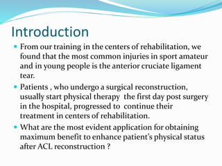
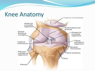
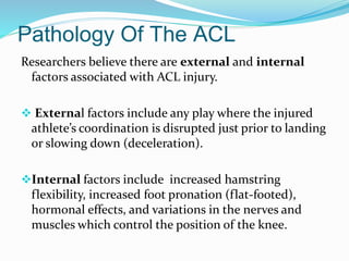
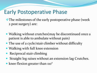
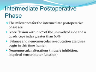
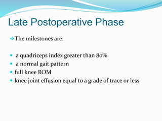
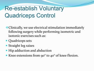
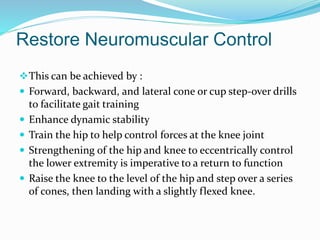
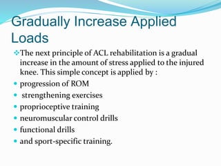
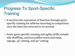
Introduction
 From our training in the centers of rehabilitation, we
found that the most common injuries in sport amateur
and in young people is the anterior cruciate ligament
tear.
 Patients , who undergo a surgical reconstruction,
usually start physical therapy the first day post surgery
in the hospital, progressed to continue their
treatment in centers of rehabilitation.
 What are the most evident application for obtaining
maximum benefit to enhance patient’s physical status
after ACL reconstruction ?
Knee Anatomy
 The knee is one of the largest and most complex joints
in the body. The knee joins the thigh bone (femur) to
the shin bone (tibia). The smaller bone that runs
alongside the tibia (fibula) and the kneecap (patella)
are the other bones that make the knee joint.
 Tendons connect the knee bones to the leg muscles
that move the knee joint. Ligaments join the knee
bones and provide stability to the knee:
• Lateral collateral ligament(LCL)
• Medial collateral ligament(MCL)
• Anterior cruciate ligament(ACL)
• Posterior cruciate ligament(PCL)
Muscles surrounding knee joint
 Quadriceps
 Hamstring
 Tensor fascia lata
 Hip adductors
 Sartorius
 Gracilis
Pathology Of Knee Joint
Intra-articular :
 Meniscal tear
 ACL and PCL injuries
 Osteoarthritis
 Spondiloarthropathy
Extra- articular:
 Pre-patellar bursitis
 Patello femoral syndrome
 Collateral ligament injuries
 iliotibial band syndrome
 baker’s cyst
 osteonecrosis
Anatomy Of ACL
 The ACL is a band of dense connective tissue that connects the femur and the
tibia. The ligament originates at the medial side of the lateral femoral condyle
and runs an oblique course through the intercondylar fossa distal–anterior–
medial to the insertion at the medial tibial eminence.
Where its :
• Length of 38mm ( range 25 to 41mm)
• Width of 10 mm(range 7 to 12mm
• Made up of multiple collagen fascicles
• Microscopically: interlacing fibrils (150 to 250 nanometer in diameter)
• Grouped into fibers (1 to 20 in diameter)
• Synovial membrane envelope
• Infiltrates the capsule posteriorly
• Receives its innervation from tibial nerve
• Golgi tendon receptors
ACL Biomechanics
 The biomechanical function of the anterior cruciate
ligament is complex for it provides both mechanical
stability and proprioceptive feedback to the knee .
In its stabilizing role, it has 4 main function:
• Restrains anterior translation of the tibia
• Prevents hyperextension of the knee
• Acts as a secondary stabilizer to stress ,reinforcing the
medial collateral ligament
• Control rotation of the tibia on the femur in femoral
extensions of 0 ̊ - 30 ̊(4)
Pathology Of The ACL
Researchers believe there are external and internal
factors associated with ACL injury.
 External factors include any play where the injured
athlete’s coordination is disrupted just prior to landing
or slowing down (deceleration).
Internal factors include increased hamstring
flexibility, increased foot pronation (flat-footed),
hormonal effects, and variations in the nerves and
muscles which control the position of the knee.
ACL Clinical Test
 Lachman Test
Patient lie supine and flex the knee to 20º-30º. The
examiner stabilizes the femur with one hand and pulls
the tibia forward with the other hand, while
estimating the amount of anterior movement. A
distinct endpoint at which forward displacement stops
suggests that the ACL is intact.
Lachman Test
 Anterior drawer test
Have the patient's tested leg bent to about 90 degrees of
flexion. The examiner should sit on the foot of the
patient's leg. Place a hand along each side of the
patient's knee, while palpating the joint line. Apply a
posterior-to-anteriorly directed force through the
superior tibia. Compare the involved side to the non-
involved side. A positive test includes the lack of an
end-feel or excessive translation.
Anterior Drawer Test
Surgical Treatment
Allograft is most commonly used recreational athletes
(as opposed to competitive athletes).
 Patellar tendon graft used because it closely
resembles the torn ACL.
Hamstring tendons graft are used in ACL surgery,
two of the tendons of these muscles are removed, and
"bundled" together to create a new ACL
Pre Operative physical Treatment
Main Goals :
 perturbation training and aggressive quadriceps
strengthening
 full range of motion equal to the opposite knee
 minimal joint swelling
 adequate strength and neuromuscular control
 positive state of mind
Immediate Postoperative Phase
Goals of this phase are to :
 Eliminate swelling due to activity
 Progress from partial weight bearing to full weight
bearing exercise
 Regain normal range of motion
 Increase quadriceps strength
 Increase Hamstring strength
Early Postoperative Phase
The milestones of the early postoperative phase (week
2 post surgery) are:
 Walking without crutches(may be discontinued once a
patient is able to ambulate without pain)
 The use of a cycle/stair climber without difficulty
 Walking with full knee extension
 Reciprocal stair climbing
 Straight leg raises without an extension lag Crutches.
 knee flexion greater than 110°
Intermediate Postoperative
Phase
The milestones for the intermediate postoperative
phase are
 knee flexion within 10° of the uninvolved side and a
quadriceps index greater than 60%.
 Balance and neuromuscular re-education exercises
begin in this time frame).
 Neuromuscular alterations (muscle inhibition,
impaired sensorimotor function)
Late Postoperative Phase
The milestones are:
 a quadriceps index greater than 80%
 a normal gait pattern
 full knee ROM
 knee joint effusion equal to a grade of trace or less
Modalities and techniques
Full Passive Knee Extension
Therefore, two of our goals are:
 Achieve some degree of hyperextension during the first few days
after surgery
 Eventually to work to restore symmetrical motion.
Specific exercises include PROM exercises performed by the
rehabilitation specialist:
1) Supine hamstring stretches with a wedge under the heel, and
gastrocnemius stretches with a towel.
2)Passive overpressure just proximal to the patella may be used for
a low-load , long-duration stretch as needed
Restore Patellar Mobility
 Mobilizations are performed by the rehabilitation spe-
cialist in the clinic and independently by patients
during their home exercise program
 Mobilizations are performed in the medial/lateral and
superior/inferior directions, especially for those with a
patellar tendon autograft, to restore the patella’s ability
to tilt, especially in the superior direction.
Reduce Postoperative
Inflammation
Pain and swelling after surgery can be reduced through
the use of:
 Cryo therapy
 Analgesic medication
 Electrical stimulation
 PROM
 Lasers to aid in the healing response.
Range Of Motion
 Flexion ROM is also gradually progressed during the
first week. Patient should exhibit 0° to 90° of knee
ROM 5 to 7 days after surgery and 0° to 100° of knee
ROM 7 to 10 days after surgery
 Thus, the primary focus at this time is on obtaining
full knee extension. Over the course of the following
month, flexion ROM may be progressed by approxi-
mately 10° per week
Re-establish Voluntary
Quadriceps Control
Clinically, we use electrical stimulation immediately
following surgery while performing isometric and
isotonic exercises such as:
 Quadriceps sets
 Straight leg raises
 Hip adduction and abduction
 Knee extensions from 90° to 40° of knee flexion.
Restore Neuromuscular Control
This can be achieved by :
 Forward, backward, and lateral cone or cup step-over drills
to facilitate gait training
 Enhance dynamic stability
 Train the hip to help control forces at the knee joint
 Strengthening of the hip and knee to eccentrically control
the lower extremity is imperative to a return to function
 Raise the knee to the level of the hip and step over a series
of cones, then landing with a slightly flexed knee.
Gradually Increase Applied
Loads
The next principle of ACL rehabilitation is a gradual
increase in the amount of stress applied to the injured
knee. This simple concept is applied by :
 progression of ROM
 strengthening exercises
 proprioceptive training
 neuromuscular control drills
 functional drills
 and sport-specific training.
 It involves the restoration of function through sport-
specific training for athletes returning to competition
once the knee has returned to its normal.
 Some sport-specific running and agility drills include
side shuffling ,cariocas,sudden starts and stops,
zigzags, 45° cutting, and 90° cutting
Progress To Sport-Specific
Training
Conclusion
 Rehabilitation protocols provide basic guidelines
through which effective outcomes can be achieved.
However, the rate and extent of recovery will depend
on many patient and external factors.
 It is still questionable whether full recovery, or return
to normality can be. The complex neuromuscular
motor patterning, strength and control which are
affected by the injury ,should return after the surgery .
Isokinetic testing is reliable and reproducible method
of evaluating muscle strength, endurance and
antagonist/agonist balance.
References
1. Brown CH Jr, Carson EW: Revision anterior cruciate ligament surgery.
Clin Sports Med 18:109-171, 1999
2.Reiman PR, Jackson DW: Anatomy of the anterior cruciate ligament, in
Jackson DW, DrezD (eds): The Anterior Cruciate Deficient Knee. St.Louis, CV Mosby & Co, 1987, pp 17-26
3.Dienst M, Burks RT, Greis PE: Anatomy and biomechanics of the anteriorcruciate ligament. Orthop Clin North Am
33:605-620, 2002
4.Cross.MJ.Anterior Cruciate Ligament injuries :Treatment and Rehabilitation. Sydney, Australia :North Sydney
Orthopedic and Sports Medicine Center.
5.Bradley JB , Klimkiewicz JJ , rytel MJ , Powell JW Anterior Cruciate Ligament Injuries In The National Football League
6.Root ML , Weed JH , Sgarlato TE , et al : Axis of Motion Of The Subtalar Joint . J Am Podiatr Med Assoc 56:149-155 , 1966.
7. Shelbourne KD, Wilckens JH, Mollabashy A, DeCarlo M. Arthrofibrosis in acute anterior cruciate ligament
reconstruction. The effect of timing of reconstruction and rehabilitation. Am J Sports Med. 1991;19:332–336. [PubMed]
8. Chmielewski TL, Stackhouse S, Axe MJ, Snyder-Mackler L. A prospective analysis of incidence and severity of quadriceps
inhibition in a consecutive sample of 100 patients with complete acute anterior cruciate ligament rupture. J Orthop
Res. 2004;22:925–930. [PubMed]

Acl ppt

  • 2.
    Introduction  From ourtraining in the centers of rehabilitation, we found that the most common injuries in sport amateur and in young people is the anterior cruciate ligament tear.  Patients , who undergo a surgical reconstruction, usually start physical therapy the first day post surgery in the hospital, progressed to continue their treatment in centers of rehabilitation.  What are the most evident application for obtaining maximum benefit to enhance patient’s physical status after ACL reconstruction ?
  • 3.
  • 4.
     The kneeis one of the largest and most complex joints in the body. The knee joins the thigh bone (femur) to the shin bone (tibia). The smaller bone that runs alongside the tibia (fibula) and the kneecap (patella) are the other bones that make the knee joint.  Tendons connect the knee bones to the leg muscles that move the knee joint. Ligaments join the knee bones and provide stability to the knee: • Lateral collateral ligament(LCL) • Medial collateral ligament(MCL) • Anterior cruciate ligament(ACL) • Posterior cruciate ligament(PCL)
  • 5.
    Muscles surrounding kneejoint  Quadriceps  Hamstring  Tensor fascia lata  Hip adductors  Sartorius  Gracilis
  • 6.
    Pathology Of KneeJoint Intra-articular :  Meniscal tear  ACL and PCL injuries  Osteoarthritis  Spondiloarthropathy Extra- articular:  Pre-patellar bursitis  Patello femoral syndrome  Collateral ligament injuries  iliotibial band syndrome  baker’s cyst  osteonecrosis
  • 7.
    Anatomy Of ACL The ACL is a band of dense connective tissue that connects the femur and the tibia. The ligament originates at the medial side of the lateral femoral condyle and runs an oblique course through the intercondylar fossa distal–anterior– medial to the insertion at the medial tibial eminence. Where its : • Length of 38mm ( range 25 to 41mm) • Width of 10 mm(range 7 to 12mm • Made up of multiple collagen fascicles • Microscopically: interlacing fibrils (150 to 250 nanometer in diameter) • Grouped into fibers (1 to 20 in diameter) • Synovial membrane envelope • Infiltrates the capsule posteriorly • Receives its innervation from tibial nerve • Golgi tendon receptors
  • 8.
    ACL Biomechanics  Thebiomechanical function of the anterior cruciate ligament is complex for it provides both mechanical stability and proprioceptive feedback to the knee . In its stabilizing role, it has 4 main function: • Restrains anterior translation of the tibia • Prevents hyperextension of the knee • Acts as a secondary stabilizer to stress ,reinforcing the medial collateral ligament • Control rotation of the tibia on the femur in femoral extensions of 0 ̊ - 30 ̊(4)
  • 9.
    Pathology Of TheACL Researchers believe there are external and internal factors associated with ACL injury.  External factors include any play where the injured athlete’s coordination is disrupted just prior to landing or slowing down (deceleration). Internal factors include increased hamstring flexibility, increased foot pronation (flat-footed), hormonal effects, and variations in the nerves and muscles which control the position of the knee.
  • 10.
    ACL Clinical Test Lachman Test Patient lie supine and flex the knee to 20º-30º. The examiner stabilizes the femur with one hand and pulls the tibia forward with the other hand, while estimating the amount of anterior movement. A distinct endpoint at which forward displacement stops suggests that the ACL is intact.
  • 11.
  • 12.
     Anterior drawertest Have the patient's tested leg bent to about 90 degrees of flexion. The examiner should sit on the foot of the patient's leg. Place a hand along each side of the patient's knee, while palpating the joint line. Apply a posterior-to-anteriorly directed force through the superior tibia. Compare the involved side to the non- involved side. A positive test includes the lack of an end-feel or excessive translation.
  • 13.
  • 14.
    Surgical Treatment Allograft ismost commonly used recreational athletes (as opposed to competitive athletes).  Patellar tendon graft used because it closely resembles the torn ACL. Hamstring tendons graft are used in ACL surgery, two of the tendons of these muscles are removed, and "bundled" together to create a new ACL
  • 15.
    Pre Operative physicalTreatment Main Goals :  perturbation training and aggressive quadriceps strengthening  full range of motion equal to the opposite knee  minimal joint swelling  adequate strength and neuromuscular control  positive state of mind
  • 16.
    Immediate Postoperative Phase Goalsof this phase are to :  Eliminate swelling due to activity  Progress from partial weight bearing to full weight bearing exercise  Regain normal range of motion  Increase quadriceps strength  Increase Hamstring strength
  • 17.
    Early Postoperative Phase Themilestones of the early postoperative phase (week 2 post surgery) are:  Walking without crutches(may be discontinued once a patient is able to ambulate without pain)  The use of a cycle/stair climber without difficulty  Walking with full knee extension  Reciprocal stair climbing  Straight leg raises without an extension lag Crutches.  knee flexion greater than 110°
  • 18.
    Intermediate Postoperative Phase The milestonesfor the intermediate postoperative phase are  knee flexion within 10° of the uninvolved side and a quadriceps index greater than 60%.  Balance and neuromuscular re-education exercises begin in this time frame).  Neuromuscular alterations (muscle inhibition, impaired sensorimotor function)
  • 19.
    Late Postoperative Phase Themilestones are:  a quadriceps index greater than 80%  a normal gait pattern  full knee ROM  knee joint effusion equal to a grade of trace or less
  • 20.
  • 21.
    Full Passive KneeExtension Therefore, two of our goals are:  Achieve some degree of hyperextension during the first few days after surgery  Eventually to work to restore symmetrical motion. Specific exercises include PROM exercises performed by the rehabilitation specialist: 1) Supine hamstring stretches with a wedge under the heel, and gastrocnemius stretches with a towel. 2)Passive overpressure just proximal to the patella may be used for a low-load , long-duration stretch as needed
  • 22.
    Restore Patellar Mobility Mobilizations are performed by the rehabilitation spe- cialist in the clinic and independently by patients during their home exercise program  Mobilizations are performed in the medial/lateral and superior/inferior directions, especially for those with a patellar tendon autograft, to restore the patella’s ability to tilt, especially in the superior direction.
  • 23.
    Reduce Postoperative Inflammation Pain andswelling after surgery can be reduced through the use of:  Cryo therapy  Analgesic medication  Electrical stimulation  PROM  Lasers to aid in the healing response.
  • 24.
    Range Of Motion Flexion ROM is also gradually progressed during the first week. Patient should exhibit 0° to 90° of knee ROM 5 to 7 days after surgery and 0° to 100° of knee ROM 7 to 10 days after surgery  Thus, the primary focus at this time is on obtaining full knee extension. Over the course of the following month, flexion ROM may be progressed by approxi- mately 10° per week
  • 25.
    Re-establish Voluntary Quadriceps Control Clinically,we use electrical stimulation immediately following surgery while performing isometric and isotonic exercises such as:  Quadriceps sets  Straight leg raises  Hip adduction and abduction  Knee extensions from 90° to 40° of knee flexion.
  • 26.
    Restore Neuromuscular Control Thiscan be achieved by :  Forward, backward, and lateral cone or cup step-over drills to facilitate gait training  Enhance dynamic stability  Train the hip to help control forces at the knee joint  Strengthening of the hip and knee to eccentrically control the lower extremity is imperative to a return to function  Raise the knee to the level of the hip and step over a series of cones, then landing with a slightly flexed knee.
  • 27.
    Gradually Increase Applied Loads Thenext principle of ACL rehabilitation is a gradual increase in the amount of stress applied to the injured knee. This simple concept is applied by :  progression of ROM  strengthening exercises  proprioceptive training  neuromuscular control drills  functional drills  and sport-specific training.
  • 28.
     It involvesthe restoration of function through sport- specific training for athletes returning to competition once the knee has returned to its normal.  Some sport-specific running and agility drills include side shuffling ,cariocas,sudden starts and stops, zigzags, 45° cutting, and 90° cutting Progress To Sport-Specific Training
  • 29.
    Conclusion  Rehabilitation protocolsprovide basic guidelines through which effective outcomes can be achieved. However, the rate and extent of recovery will depend on many patient and external factors.  It is still questionable whether full recovery, or return to normality can be. The complex neuromuscular motor patterning, strength and control which are affected by the injury ,should return after the surgery . Isokinetic testing is reliable and reproducible method of evaluating muscle strength, endurance and antagonist/agonist balance.
  • 30.
    References 1. Brown CHJr, Carson EW: Revision anterior cruciate ligament surgery. Clin Sports Med 18:109-171, 1999 2.Reiman PR, Jackson DW: Anatomy of the anterior cruciate ligament, in Jackson DW, DrezD (eds): The Anterior Cruciate Deficient Knee. St.Louis, CV Mosby & Co, 1987, pp 17-26 3.Dienst M, Burks RT, Greis PE: Anatomy and biomechanics of the anteriorcruciate ligament. Orthop Clin North Am 33:605-620, 2002 4.Cross.MJ.Anterior Cruciate Ligament injuries :Treatment and Rehabilitation. Sydney, Australia :North Sydney Orthopedic and Sports Medicine Center. 5.Bradley JB , Klimkiewicz JJ , rytel MJ , Powell JW Anterior Cruciate Ligament Injuries In The National Football League 6.Root ML , Weed JH , Sgarlato TE , et al : Axis of Motion Of The Subtalar Joint . J Am Podiatr Med Assoc 56:149-155 , 1966. 7. Shelbourne KD, Wilckens JH, Mollabashy A, DeCarlo M. Arthrofibrosis in acute anterior cruciate ligament reconstruction. The effect of timing of reconstruction and rehabilitation. Am J Sports Med. 1991;19:332–336. [PubMed] 8. Chmielewski TL, Stackhouse S, Axe MJ, Snyder-Mackler L. A prospective analysis of incidence and severity of quadriceps inhibition in a consecutive sample of 100 patients with complete acute anterior cruciate ligament rupture. J Orthop Res. 2004;22:925–930. [PubMed]