
Recommended
Recommended
More Related Content
What's hot
What's hot (19)
Viewers also liked
Viewers also liked (20)
Similar to Guided Surgery using Endoscopic Multimodal Imaging by Stavros Demos
Similar to Guided Surgery using Endoscopic Multimodal Imaging by Stavros Demos (20)
More from Industrial Partnerships Office
More from Industrial Partnerships Office (17)
Recently uploaded
Recently uploaded (20)
Guided Surgery using Endoscopic Multimodal Imaging by Stavros Demos
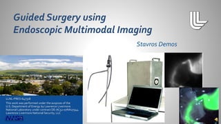
- 1. LLNL-PRES-647318 This work was performed under the auspices of the U.S. Department of Energy by Lawrence Livermore National Laboratory under contract DE-AC52-07NA27344. Lawrence Livermore National Security, LLC Stavros Demos
- 2. • Detect locations of disease • Verify complete removal after surgery • Detect and preserve anatomical features of importance Lawrence Livermore National Laboratory LLNL-PRES-647318 2 Intraoperative imaging using targeting fluorescing contrast agents has been shown to be an effective navigation tool
- 3. Lawrence Livermore National Laboratory LLNL-PRES-647318 3 …and is adaptable to any conventional endoscope system
- 4. Lawrence Livermore National Laboratory Key advantages of our system • It is easy to handle (low weight, compact) • Simultaneous image acquisition • Optimal sensitivity • It can be multiplexed to use multiple contrast agents • It can be adapted to any endoscope type system for multi-purpose use in the OR • It is relatively inexpensive to manufacture LLNL-PRES-647318 4 Hamamatsu system, Aoki et.Al., J Hepatobiliary Pancreat Sci., 2010 Intuitive Surgical system, Robotic system, Spinoglio et. Al. Surg Endosc, Dec 2012
- 5. Lawrence Livermore National Laboratory LLNL-PRES-647318 5 Competition: One camera acquires alternately color and spectral images Our approach: Separate image-coupled cameras acquire images in parallel Our approach provides orders of magnitude higher sensitivity
- 6. Enhancing the visualization of the biliary system during laparoscopic surgery • 750,000 patients undergo laparoscopic Lawrence Livermore National Laboratory LLNL-PRES-647318 6 Biliary System cholecystectomy/year USA • Common Bile Duct (CBD) injury occurs in 1/200 operations (4000/yr) • Multiple reconstructive operations and procedures, increased mortality, and lifelong disability • Liability/Medical costs: $250k-$1M per occurrence • Most significant injury occurs when surgeon misidentifies CBD as cystic duct and cuts this structure The concepts are directly applicable to other clinical needs such as cancer detection and the vascular system visualization
- 7. • Cannulation of the cystic duct and associated complications (which can include: Infection, bleeding, pancreas inflammation, damage to the common bile duct) • 10-20 minutes of OR time • Exposure to radiation • Live image is not available during the dissection phase • Anatomy is not directly overlaid onto the surgical field • Operator dependent interpretation • Cost/benefit questionable as a routine procedure Lawrence Livermore National Laboratory LLNL-PRES-647318 7 • 97% of errors due to visual perceptual illusion • Faults in technical skill only 3% of injuries
- 8. • ICG is administered intravenously before surgery • ICG is nearly exclusively eliminated by the liver into the bile • Intraoperative fluorescent cholangiography using ICG was first demonstrated in 2009 by Ishizawa et. Al., J Am Coll Surg. 208 (see left, open surgery) Lawrence Livermore National Laboratory LLNL-PRES-647318 • A large number of publications have been produced in the last 4 years on this topic, highlighting the clinical importance of this method 8
- 9. ICG emission image RGB conventional image Lawrence Livermore National Laboratory LLNL-PRES-647318 9 Imaging the gallbladder and bile ducts using ICG Ex vivo images using animal model
- 10. ICG image of tube Conventional image ICG emission image Lawrence Livermore National Laboratory LLNL-PRES-647318 10 3 mm plastic tube containing ICG solution 5-mm thick bovine muscle tissue covering the tube ICG fluorescence image through the 5-mm tissue
- 11. Lawrence Livermore National Laboratory LLNL-PRES-647318 11 Conventional image ICG fluorescence image Composite image Bile duct covered by two layers of intestinal wall ICG image visualize subsurface bile duct Image superposition can assist real time navigation
- 12. Lawrence Livermore National Laboratory LLNL-PRES-647318 12
- 13. Our system design offers optimized sensitivity and functionality for endoscopic/laparoscopic NIR fluorescent subsurface imaging This design could provide low-cost and real-time surgical navigation capability for minimally invasive surgery • Common bile duct • Blood vessels • Cancer (Sentinel lymph node biopsy) • Any application with a fluorescent contrast agent Lawrence Livermore National Laboratory LLNL-PRES-647318 13
- 14. Lawrence Livermore National Laboratory LLNL-PRES-647318 14 Genaro Mempin mempin1@llnl.gov