Call Girls Whitefield Just Call 7001305949 Top Class Call Girl Service Available
Art 3 a10.1007-2fs11605-012-2123-z
1. J Gastrointest Surg (2013) 17:668–674
DOI 10.1007/s11605-012-2123-z
ORIGINAL ARTICLE
Routine Intraoperative Cholangiography During Single-Incision
Laparoscopic Cholecystectomy: a Review of 196
Consecutive Patients
Norihiro Sato & Kazunori Shibao & Yasuki Akiyama &
Yuzuru Inoue & Yasuhisa Mori & Noritaka Minagawa &
Aiichiro Higure & Koji Yamaguchi
Received: 2 October 2012 / Accepted: 10 December 2012 / Published online: 22 December 2012
# 2012 The Society for Surgery of the Alimentary Tract
Abstract
Background Single-incision laparoscopic cholecystectomy (SILC) has been increasingly performed as a potentially less
invasive alternative to standard laparoscopic cholecystectomy. However, recent evidences suggest a higher incidence of
complications, notably bile duct injuries, in SILC. We reviewed our experiences with routine intraoperative cholangiography
(IOC) during SILC to investigate its feasibility and usefulness.
Methods Among 228 patients who underwent SILC at our institution from September 2009 to July 2012, a total of 196
patients in which an IOC was attempted were retrospectively reviewed.
Results IOC was successful in 178 of 196 patients, yielding a success rate of 90.8 %. There were no IOC-related
complications. Common bile duct (CBD) stones were detected by IOC in 16 patients (8.2 %), all of which were treated
by subsequent single-incision laparoscopic CBD exploration or postoperative endoscopic retrograde cholangiopancreatography with stone extraction. In addition, IOC revealed filling defects in the cystic duct (four patients) and poor passage of
contrast medium into the duodenum (one patient). In one patient with severe acute cholecystitis, cholangiography via an
endoscopic nasobiliary drainage tube revealed misinterpretation of CBD as cystic duct.
Conclusions We, thus, conclude that routine IOC during SILC is feasible and useful to detect biliary stones and to gain an
accurate picture of biliary anatomy.
Keywords Single-incision laparoscopic cholecystectomy .
Intraoperative cholangiography . Choledocholithiasis . Bile
duct injury
Introduction
In recent years, single-incision laparoscopic cholecystectomy (SILC) has been developed to further minimize the
invasiveness of laparoscopic cholecystectomy (LC).
Although SILC remains technically challenging for most
surgeons, it can, in theory, offer potential advantages, including less postoperative pain, shorter recovery time,
N. Sato (*) : K. Shibao : Y. Akiyama : Y. Inoue : Y. Mori :
N. Minagawa : A. Higure : K. Yamaguchi
Department of Surgery 1, School of Medicine,
University of Occupational and Environmental Health,
Kitakyushu 807-8555, Japan
e-mail: norisato@med.uoeh-u.ac.jp
improved cosmetic outcome, and higher patient satisfaction.
Recent randomized controlled trials showed that SILC is a
safe procedure with better cosmetic results as compared to
conventional LC.1–6
However, one of the concerns related to SILC is its
higher incidence of postoperative complications as compared to conventional LC.4 Importantly, it has been suggested that SILC is associated with a higher rate of bile duct
injuries.7–9 Therefore, standardization of SILC still requires
an established protocol to ensure safe dissection during the
procedure and minimize the intra- and postoperative complications. Currently employed techniques for intraoperative
assessment of biliary anatomy and prevention of bile duct
injuries include critical view of safety (CVS) approach,
laparoscopic ultrasound, and intraoperative cholangiography (IOC).10
IOC has been routinely performed during conventional
LC to obtain critical information about biliary anatomy and
to minimize the rate of biliary injuries. The use of IOC
2. J Gastrointest Surg (2013) 17:668–674
during SILC is, however, limited by the technical difficulties
of the procedure through a single incision or, in some IOC
systems, requirement of additional skin incision. In fact,
only a few studies have addressed the significance of routine
IOC during SILC in only a limited number of patients.11–13
In September 2009, we introduced a program of SILC
including routine IOC for cholelithiasis at our institution.
We are now expanding the indication of SILC to patients
with acute cholecystitis, which require more strict protocol
to prevent intraoperative bile duct injuries. In an attempt to
evaluate the feasibility and usefulness of routine IOC during
SILC, we retrospectively reviewed our experience with
SILC with routine IOC in a consecutive series of 196
patients. To our knowledge, this is the largest series to date
of SILC with routine IOC.
Patients and Methods
Patients
Between September 2009 and July 2012, a total of 228
patients with gallbladder diseases underwent SILC at our
institution. The indications for SILC included symptomatic
cholelithiasis, acute cholecystitis, cholelithiasis associated
with common bile duct stones, gallbladder polyps, and suspected gallbladder cancer (gallbladder tumor or partial wall
thickening of the gallbladder) (Table 1). This is our institution’s initial experience with SILC. All procedures were performed by a total of 21 surgeons (including 11 staff surgeons
and ten surgical residents). As a teaching hospital, we have
attempted SILC even in challenging cases, including those
with severe acute cholecystitis. Therefore, there were no exclusion criteria for performing SILC during the study period.
Patients who required conversion to the conventional fourport LC (one patient) or open cholecystectomy (one patient)
were excluded from this study. IOC was attempted in 196 of
these 228 patients but was not performed in the remaining 32
patients. The reasons that precluded the IOC attempts included suspicion of gallbladder cancer (in which spilled bile from
the puncture site may cause cancer dissemination) in nine
patients, accidental cystic duct injury during surgery (which
may result in leakage of contrast medium in the Kumar
cholangiography system described below) in seven patients,
patient’s allergy to contrast medium in four patients, stone
compaction in the gallbladder neck leaving no appropriate
space for needle puncture in three patients, and bleeding from
the cystic duct wall in one patient. In eight patients, IOC was
not attempted with no specific reason described in the operation records, despite our departmental rule of routine IOC.
Clinical charts and operative records were then retrospectively
reviewed for these 196 patients undergoing attempted SILC
and routine IOC.
669
Table 1 Indications for SILC in our present series
Diseases
Number of
patients (%)
Symptomatic cholelithiasis
Acute cholecystitis
Cholelithiasis associated with common bile duct stones
Gallbladder polyps (over 1 cm in diameter)
Suspected gallbladder cancer (gallbladder tumor
or partial wall thickening of the gallbladder)
169 (74 %)
30 (13 %)
14 (6 %)
10 (4 %)
5 (2 %)
SILC single-incision laparoscopic cholecystectomy
Operative Procedure
Basically, our technique for SILC is three-trocar approach
through a single umbilical incision. Under general anesthesia, patients were placed in the supine position with their
legs apart. A single 2.5-cm vertical incision was made
directly on the umbilicus, through which a 5-mm trocar
(Endopath Xcel, Ethicon Endo-Surgery, Cincinnati, OH,
USA) was introduced for pneumoperitoneum and a laparoscope (EndoEye camera system, Olympus Medical System,
Tokyo, Japan). After exposing the abdominal fascia under
the skin flap of the umbilical incision, a grasper for gallbladder retraction was inserted without a trocar by making a
pinhole on the fascia with a needle. Then, two 5-mm trocars
(Endopath Xcel, Ethicon Endo-Surgery, or EZ trocar, Hakko
Co., Nagano, Japan) for operator’s manipulation were
inserted into the abdominal cavity through the single umbilical incision.
In some cases, a small wound retractor (Alexis wound
retractor, Applied Medical, Rancho Santa Margarita, CA,
USA) and a surgical glove or a minilaparotomy wound
protector (Lap-Protector, Hakko) and a silicon rubber cap
(EZ Access, Hakko) were attached to the umbilical incision
and used as a multichannel port.
Our initial attempt was to perform all procedures using
the three trocars and a grasper via the single umbilical
incision. However, in cases with difficult gallbladder retraction and exposure, additional one or two ports were placed
as appropriate in the right lateral and/or subcostal region.
Dissection of Calot’s triangle was performed carefully
according to the CVS approach. After confirming that the
cystic artery and cystic duct are the only two tubular structures remaining between the gallbladder and the hepatoduodenal ligament, an IOC was routinely attempted. We thus
use both the CVS technique and IOC to further ensure the
safe dissection.
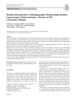
In most cases, IOC was performed using the Kumar
cholangiography system (Nashville Surgical Instruments,
Nashville, USA).14 This system consists of a 5-mm grasper
(Kumar Clamp) which is applied across the gallbladder just
3. 670
above the Hartmann’s pouch and divides the gallbladder
into a medial and lateral compartment (Fig. 1a). A catheter
carrying a short 23-gauge needle (Interject, Boston
Scientific, Spencer, IN, USA) was then introduced through
the side channel of the clamp (Fig. 1b), puncturing the
Hartmann’s pouch or the cystic duct close to the gallbladder
by advancing the needle for aspiration, followed by contrast
injection (Fig. 1c).
In cases with choledocholithiasis suspected or proven by
preoperative imaging studies (MRCP, CT, or endoscopic
retrograde cholangiopancreatography (ERCP)), a small indwelling feeding tube (Atom tube, Atom Medical
Corporation, Tokyo, Japan) was inserted into the cystic duct
and used for IOC. This tube was usually fixed and kept to be
placed postoperatively for biliary decompression and repeated cholangiography for confirmation of biliary clearance. In
some patients with severe acute cholangitis, IOC was performed via an endoscopic nasobiliary drainage (ENBD)
tube or percutaneous transhepatic gallbladder drainage
(PTGBD) tube placed preoperatively.
J Gastrointest Surg (2013) 17:668–674
After completion of IOC, the cystic duct and cystic artery
were doubly clipped with a 5-mm disposable clip applier
and then divided. The gallbladder was then dissected from
the liver bed using a hook electrocautery or Harmonic ACE
(Johnson & Johnson, Cincinnati, OH, USA). The gallbladder was then collected in a bag and removed through the
umbilical incision, usually by enlarging the fascial opening
as required.
When common bile duct (CBD) stones were detected
by IOC, subsequent laparoscopic CBD exploration was
performed via the single umbilical incision in cases with
a CBD diameter of 10 mm or larger and postoperative
ERCP was planned in cases with a CBD diameter of less
than 10 mm. The procedure of single-incision laparoscopic CBD exploration was described elsewhere in detail.
Briefly, choledochotomy was made in the suprapancreatic region, and stone extraction was performed using a
combination of different techniques (i.e., stone forceps,
saline flushing, basket catheter, and balloon catheter under choledochoscopic guidance). In any case, choledochoscopy was used to confirm that no residual stones
remained. The choledochotomy was then closed with 3-0
Vicryl sutures using a flexible manual manipulator. A Ctube or T-tube was routinely inserted into the CBD and
fixed for biliary decompression after exploration. All the
procedures were done through the single umbilical
incision.
The fascial defect in the umbilicus was closed using
absorbable monofilament suture and the skin was closed
subcuticularly with a 4-0 absorbable monofilament suture.
Data analyzed included patient demographics, operative
approach (completion with single incision or requirement
of additional port(s) placement), operative time, intraoperative blood loss, results of attempted IOC, postoperative
length of stay, and complications.
Results
Fig. 1 Kumar cholangiography system consists of a 5-mm grasper (a)
with a catheter carrying a short 23 gauge needle (b), which is applied
across the gallbladder just above the Hartmann’s pouch, followed by
needle puncturing the Hartmann’s pouch or the cystic duct close to the
gallbladder for contrast injection (c)
The outcome of study population is summarized in Fig. 2.
Among 228 patients with gallbladder diseases undergoing
SILC (excluding cases required conversion to open cholecystectomy or four-port LC), those patients in which an IOC
was attempted were included in this study. The study group
consisted of 196 patients (80 males and 116 females) with a
mean age of 61 years (range, 16 to 91 years). The mean
body mass index was 23.4 (range, 14.8 to 40.7).
In most cases (173 patients, 88.3 %), all surgical procedures (including IOC) could be completed via the single
incision. However, 23 patients (14 %) required placement of
additional one or two port(s) in the subcostal or the right
lateral region. There was no case requiring additional port
placement simply for the purpose of IOC.
4. J Gastrointest Surg (2013) 17:668–674
An IOC was attempted using the Kumar cholangiography
system in 176 patients, a feeding tube in 15 patients, an
ENBD tube in 4 patients, and a PTGBD tube in 1 patient.
Overall, an IOC was successful in 178 of 196 patients,
yielding a success rate of 90.8 %. When cholangiograms
via an ENBD or PTGBD were excluded, the success rate
was 88.3 % (173/191). The major reasons for failed/incomplete IOC included winding cystic duct, stone compaction in
the gallbladder neck, and extravasation of contrast medium
from the initial puncture site or from the injured cystic duct.
When the initial IOC attempt with the Kumar system did not
work, the conventional IOC through a cystic ductotomy was
not attempted. There was no intraoperative complication
related to IOC (such as injury of the cystic duct).
Overall, IOC detected abnormalities in the biliary system
in 21 patients (10.7 %). These included CBD stones (16
patients), filling defects in the cystic duct (four patients),
and poor passage of contrast medium into the duodenum
(one patient). The preoperative diagnosis of CBD stone was
not obtained in 2 of the 16 patients with CBD stones
detected by IOC. Of the 16 patients with documented
CBD stones on IOC, 11 patients were treated by laparoscopic CBD exploration with stone extraction via the single
umbilical incision (Shibao et al., manuscript in submission).
The remaining five patients underwent postoperative ERCP
for biliary clearance. In four patients who were found to
have filling defects in the cystic duct, the stone/sludge was
removed by making an opening in the cystic duct and
milking the duct by forceps or the cystic duct was divided
at a position proximal (CBD side) to the defects so as to
eliminate the retained biliary calculus. In one patient in
which a poor passage of contrast medium into the duodenum was revealed by IOC, the cystic duct cannulation tube
was left placed postoperatively for biliary decompression.
Fig. 2 The outcome of study population. SILC single-incision laparoscopic cholecystectomy, IOC intraoperative cholangiography, CBD
common bile duct
671
In one patient with severe acute cholecystitis, cholangiography via an ENBD tube placed preoperatively revealed
false recognition of the CBD as the cystic duct (Fig. 3a).
Further dissection towards the gallbladder and repeated
cholangiogram then identified the cystic duct (Fig. 3b),
which was subsequently clipped and divided. In this case,
therefore, an accurate biliary anatomy obtained by cholangiography enabled us to prevent the injury of the CBD.
The early postoperative complications (occurring before
the seventh postoperative day) were found in seven patients
(3.6 %). These included pulmonary complications (aspiration pneumonia and bronchitis) (four patients), wound infection (two patients), and paralytic ileus (one patient).
None of the patients developed bile leakage. The late postoperative complication (on and after the seventh postoperative day) was found in one patient (0.5 %) who developed
delayed intraabdominal abscess due to spilled gallstones.
The mean length of postoperative hospital stay was 6 days
(range, 2 to 27 days). The length was significantly longer in
patients undergoing SILC with CBD exploration than in those
undergoing SILC without CBD exploration (14.1 versus
5.5 days, P<0.001). The length of stay (5.5 days) in patients
undergoing SILC without CBD exploration was significantly
shorter than that (6.7 days) in 112 patients undergoing conventional LC (without simultaneous CBD exploration) at our
institution before the study period (P=0.0015).
Fig. 3 Cholangiogram via an endoscopic nasobiliary drainage tube
revealed a misinterpretation of the common bile duct (clamped by forceps) as the cystic duct (a). Repeated cholangiogram after further dissection identified the cystic duct for division (clamped by forceps) (b)
5. 672
Discussion
Since its first description in 1997 by Navarra et al.,15 SILC
has emerged as a potentially less invasive alternative to
standard LC. With improved surgical skills and advanced
technologies, SILC has recently been disseminating quite
rapidly. At our institution, we introduced SILC for selected
patients with gallbladder diseases in September 2009. Since
then, we have continued to perform routine IOC to enhance
the safety of SILC and minimize the intraoperative complications. In this study, we retrospectively reviewed our initial
experience of SILC with routine IOC in a consecutive series
of 196 patients. The major findings of our present study
were as follows: (1) IOC during SILC was successful in
90.8 % with no procedure-related complication; (2) IOC
detected choledocholithiasis, cystic duct stones, and bile
stasis in a significant proportion of patients, leading to
appropriate management; and (3) cholangiogram through
an ENBD tube revealed misinterpretation of biliary anatomy
and enabled us to prevent bile duct injury in one patient with
acute cholecystitis. These findings suggest the feasibility
and usefulness of routine IOC during SILC.
According to previous studies, the incidence of CBD
stones at the time of LC, as detected by IOC, has been
reported to be between 3 and 12 %.16–18 In this study, IOC
detected CBD stones in 16 patients (8 %), all of which were
successfully treated by laparoscopic CBD exploration
(Shibao et al., manuscript in submission) or postoperative
ERCP with stone clearance. Importantly, CBD stones were
newly diagnosed on IOC in 2 of these 16 patients, raising a
possibility of false-negative findings by preoperative imaging studies or stone passage from the gallbladder into the
CBD during an interval between the preoperative imaging
studies and surgery. Consistent with our present results, it
has been reported that in 109 patients without CBD stones
on preoperative ERCP, nine patients (8.3 %) were found to
have CBD stones on IOC during LC.19 In this regard,
routine IOC should be considered even in patients with no
suspicion of CBD stones on preoperative imaging studies,
including ERCP. Furthermore, IOC also detected cystic duct
stones in four patients, leading to intraoperative clearance of
these stones. Because retained gallbladder and cystic duct
calculi can be a source of recurrent biliary pain,20 efforts
should be made to detect and remove the cystic duct stones.
With increasing cases of SILC reported, a concern has
raised for a propensity for its higher incidence of postoperative complications. A meta-analysis of randomized controlled trials showed a higher incidence of postoperative
complications (including bile duct injuries, bile leakage,
biliary collection or abscess, retained choledocholithiasis,
port-site bleeding, and wound complications) in SILC
(16.0 %, 56/349) than in conventional LC (12.3 %, 38/
310), though the difference was not statistically significant.4
J Gastrointest Surg (2013) 17:668–674
Recently, a comprehensive database search demonstrated a
higher rate of bile duct injuries in SILC (0.72 %) as compared to the accepted historic rate of 0.4–0.5 % for standard
LC.9 Because unfavorable results are less likely to be
reported, this incidence of bile duct injuries associated with
SILC might be underestimated. Considering that the main
benefit of SILC appears to be improved cosmesis, this
incremental increase in bile duct injury is not justified. It
is, therefore, critically important to maintain safe dissection
principles in order to avoid an increase in bile duct injuries
during SILC.
Among the techniques to prevent bile duct injuries during
LC, IOC is the most frequently applied technique for intraoperative assessment of the biliary anatomy.10 Although the
debate whether to perform routine or selective IOC has not
yet been concluded, population-based studies have shown
beneficial roles of IOC in the prevention or detection of bile
duct injuries during cholecystectomy.21–24 As a result, routine IOC is recommended for prevention of bile duct
injury.10,25 The use of IOC during SILC is, however, limited
probably by the technical difficulties in the cystic duct
cannulation through a single incision or, in some IOC systems, requirement of additional skin incision. In fact, a
recent database search revealed that IOC was utilized in
only 13.4 % of a total of 2,626 reported SILC procedures.9
Only a few studies have addressed the significance of routine IOC during SILC.11–13 They demonstrated a success
rate of 88–95 % by the use of the needle puncture techniques or conventional IOC system that requires partial cystic
ductotomy and tube cannulation.11–13 Since one of the adverse opinions against routine IOC is a possible bile duct
injury by the cholangiogram itself, the IOC system should
be a safe procedure with a minimal risk of unexpected cystic
duct injury. In this series, we mainly used the Kumar cholangiography system to achieve a success rate of 90.8 %
(178/196) without the procedure-related complications. In
this system, puncturing the gallbladder (usually Hartmann’s
pouch) with a small needle keeps the cystic duct free from
the ductotomy, thereby minimizing the bile spillage and
avoiding the procedure-related bile duct injury. In our present series, cholangiogram revealed misinterpretation of
CBD as cystic duct and thus enabled us to prevent bile duct
injury, highlighting the importance of IOC to prevent bile
duct injuries during SILC. It should be noted, however, that
there are a certain percentage of cases in which attempted
IOC was failed due to technical or anatomical problems. In
such cases, achievement of CVS is mandatory to maintain
the quality of safe dissection.
Despite increasing number of reports, benefits and drawbacks of SILC still remain controversial. According a recent
meta-analysis of randomized controlled trials,4 SILC had
significantly favorable cosmetic scoring compared to conventional LC, whereas the operating time was significantly
6. J Gastrointest Surg (2013) 17:668–674
longer in SILC. In addition, SILC does not confer any
benefit in postoperative pain and hospital stay as compared
to conventional LC.4 Regarding the cost-effectiveness, a
prospective randomized blinded comparison showed that
SILC has higher cost than conventional LC,26 while the
other studies showed no such difference.27,28 In the present
study, the mean length of stay in patients undergoing SILC
without simultaneous CBD exploration was 5.5 days. This
extended stay was unlikely to be related to the procedure
(SILC) itself, because the length of stay in patients undergoing conventional LC at our institution was even longer
(6.7 days). In general, the length of stay is longer in Japan as
compared to other Western countries, primarily due to the
differences in the health insurance systems and the actual
medical costs charged to the patients. Because the medical
insurance in Japan covers the complete cost of hospitalization, most patients tend to stay longer in hospital until they
recover completely from surgery. We are now making various efforts to shorten the length of stay, for example, by
using clinical pathways.
In summary, our findings suggest that routine IOC during
SILC is technically feasible and useful to detect biliary
stones and to gain an accurate picture of biliary anatomy.
Our study is limited by the fact that it is retrospective in
nature. Therefore, in order to precisely determine the clinical
value of routine IOC during SILC, a prospective randomized trial should be performed in the future.
673
7.
8.
9.
10.
11.
12.
13.
14.
15.
References
16.
1. Tsimoyiannis, E.C., Tsimogiannis, K.E., Pappas-Gogos, G.,
Farantos, C., Benetatos, N., Mavridou, P., and Manataki, A.
2010. Different pain scores in single transumbilical incision
laparoscopic cholecystectomy versus classic laparoscopic cholecystectomy: a randomized controlled trial. Surg Endosc
24:1842-1848.
2. Cao, Z.G., Cai, W., Qin, M.F., Zhao, H.Z., Yue, P., and Li, Y. 2011.
Randomized clinical trial of single-incision versus conventional
laparoscopic cholecystectomy: short-term operative outcomes.
Surg Laparosc Endosc Percutan Tech 21:311-313.
3. Marks, J., Tacchino, R., Roberts, K., Onders, R., Denoto, G.,
Paraskeva, P., Rivas, H., Soper, N., Rosemurgy, A., and Shah, S.
2011. Prospective randomized controlled trial of traditional laparoscopic cholecystectomy versus single-incision laparoscopic cholecystectomy: report of preliminary data. Am J Surg 201:369-372;
discussion 372-363
4. Garg, P., Thakur, J.D., Garg, M., and Menon, G.R. 2012. SingleIncision Laparoscopic Cholecystectomy vs. Conventional Laparoscopic Cholecystectomy: a Meta-analysis of Randomized Controlled Trials. J Gastrointest Surg. 16(8):1618–1628
5. Markar, S.R., Karthikesalingam, A., Thrumurthy, S., Muirhead, L.,
Kinross, J., and Paraskeva, P. 2012. Single-incision laparoscopic
surgery (SILS) vs. conventional multiport cholecystectomy: systematic review and meta-analysis. Surg Endosc 26:1205-1213.
6. Phillips, M.S., Marks, J.M., Roberts, K., Tacchino, R., Onders, R.,
DeNoto, G., Rivas, H., Islam, A., Soper, N., Gecelter, G., et al.
17.
18.
19.
20.
21.
22.
2012. Intermediate results of a prospective randomized controlled
trial of traditional four-port laparoscopic cholecystectomy versus
single-incision laparoscopic cholecystectomy. Surg Endosc
26:1296-1303.
Lau, K.N., Sindram, D., Agee, N., Martinie, J.B., and Iannitti,
D.A. 2010. Bile duct injury after single incision laparoscopic
cholecystectomy. JSLS 14:587-591.
Garg, P., Thakur, J.D., Singh, I., Nain, N., Mittal, G., and Gupta, V.
2012. A Prospective Controlled Trial Comparing Single-incision
and Conventional Laparoscopic Cholecystectomy: Caution Before
Damage Control. Surg Laparosc Endosc Percutan Tech 22:220225.
Joseph, M., Phillips, M.R., Farrell, T.M., and Rupp, C.C. 2012.
Single incision laparoscopic cholecystectomy is associated with a
higher bile duct injury rate: a review and a word of caution. Ann
Surg 256:1-6.
Buddingh, K.T., Nieuwenhuijs, V.B., van Buuren, L., Hulscher,
J.B., de Jong, J.S., and van Dam, G.M. 2011. Intraoperative
assessment of biliary anatomy for prevention of bile duct injury:
a review of current and future patient safety interventions. Surg
Endosc 25:2449-2461.
Rawlings, A., Hodgett, S.E., Matthews, B.D., Strasberg, S.M.,
Quasebarth, M., and Brunt, L.M. 2010. Single-incision laparoscopic cholecystectomy: initial experience with critical view of safety
dissection and routine intraoperative cholangiography. J Am Coll
Surg 211:1-7.
Bagloo, M.B., Dakin, G.F., Mormino, L.P., and Pomp, A. 2011.
Single-access laparoscopic cholecystectomy with routine intraoperative cholangiogram. Surg Endosc 25:1683-1688.
Yeo, D., Mackay, S., and Martin, D. 2012. Single-incision laparoscopic cholecystectomy with routine intraoperative cholangiography and common bile duct exploration via the umbilical port. Surg
Endosc 26:1122-1127.
Kumar, S.S. 1992. Laparoscopic cholangiography: a new method
and device. J Laparoendosc Surg 2:247-254.
Navarra, G., Pozza, E., Occhionorelli, S., Carcoforo, P., and
Donini, I. 1997. One-wound laparoscopic cholecystectomy. Br J
Surg 84:695.
Traverso, L.W., Hauptmann, E.M., and Lynge, D.C. 1994. Routine
intraoperative cholangiography and its contribution to the selective
cholangiographer. Am J Surg 167:464-468.
Koo, K.P., and Traverso, L.W. 1996. Do preoperative indicators
predict the presence of common bile duct stones during laparoscopic cholecystectomy? Am J Surg 171:495-499.
Ludwig, K., Bernhardt, J., and Lorenz, D. 2002. Value and consequences of routine intraoperative cholangiography during cholecystectomy. Surg Laparosc Endosc Percutan Tech 12:154-159.
Pierce, R.A., Jonnalagadda, S., Spitler, J.A., Tessier, D.J., Liaw,
J.M., Lall, S.C., Melman, L.M., Frisella, M.M., Todt, L.M., Brunt,
L.M., et al. 2008. Incidence of residual choledocholithiasis
detected by intraoperative cholangiography at the time of laparoscopic cholecystectomy in patients having undergone preoperative
ERCP. Surg Endosc 22:2365-2372.
Walsh, R.M., Ponsky, J.L., and Dumot, J. 2002. Retained gallbladder/cystic duct remnant calculi as a cause of postcholecystectomy
pain. Surg Endosc 16:981-984.
Z'Graggen, K., Wehrli, H., Metzger, A., Buehler, M., Frei, E., and
Klaiber, C. 1998. Complications of laparoscopic cholecystectomy
in Switzerland. A prospective 3-year study of 10,174 patients.
Swiss Association of Laparoscopic and Thoracoscopic Surgery.
Surg Endosc 12:1303-1310.
Fletcher, D.R., Hobbs, M.S., Tan, P., Valinsky, L.J., Hockey, R.L.,
Pikora, T.J., Knuiman, M.W., Sheiner, H.J., and Edis, A. 1999.
Complications of cholecystectomy: risks of the laparoscopic approach and protective effects of operative cholangiography: a
population-based study. Ann Surg 229:449-457.
7. 674
23. Flum, D.R., Dellinger, E.P., Cheadle, A., Chan, L., and Koepsell,
T. 2003. Intraoperative cholangiography and risk of common bile
duct injury during cholecystectomy. JAMA 289:1639-1644.
24. Waage, A., and Nilsson, M. 2006. Iatrogenic bile duct injury: a
population-based study of 152 776 cholecystectomies in the Swedish
Inpatient Registry. Arch Surg 141:1207-1213.
25. Massarweh, N.N., and Flum, D.R. 2007. Role of intraoperative
cholangiography in avoiding bile duct injury. J Am Coll Surg
204:656-664.
26. Leung, D., Yetasook, A.K., Carbray, J., Butt, Z., Hoeger, Y.,
Denham, W., Barrera, E., and Ujiki, M.B. 2012. Single-Incision
J Gastrointest Surg (2013) 17:668–674
Surgery Has Higher Cost with Equivalent Pain and Quality-of-Life
Scores Compared with Multiple-Incision Laparoscopic Cholecystectomy: A Prospective Randomized Blinded Comparison. J Am
Coll Surg 215(5):702–708
27. Love, K.M., Durham, C.A., Meara, M.P., Mays, A.C., and Bower,
C.E. 2011. Single-incision laparoscopic cholecystectomy: a cost
comparison. Surg Endosc 25:1553-1558.
28. Beck, C., Eakin, J., Dettorre, R., and Renton, D. 2012. Analysis of
perioperative factors and cost comparison of single-incision and
traditional multi-incision laparoscopic cholecystectomy. Surg
Endosc. doi:10.1007/s00464-012-2428-8