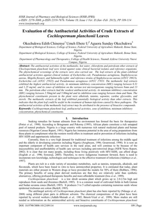
Evaluation of the Antibacterial Activities of Crude Extracts of Cochlospermum planchonii Leaves

The antibacterial activities of the methanolic, hot water, chloroform and petroleum ether extracts of Choclospermum planchonii leaf were tested against some clinical bacterial isolates and reference organisms. The phytochemical screenings of the extracts were also carried out. The leaf extracts of the plant exhibited antibacterial activities against clinical isolates of Escherichia coli, Pseudomonas aeruginosa, Staphylococcus aureus, Shigella flexneri, and Salmonella typhii and reference strains of Staphylococcus aureus (ATCC 28923), Escherichia coli (ATCC 25922) and Pseudomonas aeruginosa (ATCC 27853. The methanolic leaf extracts exhibited the highest antibacterial activity, its minimum inhibitory concentration (MIC) ranging between 0.31 and 1.25 mg/ml; and its zones of inhibition on the various test microorganisms ranging between 8mm and 13 mm. The petroleum ether extracts had the weakest antibacterial activity, its minimum inhibitory concentration (MIC) ranging between 2.50mg/ml and 5.00mg/ml and its inhibition zone ranging between 4mm and 8mm. The phytochemical constituents present in the plant were alkaloids, tannins, saponins, cardiac glycosides, and sterols. The methanolic extracts of the leaf exhibited potentially biological activity than other extracts. This indicates that the plant leaf could be useful in the treatment of human infections caused by these pathogens. The antibacterial activities of the methanolic leaf extract may be attributed to the presence of bioactive compounds.

Recommended
Recommended
More Related Content
What's hot
What's hot (18)
Similar to Evaluation of the Antibacterial Activities of Crude Extracts of Cochlospermum planchonii Leaves
Similar to Evaluation of the Antibacterial Activities of Crude Extracts of Cochlospermum planchonii Leaves (20)
More from iosrjce
More from iosrjce (20)
Recently uploaded
Recently uploaded (20)
Evaluation of the Antibacterial Activities of Crude Extracts of Cochlospermum planchonii Leaves
- 1. IOSR Journal of Pharmacy and Biological Sciences (IOSR-JPBS) e-ISSN: 2278-3008, p-ISSN:2319-7676. Volume 10, Issue 1 Ver. II (Jan -Feb. 2015), PP 109-114 www.iosrjournals.org DOI: 10.9790/3008-1012109114 www.iosrjournals.org 109 | Page Evaluation of the Antibacterial Activities of Crude Extracts of Cochlospermum planchonii Leaves Okechukwu Edith Chinenye1 Umeh Ebere U2 Aguora Sunday Okechukwu3 Department of Biological Sciences, College of Science, Federal University of Agriculture Makurdi, Benue State, Nigeria. Department of Biological Sciences, College of Science, Federal University of Agriculture Makurdi, Benue State, Nigeria. Department of Pharmacology and Therapeutics, College of Health Sciences, Nnamdi Azikiwe University Nnewi Campus, Nigeria. Abstract: The antibacterial activities of the methanolic, hot water, chloroform and petroleum ether extracts of Choclospermum planchonii leaf were tested against some clinical bacterial isolates and reference organisms. The phytochemical screenings of the extracts were also carried out. The leaf extracts of the plant exhibited antibacterial activities against clinical isolates of Escherichia coli, Pseudomonas aeruginosa, Staphylococcus aureus, Shigella flexneri, and Salmonella typhii and reference strains of Staphylococcus aureus (ATCC 28923), Escherichia coli (ATCC 25922) and Pseudomonas aeruginosa (ATCC 27853. The methanolic leaf extracts exhibited the highest antibacterial activity, its minimum inhibitory concentration (MIC) ranging between 0.31 and 1.25 mg/ml; and its zones of inhibition on the various test microorganisms ranging between 8mm and 13 mm. The petroleum ether extracts had the weakest antibacterial activity, its minimum inhibitory concentration (MIC) ranging between 2.50mg/ml and 5.00mg/ml and its inhibition zone ranging between 4mm and 8mm. The phytochemical constituents present in the plant were alkaloids, tannins, saponins, cardiac glycosides, and sterols. The methanolic extracts of the leaf exhibited potentially biological activity than other extracts. This indicates that the plant leaf could be useful in the treatment of human infections caused by these pathogens. The antibacterial activities of the methanolic leaf extract may be attributed to the presence of bioactive compounds. Keywords: Cochlospermum planchonii leaf, antibacterial activities, zone of inhibition, minimum inhibitory concentration, phytochemical constituents. I. Introduction Seeking remedies for human ailments from the environment has formed the basis for therapeutics (Potier et al., 1990). According to Bringmann and Pokorny (1995), African plants constitute a rich untapped pool of natural products. Nigeria is a large country with numerous rich natural medicinal plants and human resources (Nigerian Census Report, 1991). Nigeria has immense potential in the area of using preparations from these plants to complement what the western world offers in treatment and/or prevention of infections including HIV/AIDS and opportunistic infections Currently, there is a very high demand for traditional treatment of infections among the middle aged and the elderly in developing countries including Nigeria (Aregbeyen, 1996; Greenwood, 1998). It is now an important component of health care services in the rural areas, and will continue to be because of the inaccessibility and unaffordability of health care services including anti-retroviral drugs (NIFR News Report, 2002). It is clear that not many people, including those living positively with HIV/AIDS, can afford drugs (English et al., 1996; Enwereji, 2005). Therefore, to move indigenous treatment forward, there is need to incorporate new knowledge, technologies and techniques in the effective treatment of infections (Adebayo et al., 1997). Plants are rich in a wide variety of secondary metabolites, such as tannins, terpenoids, alkaloids, and flavonoids, which have been found in vitro to have antimicrobial properties. It is estimated that today, plant materials are either present in Western drugs or have provided the models for 50% of them (Robbers, 1996). The primary benefits of using plant derived medicines are that they are relatively safer than synthetic alternatives, offering profound therapeutic benefits and more affordable treatment (Iwu et al., 1999). Cochlospermum planchonii is a low shrub savanna plant which grows up to 2.0-2.5m high and reproduces from seeds and rhizomes (Burkill, 1985). It is a common weed of cultivated fields in both Guinea and Sudan savanna zones (Burkill, 1985). It produces 3 to 5 celled capsules containing numerous seeds whose epidermal trichomes are cotton (Burkill, 1985). The antifungal activity of Cochlospermum planchonii plant has also been reported by (Nduagu et al., 2008) using different plant parts in different solvents. The chemical compositions of the plant have been reported by several workers (Addeh-Mensah et al., 1985; Benoit-Vical et al., 1999). More studies are still needed as information on the antimicrobial activity and bioactive constituents of Cochlospermum planchonii
- 2. Evaluation Of The Antibacterial Activities Of Crude Extracts Of Cochlospermum planchonii Leaves DOI: 10.9790/3008-1012109114 www.iosrjournals.org 110 | Page leaf is scanty. The study was designed to evaluate the antimicrobial activities and phytochemical constituents of the crude extracts of Cochlospermum planchonii leaf. II. Materials And Methods 2.1 Test plant The leaves of Cochlospermum planchonii were collected in August 2008 from the farmland behind Block B, South-Core of Federal University of Agriculture Makurdi. The plant was identified by Mr. P.O. Ekwuno of the Department of Forestry, University of Agriculture Makurdi, Benue State, Nigeria. 2.2 Test Organisms The microorganisms used were clinical strains of Staphylococcus aureus, Escherichia coli, Salmonella typhii, Pseudomonas aeruginosa and Shigella flexneri; and reference strains of Staphylococcus aureus (ATCC 28923), Escherichia coli (ATCC 25922) and Pseudomonas aeruginosa (ATCC 27853) used as controls. 2.3 Extraction procedure The leaves of the plant Cochlospermum planchonii were collected, sun dried and pulverized by pounding. One hundred grams (100g) of the powdered leaves of Cochlospermum planchonii were placed in a corked bottle, and 500 ml of solvent (methanol, petroleum ether, chloroform or hot water) were added in the cold (cold extraction). The resulting suspension was allowed to stand in a tightly covered bottle for 48 hours at room temperature after which it was filtered using Whatman’s No.1 filter paper into a round bottom flask. The flask containing the filtrate was placed in the water bath and allowed to evaporate off the extraction solvent to obtain the crude extract. The crude extract was placed in sterile sample bottles and labeled appropriately. The yield was weighed and recorded in grams. 2.4 Determination of minimum inhibitory concentration (MIC): The microdilution assay and disc diffusion were carried out using the Ciprofloxacin against the test organisms both the reference strains and clinical isolates to confirm the sensitivity of Ciprofloxacin against the test organisms. 2.5 Microdilution Assay Technique The antibacterial activities of the various extracts were assayed using modified microdilution techniques of Drummond and Waigh (2000), as described by Satyajit et al. (2007). Micro titre plates were prepared under aseptic conditions. A sterile 96-well microtitre plate was labeled. One hundred microlitres (100 µL) of test material in 10%(vv) dimethylsulpoxide (a stock concentration of 10mg/ml) was pipetted into the first row of wells in triplicates. Sterile nutrient broth (50 µL) was added to all other wells. The extract was serially diluted two-folds using a multichannel pipette. Tips were discarded after use so that each well had 50 µL of the test material in serially descending concentrations. To each well 10 µL of resazurin indicator dye solution in sterile water was added. Bacterial suspension (10 µL) in nutrient broth (5x10⁶ cfu/ml) was added to each well to achieve a concentration of 5x10⁵ cfu/ml. Two columns were used as controls: positive control (comprising broad-spectrum antibiotic Ciprofloxacin) that prevented bacterial growth; and negative control containing bacterial suspension, resazurin indicator dye and 10%v/v dimethylsulphoxide without the test extract. Finally each plate was sealed with paraffin film to ensure that the bacteria did not become dehydrated. Plates were incubated at 33°C for 18-22hrs. The plates were then observed macroscopically for colour change and turbidity under the reading mirror. The minimum inhibitory concentration, which is the lowest concentration of the extract that caused bacterial growth inhibition, was read off. 2.6 Minimum bactericidal concentration The minimum bactericidal concentration was determined by sub culturing the minimum inhibitory concentrations onto a sterile surface of drug free Mueller-Hinton agar plates. The plates were incubated at 330 C for 18-24hours. The plates were observed for colonies and non colonies. Plates without growth colonies indicated minimum bactericidal concentration. 2.7 Disc Diffusion Assay Technique Antimicrobial screening using the disk diffusion method as described by the NCCLS (2004) was carried out. Twenty milliliters (20 ml) of molten sterile Mueller-Hinton agar was poured into sterile Petri dishes and was allowed to solidify. A loopful of inoculum containing 5x10⁵ cfuml bacterial suspension was uniformly streaked on the surface of the agar. Pre-sterilized filter paper discs of 3 mm diameter were impregnated with the different concentrations of extracts, and were placed on the seeded agar. Ciprofloxacin discs (10µg) was used
- 3. Evaluation Of The Antibacterial Activities Of Crude Extracts Of Cochlospermum planchonii Leaves DOI: 10.9790/3008-1012109114 www.iosrjournals.org 111 | Page as a positive control and discs saturated with sterile water (negative control) were placed at the center of the seeded plates and incubated for 18-22hrs at 33 to 37o C. At the end of incubation period, diameter of inhibition zones in all three replicates were measured in millimeters using measuring slide and the mean of the three was determined. 2.8 Phytochemical Test Phytochemical test was carried out on the extracts for the following bioactive constituents: tannins, saponins, cardiac glycosides, alkaloids, sterols and flavonoids. Chemical tests were carried out on the extracts of the leaf using standard procedures to identify the constituents as described by Sofowara (1993), Trease and Evans (2002), and Harborne (1998). 2.8.1 Test for Tannins: Small portion of extracts of leaf each was stirred with 1ml absolute methanol and FeCl3 (aq) added in a test tube. A blue black precipitate is indicative of the presence of tannins. 2.8.2 Test for Saponins: i. Frothing test; Small portion of extracts of leaf each was shaken with water in a test tube. Frothing which persisted on warming is indicative of the presence of saponins. ii. Emulsion test ; Five drops of olive oil was added to 3ml of extracts of leaf in a test tube and the mixture was vigorously shaken to form stable emulsion. Formation of stable emulsion indicates the presences of saponins. 2.8.3 Test for Flavonoids: 2ml of solution of extracts was added to Magnesium chip and few drops of H2SO4 in a test tube. Light yellow precipitate in brown solution is indicative of flavonoids. 2.8.4 Test for Alkaloids: i). Small portion of the extracts of leaf was dissolved in 2ml 0.1 HCl (aq) in a test tube. To this was added 2 drops of Mayer’s reagent, a light yellowish white precipitate indicates the presence of alkaloids. ii). Dragendroff’s reagent, 0.85g basic bismuth nitrate in 10ml glacial acetic acid into 40ml water was added to small portion of extracts of leaf in a test tube and the mixture was heated for 2minutes. Faint yellowish orange precipitate indicates the presence of alkaloids. 2.8.5 Test for Cardiac glycosides: Keller-Killani test; To a small portion of solution of extracts in glacial acetic acid containing 2 drops of FeCl3 (aq) was added 1.5ml conc. H2SO4. Lower conc. H2SO4 layer that is colourless, with upper acetic acid layer is indicative of the presence of glycosides. 2.8.6 Test for Sterols: The extracts were dissolved in 2ml of acetic anhydride and cooled ice conc. H2SO4 was carefully added. A colour change from violet to blue then to green indicates the presence of sterols. III. Results The results of the minimum inhibitory concentrations of the leaf extracts of C. planchonii using modified indicator based microdilution technique are shown on table 1. The MIC of methanolic leaf extracts was in the range of 0.31 and 1.25 mg/ml. Methanolic leaf extracts exhibited the highest inhibitory effect on all the test organisms, for example E.coli ATCC 25922 was 0.31mg/ml, P. aeruginosa ATCC 27853 1.25mg/ml and for S aureus ATCC 28923 was 0.63mg/ml. S. typhii and P. aeruginosa 0.625mg/ml. S. aureus, S. flexneri and E. coli for 1.25mg/ml. Petroleum ether leaf extracts had the weakest antibacterial activity: the MIC on the organisms ranging from 2.50 to 5.00mg/ml ; E.coli ATCC 25922 was 2.5mg/ml and S. aureus ATCC 28923 was 5.0mg/ml. S. typhii, P. aeruginosa, S. aureus, S. flexneri and E. coli were 5.0mg/ml. The results of the antibacterial susceptibility test of the plant extracts using the disc diffusion method are shown in Table 2. The extracts of different solvent screened showed varying inhibitory effects; methanolic extracts (9mm-13mm), chloroform extracts (6mm-10mm), petroleum ether extracts (5mm-8mm) and hot water (6mm-10mm) against the test organisms. Ciprofloxacin served as the positive control (11mm-15mm) exhibited the largest zone of inhibition. The viability controls in the last column showed that the organisms were not inhibited as they were cultured without any extracts or antimicrobial agents in Tables 1 and 2. The results of the phytochemical constituents of the leaf extracts of Choclospermum planchonii are presented in table 3. The phytochemical investigation revealed the presence of alkaloids, saponins, tannins glycosides and sterols in the leaf extracts of Choclospermum planchonii . The zone of inhibitions and minimum inhibitory concentrations of the leaf extracts are shown in Fig.2 and Fig.3 respectively. In fig.2 the results showed that methanolic leaf extract of Choclospermum planchonii was the highest ranging between 11mm - 13mm as observed for the reference organisms and 9mm -13mm for the clinical isolates. Petroleum ether leaf
- 4. Evaluation Of The Antibacterial Activities Of Crude Extracts Of Cochlospermum planchonii Leaves DOI: 10.9790/3008-1012109114 www.iosrjournals.org 112 | Page extract was the weakest with 5mm to 8mm as observed for the reference organisms and 5mm-8mm for clinical isolates respectively. In fig.3 the methanolic leaf extract showed the strongest antibacterial activity with MICs ranging between 0.31mg/ml-1.25mg/ml as observed for reference organisms and 0.63mg/ml-1.25mg/ml for clinical isolates. The minimum inhibitory concentrations of the petroleum ether leaf extracts had the weakest MICs ranging between 2.5mg/ml-5.0mg/ml for reference strains and 5.0mg/ml for clinical isolates. Table 1: Minimum Inhibitory Concentrations (mg mL-1 ) of Leaf Extract obtained by the Microdilution Technique Organisms Methanol Hot Water Chloroform Pet. Ether Ciproflo- xacin Viability Control Reference Strains Staphylococcus aureus ATCC 28923 0.63 5.00 5.00 5.00 + - Escherichia coli ATCC 25922 0.31 5.00 5.00 2.50 + - Pseudomonas aeruginosa ATCC 27853 1.25 2.50 5.00 5.00 + - Clinical Isolates Staphylococcus aureus 1.25 2.50 5.00 5.00 + Escherichia coli 1.25 2.50 5.00 5.00 + - Pseudomonas aeruginosa 0.63 2.50 5.00 5.00 + - Salmonella typhi 0.63 1.25 2.50 5.00 + - Shigella flexneri 1.25 1.25 2.50 5.00 + - Key: + = bacterial growth inhibited; - bacterial growth uninhibited. Table 2 : Zone of inhibition diameter (mm) of bacterial growth by Leaf Extracts Organisms Methanol Hot Water Chloroform Pet. Ether Ciprofloxacin Viability Control Reference Strains Staphylococcus aureus ATCC 28923 11 6 6 5 13 - Escherichia coli ATCC 25922 13 7 7 7 15 - Pseudomonas aeruginosa ATCC 27853 11 6 6 5 12 - Clinical Isolates Staphylococcus aureus 9 9 7 5 11 - Escherichia coli 12 9 9 7 14 - Pseudomonas aeruginosa 11 8 8 8 12 - Salmonella typhi 13 10 10 7 14 - Shigella flexneri 12 8 8 8 13 - Note: Each value represent mean of three different observations; + means uninhibited growth Table 3: Phytochemical Constituents of Leaf Extracts Test Methanolic Extract Chloroform Extract Pet. Ether Extract Hot Water Extract Alkaloid + - - + Saponin + - + - Tannins + + - - Flavonoids - - - - Cardiac glycosides + - + - Steroids + + - - +: Presence of constituent; -: Absence of constituent Fig.1 Cochlospermum planchonii leaf
- 5. Evaluation Of The Antibacterial Activities Of Crude Extracts Of Cochlospermum planchonii Leaves DOI: 10.9790/3008-1012109114 www.iosrjournals.org 113 | Page Fig. 2 Pattern of zone of inhibition diameter of leaf extracts Fig.3 Pattern of Minimum Inhibitory Concentration of leaf extracts. IV. Discussion The plant C. planchonii growing locally within the university campus was screened for antibacterial activity and was found to exhibit antibacterial activity against some clinical isolates of E. coli and P. aeruginosa and their reference strains. This finding agrees with the previous antibacterial studies related to this plant and reported by Ouattara et al. (2007). The significant antibacterial activity of the plant could be attributed to the presence of bioactive compounds in the plant. This result suggests that the plant could be used as potential broad spectrum antibacterial agent for treatment of bacterial infections. Antibacterial activity and susceptibility assay of the plant varied according to extraction solvents. This indicates that the minimum inhibitory concentration and zone of inhibition diameter of the leaf extracts were affected by the solvent used for extraction. For example the methanolic extract showed the highest antibacterial activity amongst other extracts, the variation may probably be due to the type of bioactive compounds present in the different extracts as suggested by Abiodun et al. (2007). Although the inhibitory effects of aqueous and methanolic extracts of medicinal plants are usually reported (Olayinka et al., 1992; Omer et al., 1998), our findings show that methanolic leaf extract of the plant studied had more inhibitory effect than extracts of petroleum ether, chloroform and hot water. For example the value of minimum inhibitory concentration of the methanolic leaf extract was lower than those of other extracts and the zone of inhibition diameter higher than those of others. MIC of methanolic extract ranged between 0.31 and 1.25mg/ml, and its zone of inhibition diameter ranged between 8 and 13mm. These results confirm the evidence in previous studies (Ahmad et al., 1998; Cowan, 1999) that alcoholic solvents like methanol and ethanol are more suitable than other solvents such as water in extracting components of medicinal plants. This result suggests that the antibacterial constituents of the plant may be residing in higher concentration in the methanolic leaf extracts and could have contained more active constituents than other solvents. Among the bacterial strains tested Staphylococcus aureus appears to be the most resistant bacteria and this could be attributed to genetic mutation of the microorganism probably due to multidrug resistance factor (Adwan et al., 2005;
- 6. Evaluation Of The Antibacterial Activities Of Crude Extracts Of Cochlospermum planchonii Leaves DOI: 10.9790/3008-1012109114 www.iosrjournals.org 114 | Page Fridkin et al., 2005; Schito, 2006). Salmonella typhii, though a Gram-negative bacterium appeared to be the most sensitive organism to the inhibitory effect of the leaf extracts of the plant and has been reported to be sensitive to other medicinal plant such as Carica papaya (Osato et al., 1993). Therefore this scientific finding validates with the traditional use of the plant C. planchonii in the treatment of typhoid fever and diarrheal diseases (Igoli et al., 2005). V. Conclusion Cochlospermum planchonii methanolic leaf extracts that have shown significant antibacterial activities can be considered a potential antimicrobial agent in the treatment of bacterial diseases. Acknowledgement The authors sincerely acknowledge the staff of the Department of Microbiology and Biotechnology of National Institute of Pharmaceutical Research and Development (NIPRD) and the staff of the Department of Microbiology of National Veterinary Research Institute (NVRI) Vom, Plateau State, Nigeria for their assistance to make this research a dream come true. References [1]. Abiodun, F., Igwe, A. and Obasuyi O. (2007). Anti-microbial evaluaton of a herbal dental remedy: stem bark of Nauclea latifoli-family Rubiaacea. Journal of Applied Sciences 7 (18): 2696-2700. [2]. Adebayo, R.A., Sofowora,G.G., Onayemi, 0., Udoh, S.J, and Ajayim, A.A., (1997). Chloroquine induced pruritus in malaria fever, contribution of malaria parasitaemia and the effects of prednisolone niacin, and their combination, compared with antihistamine. British Journal of Clinical Pharmacology, 44 (2): 157-61. [3]. Addeh-Mensah, R. Waibel and H. Achenbach. (1985). Novel long-chain triacylbenzenes from Cochlospermum planchonii constituents of West Africa medicinal plants. Liebigs Anna.Chem. XVI,1284-1288. [4]. Adwan, K. Abu Hasan N. Adwan G. et al.,(2005). Nosocomial infection caused by methicillin-resistant Staphylococcus aureus in Palestine.Journal of Microbial Drug Resistance. 11: 75 -77. [5]. Ahmad I, Mehmood Z, and Mohammad F. (1998). Screening of some Indian medicinal plants for their antimicrobial properties. Journal of Ethnopharmacology 62: 183-193. [6]. Aregbeyen, J.B.0. (1996). Traditional herbal medicine for sustainable PHC. Indigenous and Development Monitor Vol 4. Issue (2) pp 14 - 15. [7]. Benoit-Vical, F., Valentin, A., Mallié, M., Bastide, J.M. and Bessière, J. M. (1999). In vitro antimalarial activity and cytotoxicity of Cochlospermum tinctorium and Cochlospermum planchonii extracts and essential oils. Planta Med., 65 (4): 378-81. [8]. Burkill, H. M., (1985). The Useful Plants of West Tropical Africa, 2 nd Edition, Volume 1. Royal Botanic Gardens, Kew. 960p. [9]. Bringmann, G. and Pokorny, F. (1995). The Naphthylisoquinoline alkaloids. In: The Alkaloids. Vol. 46. G. Cordell (Ed.) AcademicUniversity Press, New York, pp 127. [10]. Cowan M.M. (1999). Plant products as antimicrobial agents. Clinic Microbiol Rev 12: 564-582. [11]. Drummond,A.J. and Waigh,R.D. (2000) . The development of microbiological methods for phytochemical screening . Recent Rresearch in Development. Phytochemistry 4: 143-152. [12]. Fridkin, S. K. Hageman ,J.C., Morrison, et al.,.(2005). Active Bacterial Core Surveillance Programme of the Emerging Infections Program Network. Methicillin-resistant Staphylococcus aureus disease in three communities. N Engl J Med 352: 1436-1444. [13]. Nduagu C, Ekefan EJ and Nwankiti AO, (2008). Effect of some crude plant extracts on growth of Colletotrichum capsici (Synd) & Bisby, causal agent of pepper anthracnose. Journal of Applied Biosciences 6(2): 184-190. [14]. News Report of Nigerian Institute of Forestry Research, Umuahia ( 2002) [15]. Nigerian Census Report 1991. [16]. English, M., Marsh, V. Amukoye, E., Lowe, B., Murphy, S., and Marsh, K. (1996). Chronic Salicylate poisoning and severe malaria. Lancet: 347(9017): 1736-7. [17]. Enwereji, E. (2005). Care and support of people living positively with HIV/AIDS in South-east Nigeria. Research findings carried out for Presbyterian Church of Nigeria, in collaboration with Presbyterian Church of Canada. [18]. Greenwood,B. (1998). Traditional medicine to DNA vaccines. The advance for medical research in West Africa. Tropical Medicine and International Health 3(3): 166-76. [19]. Harborne, J.B., 1998. Phytochemical Methods: A Guide to Modern Techniques of Plant Analysis. (3rd Edition). Chapman and Hall Co., New York. 1998, 88-190p. [20]. Iwu, M.W., A.R. Duncan, and C.O. Okunji. (1999). New antimicrobials of plant origin. p. 457–462. In: J. Janick (ed.), Perspectives on new crops and new uses. ASHS Press, Alexandria, VA. [21]. NCCLS. (2004). Performance standards for antimicrobial susceptibility testing, fourteenth informational supplement. M100 – S14. NCCLS, Wayne, P. A. USA. [22]. Olayinka,D.O., Onuruv, W. O. and Lots, T.Y. (1992). Cadiovascular effects in rodents of methanolic extracts of Khaya senegalensis. Phytother Res 6: 282-284. [23]. Omer, M., E.A., AI Mabgoul,A.Z. and EI Egami,A.A.(1998). Sudanese plants used in folkloric medicine:screening for antimicrobial activity. Fitoterapia 69: 542-545 [24]. Potier, P., Mangatal, L., Le Goff, M. T., Gueritte, V. F., Guenard, O., and Lavelle, F. (1990). Relationships between the structure of taxol analogues and their antibiotic activity. J. Med Chem. 34: 992 – 998. [25]. Ouattara L.,Obame L.C.E.,Karou D.S.,Koudou J.,Traore A.,Bessiere J.M. (2007). Chemical Composition and Antibacterial activity of Cochlospermum planchonii Hook.f.ex. planch.Essential oil from Burkina Faso. Paskistan Journal of Biological Sciences 10 (22): 4177- 4179. [26]. Robbers, J., M. Speedie, and V. Tyler. (1996). Pharmacognosy and pharmacobiotechnology. Williams and Wilkins, Baltimore. p. 1–14. [27]. Satyajit, D.Sarker, S. Nahar, L. and Kumarasamy, Y. (2007). Microtitre plate-based antibacterial assay incorporating resazurin as an indicator of cell growth, and its application in the in vitro antibacterial screening of phytochemicals. Methods 42 (4): 321-324. [28]. Schito, S. (2006). The importance of development of antibiotic resistance in Staphylococcus aureus. Clin. Microbiol.Infect. 1: 3-8. [29]. Sofowara A (1993). Medicinal plants and Traditional medicine in Africa. Spectrum Books Ltd, Ibadan, Nigeria. p. 289. [30]. Trease, A., Evans, W.C., 2002. A textbook of Pharmacognosy (14th edition) Bailliere Tindall London, 76-84p.