Scaling API-first – The story of a global engineering organization
www.ijerd.com
1. International Journal of Engineering Research and Development
ISSN: 2278-067X, Volume 1, Issue 3 (June 2012), PP.33-36
www.ijerd.com
Accurate Multimodality Registration of medical images
M.V.Sruthi1, Dr.K.Soundararajan, Dr.V.Usha Sree
1
Jawaharlal Nehru technological university Anantapur
Abstract––This paper presents a method of motion correction in registering multimodal imagery. Image registration is a
vital problem in medical imaging. It has many potential applications in clinical diagnosis It is a process of aligning two
images into a common coordinate system .Thus aligning them in order to monitor subtle changes between the two.
Registration algorithms will compute transformations to set correspondence between the two images the purpose of this
paper is to provide a comprehensive review of the existing literature available on Image registration methods in different
modalities. It has been extensively shown that metrics based on the evaluation of mutual information are well suited for
overcoming the difficulties of multi-modality registration. For this we are implementing multimodality registration
algorithm which goals the motion correction.
Keywords––modality, interpolater, optimizer image registration
I. INTRODUCTION
Some of the most challenging cases of image registration arise when images of different modalities are
involved. In such cases, metrics based on direct comparison of gray levels are not applicable. Image processing methods,
which are possibly able to visualize objects inside the human body, are of special interest. Advances in computer science
have led to reliable and efficient image processing methods useful in medical diagnosis, treatment planning and medical
research. In clinical diagnosis using medical images, integration of useful data obtained from separate images is often
desired. The images need to be geometrically aligned for better observation. This procedure of mapping points from one
image to corresponding points in another image is called Image Registration. It is a spatial transform. In order to
associate the information from modality, corresponding data in each image must be successfully registered. In long range
surveillance applications the alignment function will register all objects in the scene. The reference and the referred
image could be different because were taken
At different times
Using different devices like MRI, CT, PET, SPECT etc (multi modal).
From different angles in order to have 2D or 3D perspective (multi temporal).
Image registration finds its applications in various fields remote sensing(multispectral classification),
environmental monitoring, change detection, image mosaicing, weather forecasting, creating super-resolution images,
integrating information into geographic information systems (GIS)), in medicine (combining data from different
modalities e.g. computer tomography (CT) and magnetic resonance imaging (MRI), to reatment verification, comparison
of the patient’s data with anatomical atlases ,in cartography (map updating) and in computer vision (target localization,
automatic quality control).
The concept of Mutual Information is derived from Information Theory and its application to One way to
simplify the computation of the mutual information is to normalize the statistical distribution of the two input images.
Image registration has been proposed in different forms by different groups .
II. IMPLEMENTATION
The metric requires a number of parameters to be selected, including the standard deviation of the Gaussian
kernel for the fixed image density estimate, the standard deviation of the kernel for the moving image density and the
number of samples use to compute the densities and entropy values. We should now define the number of spatial samples
to be considered in the metric computation. Image registration is the process of determining the spatial transform that
maps points from one image to homologous points on a object in the second image.
33
2. Accurate Multimodality Registration of medical images
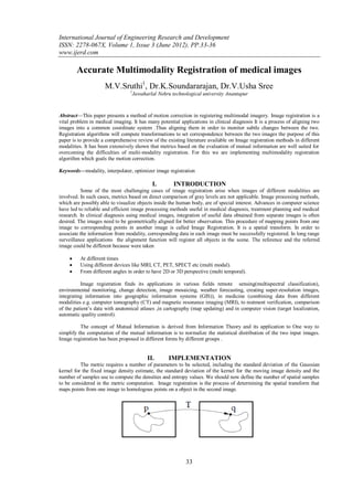
Fig 1 The basic components of the registration framework are two input images, a transform, a metric, an interpolator
and an optimizer
The components of the registration framework and their interconnections are shown in Figure.The basic input
data to the registration process are two images: one is defined as the fixed image f (X) and the other as the moving image
m(X). Where X represents a position in Ndimensional space. Registration is treated as an optimization problem with the
goal of finding the spatial mapping that will bring the moving image into alignment with the fixed image. The transform
component T(X) represents the spatial mapping of points from the fixed image space to points in the moving image
space. The interpolator is used to evaluate moving image intensities at non-grid positions. The metric component S( f ,m◦
T) provides a measure of how well the fixed image is matched by the transformed moving image. This measure forms the
quantitative criterion to be optimized by the optimizer over the search space defined by the parameters of the transform.
Fig 2: MRI (fixed image) and a proton density MRI (moving image) are provided as input to the registration method.
The normalization filters are instantiated using the fixed and moving image types as input and the internal
image type as output. The output of the readers becomes the input to the ormalization filters. The output of the
normalization filters is connected as input to the blurring filters. The input to the registration method is taken from the
blurring filters.
Fig 3: block diagram
Fixed and moving images are given to the image read and write filter. Depending upon the intensity of the pixel
ratio image is been read or write. After the images are been read the output is given to the register filter. In this filter the
two images i.e., fixed and moving images depending on the pixel intensities the two images get overlap and form a new
image with both modalities in it. It is useful for the doctor to compare both the images of the same patient and check the
results. Multimodality registration algorithm has the following steps.
34
3. Accurate Multimodality Registration of medical images
1. Feature detection: Salient and distinctive objects (closed-boundary regions,edges, contours, line
intersections, corners, etc) in both reference and sensed images are detected.
2. Feature matching: The correspondence between the features in the reference and sensed image established.
3. Transform model estimation: The type and parameters of the so-called mapping functions, aligning the
sensed image with the reference image, are estimated.
4. Image resampling and transformation: The sensed image is transformed by means of the mapping
functions.
Fig4: steps of mutimodality algorithm
Boundaries or surfaces, in medical images are many times more distinct than landmarks and hence can be used
for segmentation by locating high contrast surfaces. Surface matching algorithms are normally used for rigid body
registration. The representation of surface methods is a surface-based approach for registering multimodality brain
image. They fit a set of points extracted from contours in one image to a surface model and extracted from contours in the
other image. The image that covers the larger volume of the patient, or the image that has a higher resolution if volume
coverage is comparable, is used to generate the surface model. Another version of surface matching is to provide user
with an interactive transformation package that allows the user to translate and rotate one image with respect to the other
III. RESULTS & DISCUSSION
In this paper we have presented and analyzed a method for registering multimodality medical images . The
algorithm has given successful and reliable registration without relying on the limits. Followed by image registration in
the medical applications, segmentation is
Fig 5: Mapped moving image (left) and composition of fixed and moving images before (center) and after (right)
registration.
Needed to determine areas of interest in the image and in many cases accurate demarcation of objects yields
valuable information. Quantification is often the ultimate goal especially in medical applications. We believe that it will
be a useful document for researchers longing to implement alternative Image registration methods for specific
applications.
35
4. Accurate Multimodality Registration of medical images
IV. ACKNOWLEDGMENT
I thank Dr K .Soundararajan, Dr .V. Ushasree and S .Ismail saheb for their comments and suggestions on this manuscript.
REFERENCES
[1]. D.Ruckert, L.I.Sonoda, C.Hayes,, D.L.G.Hill ,M.O. Leach D.J.Hawkes, “Non rigid registration using free form deformations:
Application to breast MR images”, IEEE transactions on Medical imaging, 18(8), 1999, 712721
[2]. J.V. Hajnal, D.L.G. Hill, D.J. Hawkes, Medical Image Registration, CRC Press, Boca Raton London New York Washington
DC, 2001, ISBN 0-8493-0064-9.
[3]. Barbara Zitova, Jan Flusser, “Image registration methods: a survey”, Image and Vision Computing 21 (2003), 977–1000
[4]. P. A. Van Den Elsen, E. J. D. Pol and M. A. Viergever, “Medical image matching: a review with classification”, IEEE
Engineering in medicine and biology, 12(1):1993,26-39
[5]. J. B. Antoine Maintz and Max A. Viergever, “A Survey of Medical Image Registration”, Medical Image Analysis (1998)
Volume2, number 1, pp 1-36, Oxford University Press
[6]. Brown Gottesfeld L, “A survey of image Registration Techniques”, ACM Computing surveys 24, 1992, 325376
[7]. Mauer C.R., Fitzpatrick J.M., “A review of medical image registration, in: Interactive Image Guided Neurosurgery”,
Maciunas R.J. (ed), American association of Neurological Surgeons, 1993, 17-44
[8]. Letser.H, Arrige S.R., “A survey of hierarchical nonlinear medical image registration” Pattern Recognition, 1999, 129-149
[9]. Wan Rui, Prof.Li Minglu, “An Overview of Medical Image Registration”, Proceedings of the Fifth International Conference
on Computational Intelligence and Multimedia Applications (ICCIMA’03), 2003, 385-390
[10]. J.V.Chapnick, M.E.Noz, G.Q. Maguire, E.L.Kramer, J.J.Sanger, B.A.Birnbaum, A.J.Megibow, “Techniques of
Multimodality Image Registration” Proceedings of the IEEE nineteenth Annual North East Bioengineeering conference, 18-
19 March 1993, 221-222
[11]. L. Lemieux, N. D. Kitchen, S. W. Hughes and D. G. T. Thomas, “Voxel-based localization in frame-based and frameless
stereotaxy and its accuracy”. Medical physics, 21(8):1301–1310, 1994.
[12]. L. Lemieux and R. Jagoe, “Effect of fiducial marker localization on stereotactic target coordinate calculation in CT slices and
radiographs” Physics in medicine and biology, 39:1915–1928, 1994.
[13]. S. C. Strother, J. R. Anderson, X. Xu, J. Liow, D. C. Bonar and D. A. Rottenberg, “Quantitative comparisons of
image registration techniques based on high- resolution MRI of the brain” Journal of computer assisted tomography,
18(6):954–962, 1994.
[14]. T. Peters, B. Davey, P. Munger, R. Comeau, A. Evans, and A. Olivier, “Three-dimensional multimodal image-guidance for
neurosurgery”,IEEE Transactions on medical imaging, 15(2), 1996, 121–128
[15]. K. P. Gall and L. J. Verhey, “Computer-assisted positioning of radiotherapy patients using implanted radioopaque fiducials”,
Medical physics, 1993, 1152–1159
[16]. C. R. Maurer, G. B. Aboutanos, B. M. Dawant, R. A. Margolin, R. J. Maciunas and J. M. Fitzpatrick., 9“Registration
of CT and MR brain images using a combination of points and surfaces”, Medical.
36