Downloaded 581 times

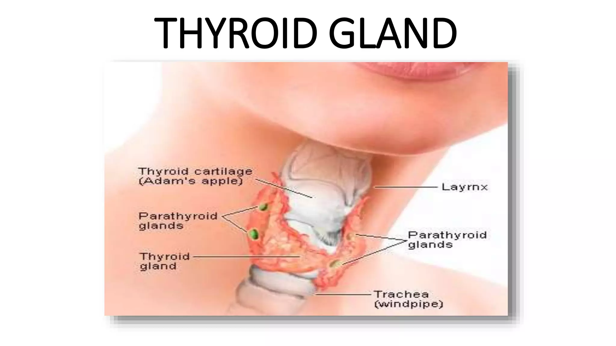
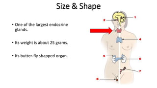
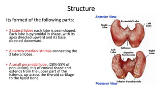
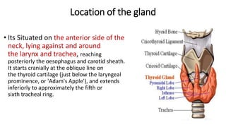
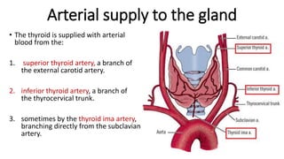
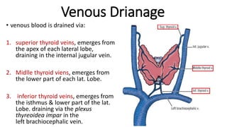
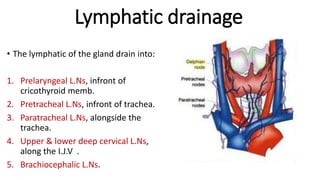

The thyroid gland is a butterfly-shaped endocrine gland located in the front of the neck. It weighs around 25 grams and is composed of two lateral lobes connected by a narrow isthmus. The gland is situated anterior to the larynx and trachea. It receives blood supply from the superior, inferior, and sometimes thyroid ima arteries, and drains into the superior, middle, and inferior thyroid veins. Lymphatic drainage occurs into the prelaryngeal, pretracheal, paratracheal, deep cervical, and brachiocephalic lymph nodes. The thyroid gland regulates metabolism in the body.