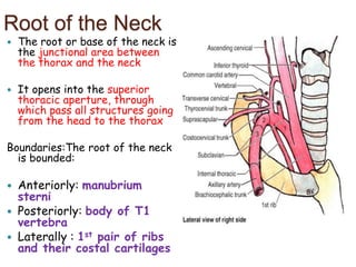
The root of the neck is the junction between the thorax and neck. It contains several important structures including arteries, veins and nerves.
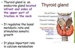
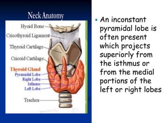
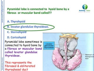
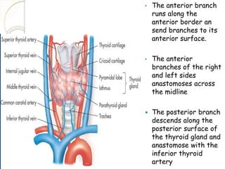
The document describes the anatomy of the thyroid gland, including its location in the neck, lobes and isthmus, relations to surrounding structures, blood supply from the superior and inferior thyroid arteries, innervation and role in hormone production and calcium metabolism.
The parathyroid glands are usually four in number, located on the posterior surface of the thyroid gland. They are supplied by branches from the inferior thyroid artery and their primary function is to regulate calcium levels through secretion of parathyroid hormone.


![Applied Anatomy
A benign enlargement of
the thyroid may
compress or displace any
of its relations; the
trachea and oesophagus
may be narrowed, with
resulting difficulty in
breathing and swallowing,
and the carotid may be
displaced posteriorly
Hypothyroidism causes
cretinism in infants and
myxoedema in adults
Any swelling of the
thyroid gland [goitre]
moves with deglutition
In partial thyroidectomy,
the posterior parts of
both lobes are left
behind to:
1. Avoid the risk of
removing the
parathyroid glands
2. Avoid postoperative
myxoedema caused by
deficiency of thyroid
hormones](https://image.slidesharecdn.com/n2ijrmmwrjistrgaakr0-thyroid-gland-230521233852-ee80f4e8/85/Thyroid_gland-pptx-30-320.jpg)


![Thyroid final [part 1]](https://cdn.slidesharecdn.com/ss_thumbnails/thyroidfinalpart1-161126043454-thumbnail.jpg?width=640&height=640&fit=bounds)


