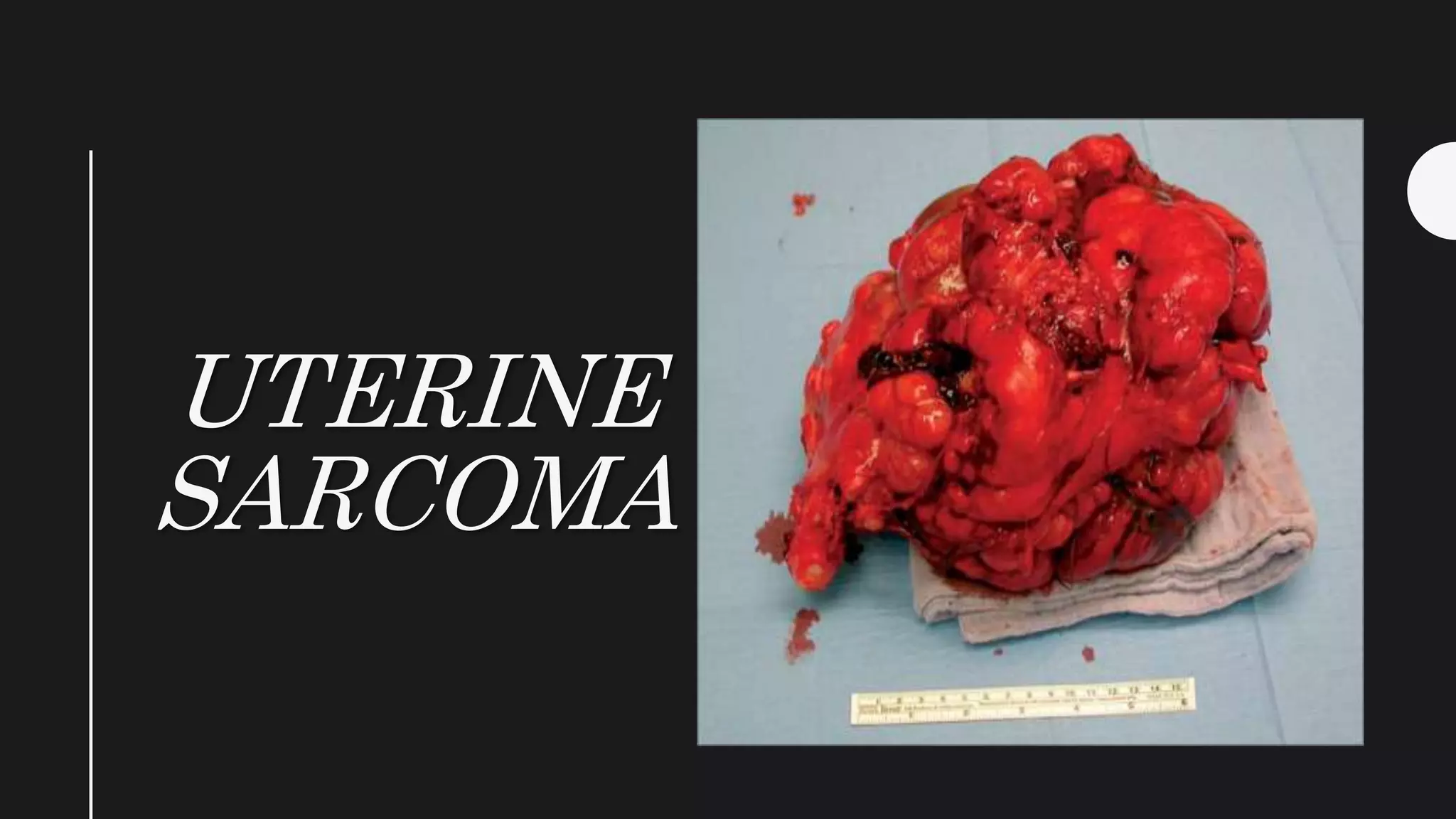
UTERINE
SARCOMA
Epidemiology
• Tumors are rare 3-7% of uterine malignancies are uterine
sarcoma. 15% of all deaths from uterine cancer
• More in Black women
TYPES • Leiomyosarcomas
• Endometrial stromal sarcomas
• Malignant mixed mullerian tumor
• Other uterine sarcomas.
Leiomyosarcomas
• Intravenous leiomyomatosis — where
benign smooth muscle grows into venous
channels within the broad ligaments, uterine
and iliac veins.
• Leiomyomatosis peritonealis disseminata –
where benign smooth muscle nodules grow
over the peritoneal surfaces.
Endometrial stromal sarcomas
• Endometrial stromal nodule (benign)
• Endolymphatic stromal myosis (low grade
Malignancy)
• Endometrial stromal sarcoma (High grade
malignancy).
• Malignant mixed müllerian tumors (MMMT) of the uterus
usually forms a large fleshy mass protruding into the uterine
cavity with a broad base.
• The commonest one arises from the intramural part.
• The consistency is soft and friable.
• The cut surface shows hemorrhage and irregular margins.
MICROSCOPIC APPEARANCE
• Uterine sarcomas may be pure (single cell type) or mixed (more than
one cell type).
• The tumor is termed homologous when the tissue elements are native
(smooth muscle) or heterologous when tissue elements are not native
(cartilage, striated muscle, bones). This is due to the totipotent nature
of endometrial stromal cells.
SPREAD
• Blood borne: This is the commonest mode of
spread. The organs involved are liver, lungs,
kidneys, brain, bones.
• Directly to the adjacent structures.
• Lymphatic spread to the regional lymph
glands.
CLINICAL FEATURES
• Patient profile: The age is usually between 40 and 60
years.
• There may be history of pelvic irradiation either for
induction of menopause or malignancy.
SYMPTOMS
• Irregular premenopausal or postmenopausal
vaginal bleeding.
• Abnormal vaginal discharge — offensive,
watery associated at times with expulsion of
fleshy necrotic mass.
• Abdominal pain — due to involvement of the
surrounding structures.
• Pyrexia, weakness and anorexia.
PELVIC EXAMINATION:
• The uterus may be enlarged and irregular. Parametrium may be
thickened and indurated.
• Speculum examination may reveal a polypoidal mass protruding out
through the external os.
Diagnosis
• Diagnosis is made usually following histological examination of the
removed uterus.
• Diagnostic uterine curettage may reveal the mucosal form of
sarcoma.

Uterine sarcoma

  • 1.
  • 2.
    Epidemiology • Tumors arerare 3-7% of uterine malignancies are uterine sarcoma. 15% of all deaths from uterine cancer • More in Black women
  • 3.
    TYPES • Leiomyosarcomas •Endometrial stromal sarcomas • Malignant mixed mullerian tumor • Other uterine sarcomas.
  • 4.
    Leiomyosarcomas • Intravenous leiomyomatosis— where benign smooth muscle grows into venous channels within the broad ligaments, uterine and iliac veins. • Leiomyomatosis peritonealis disseminata – where benign smooth muscle nodules grow over the peritoneal surfaces.
  • 5.
    Endometrial stromal sarcomas •Endometrial stromal nodule (benign) • Endolymphatic stromal myosis (low grade Malignancy) • Endometrial stromal sarcoma (High grade malignancy).
  • 6.
    • Malignant mixedmüllerian tumors (MMMT) of the uterus usually forms a large fleshy mass protruding into the uterine cavity with a broad base.
  • 7.
    • The commonestone arises from the intramural part. • The consistency is soft and friable. • The cut surface shows hemorrhage and irregular margins.
  • 8.
    MICROSCOPIC APPEARANCE • Uterinesarcomas may be pure (single cell type) or mixed (more than one cell type). • The tumor is termed homologous when the tissue elements are native (smooth muscle) or heterologous when tissue elements are not native (cartilage, striated muscle, bones). This is due to the totipotent nature of endometrial stromal cells.
  • 9.
    SPREAD • Blood borne:This is the commonest mode of spread. The organs involved are liver, lungs, kidneys, brain, bones. • Directly to the adjacent structures. • Lymphatic spread to the regional lymph glands.
  • 10.
    CLINICAL FEATURES • Patientprofile: The age is usually between 40 and 60 years. • There may be history of pelvic irradiation either for induction of menopause or malignancy.
  • 11.
    SYMPTOMS • Irregular premenopausalor postmenopausal vaginal bleeding. • Abnormal vaginal discharge — offensive, watery associated at times with expulsion of fleshy necrotic mass. • Abdominal pain — due to involvement of the surrounding structures. • Pyrexia, weakness and anorexia.
  • 12.
    PELVIC EXAMINATION: • Theuterus may be enlarged and irregular. Parametrium may be thickened and indurated. • Speculum examination may reveal a polypoidal mass protruding out through the external os.
  • 13.
    Diagnosis • Diagnosis ismade usually following histological examination of the removed uterus. • Diagnostic uterine curettage may reveal the mucosal form of sarcoma.