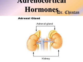
2. Adrenal Glands
The two adrenal glands, each of which weighs about 4 grams, lie at
the superior poles of the two kidneys.
each gland is composed of two distinct parts, the adrenal medulla
and the adrenal cortex.
The adrenal medulla, the central 20 per cent of the gland, is
functionally related to the sympathetic nervous system; it secretes
the hormones epinephrine and norepinephrine in response to
sympathetic stimulation.
The adrenal cortex secretes an entirely different group of hormones,
called corticosteroids. These hormones are all synthesized from the
steroid cholesterol.
3. Adrenal Glands
Adrenal cortex – mineralocorticoids, glucocorticoids
and androgenic hormones
Mineralocorticoids - they affect the electrolytes (the
“minerals”) of the ECF (Na & K)
Aldosterone - the principal mineralocorticoid
Glucocorticoids
concentration
-
increase
Cortisol - the principal glucocorticoid
blood
glucose
4. Adrenal Cortex
1. The zona glomerulosa - just underneath the
capsule - 15 % - aldosterone - enzyme aldosterone
synthase - controlled by the ECF concentrations of
angiotensin II and potassium
2. The zona fasciculata – middle layer – 75 % glucocorticoids cortisol and corticosterone - small
amounts of adrenal androgens and estrogens controlled by the hypothalamic-pituitary axis via ACTH
3. The zona reticularis - deep layer of the cortex - the
adrenal androgens dehydroepiandrosterone (DHEA)
and androstenedione - small amounts of estrogens
and some glucocorticoids - ACTH
5.
6.
7. Mineralocorticoids
• Aldosterone (very potent, accounts for about 90 per
cent of all mineralocorticoid activity)
• Desoxycorticosterone (1/30 as potent as
aldosterone, but very small quantities secreted)
• Corticosterone (slight mineralocorticoid activity)
• Cortisol (very slight mineralocorticoid activity, but
large quantity secreted)
• Cortisone (synthetic, slight mineralocorticoid
activity)
• 9a-Fluorocortisol (synthetic, slightly more potent
than aldosterone)
8. Glucocorticoids
• Cortisol (very potent, accounts for about 95 per cent
of all glucocorticoid activity)
• Corticosterone (provides about 4 per cent of total
glucocorticoid activity, but much less potent than
cortisol)
• Cortisone (synthetic, almost as potent as cortisol)
• Prednisone (synthetic, four times as potent as
cortisol)
• Methylprednisone (synthetic, five times as potent
as cortisol)
• Dexamethasone (synthetic, 30 times as potent as
cortisol)
9.
10. Adrenocortical Hormones
Cortisol: cortisol-binding globulin or transcortin
and, to a lesser extent, to albumin
long half-life of 60 to 90 minutes
Aldosterone: 60 per cent of combines with the
plasma proteins, about 40 per cent is in the free
form
short half-life of about 20 minutes
conjugated especially to glucuronic acid and, to a
lesser extent, sulfates – bile, feces, urine
11. Aldosterone
Aldosterone increases absorption of Na & simultaneously
increases secretion of K by the renal tubular epithelial cells,
especially in the principal (P) cells of the collecting tubules
and, to a lesser extent, in the distal tubules and collecting
ducts.
Therefore, aldosterone causes Na to be conserved in the
ECF while increasing K excretion in the urine
Excess Aldosterone Increases ECF Volume & BP but Has
Only a Small Effect on Plasma Sodium Concentration
pressure natriuresis and pressure diuresis - aldosterone
escape
12. Aldosterone
Total loss of adrenocortical secretion usually causes
death within 3 days to 2 weeks unless the person
receives extensive salt therapy or injection of
mineralocorticoids.
NaCl lost - Total ECF volume and blood volume
become greatly reduced – hyperkalemia
diminished cardiac output – shock like state – death
Aldosterone’s mineralocorticoid activity is about 3000
times greater than that of cortisol, but the plasma
concentration of cortisol is nearly 2000 times that of
aldosterone
13. Aldosterone
Excess aldosterone causes loss of K ions from the ECF
into the urine & also stimulates transport of K from
the ECF into most cells of the body.
Hypokalemia - normal value of 4.5 mEq/L to as low as
2 mEq/L - muscle weakness - alteration of the
electrical excitability of the nerve and muscle fiber
membranes, which prevents transmission of normal
action potentials.
aldosterone is deficient – Hyperkalemia - serious
cardiac toxicity, including weakness of heart
contraction and development of arrhythmia – diastolic
heart failure
14. Aldosterone
causes secretion of H ions in exchange for Na in the intercalated
cells of the cortical collecting tubules. This decreases the H ion
concentration in the ECF, causing a mild degree of alkalosis.
Aldosterone Stimulates Na & K Transport in Sweat
Glands, Salivary Glands - The effect on the sweat glands is
important to conserve body salt in hot environments, and the
effect on the salivary glands is necessary to conserve salt when
excessive quantities of saliva are lost
Aldosterone also greatly enhances Na absorption by the
intestines, especially in the colon, which prevents loss of sodium
in the stools - absence of aldosterone, The unabsorbed NaCl and
water then lead to diarrhea
Quick Nongenomic Actions of Aldosterone via CAMP
15. Mechanism of Aldosterone Action
lipid solubility in the cellular membranes, aldosterone diffuses
readily to the interior of the tubular epithelial cells - in the
cytoplasm of the tubular cells, aldosterone combines with a
highly specific cytoplasmic receptor protein
aldosterone-receptor complex diffuses into the nucleus – DNA,
mRNA, proteins (enzymes, transport proteins)
One of the enzymes especially increased is Na – K ATPase, which
serves as the principal part of the pump for Na & K exchange at
the basolateral membranes of the renal tubular cells.
Epithelial Na channel proteins inserted into the luminal
membrane of the same tubular cells that allows rapid diffusion
of Na ions from the tubular lumen into the cell → basolateral
membrane Na – K ATPase → 45 minutes to several hours
16. Regulation of Aldosterone
The regulation of aldosterone secretion by the zona glomerulosa
cells is almost entirely independent of the regulation of cortisol and
androgens by the zona fasciculata and zona reticularis
1. Increased K ion concentration in the ECF greatly increases
2. Increased activity of the RAS (increased levels of angiotensin II)
also greatly increases
3. Increased Na ion concentration in the ECF very slightly decreases
4. ACTH from the anterior pituitary gland is necessary for
aldosterone secretion but has little effect in controlling the rate of
secretion
17.
18.
19. • Stimuli that increase aldosterone secretion
•
•
•
•
•
Glucocorticoid secretion also increased
Surgery
Anxiety
Physical trauma
Hemorrhage
•
•
•
•
•
•
Glucocorticoid secretion unaffected
High potassium intake
Low sodium intake
Constriction of inferior vena cava in thorax
Standing
Secondary hyperaldosteronism (in some cases
congestive heart failure, cirrhosis, and nephrosis)
of
20. Glucocorticoids
Stimulation of Gluconeogenesis - formation of
carbohydrate from proteins and some other substances by
liver
1. Cortisol increases the enzymes required to convert
amino acids into glucose in the liver cells
2. Cortisol causes mobilization of amino acids from the
extra hepatic tissues mainly from muscle
marked increase in glycogen storage in the liver cells. This
effect of cortisol allows other glycolytic hormones, such as
epinephrine and glucagon, to mobilize glucose in times of
need, such as between meals
21. Carbohydrate Metabolism
Decreased Glucose Utilization by Cells –
glucocorticoids depress the oxidation of NADH to form
NAD+. Because NADH must be oxidized to allow
glycolysis.
The rise in blood glucose in turn stimulates secretion
of insulin.
high levels of glucocorticoid reduce the sensitivity of
many tissues, especially skeletal muscle and adipose
tissue, to the stimulatory effects of insulin on glucose
uptake and utilization - adrenal diabetes
22. Protein Metabolism
reduction of the protein stores in essentially all body
cells except those of the liver – muscle weakness
This is caused by both decreased protein synthesis
and increased catabolism of protein - decreased
amino acid transport into extrahepatic tissues cortisol mobilizes amino acids from the nonhepatic
tissues
enhance amino acid transport into liver cells and
enhance the liver enzymes required for protein
synthesis - Cortisol Increases Liver and Plasma
Proteins
23. Protein Metabolism
(1) increased rate of deamination of amino acids
by the liver,
(2) increased protein synthesis in the liver,
(3) increased formation of plasma proteins by the
liver,
(4) increased conversion of amino acids to glucose
24. Fat Metabolism
Mobilization of Fatty Acids - increases the concentration of
free fatty acids in the plasma, which also increases their
utilization for energy. Cortisol also seems to have a direct
effect to enhance the oxidation of fatty acids in the cells
diminished transport of glucose into the fat cells: alpha glycerophosphate, which is derived from glucose, is
required for both deposition and maintenance of
triglycerides in these cells, and in its absence the fat cells
begin to release fatty acids.
Obesity - excess deposition of fat in the chest and head
regions of the body, giving a buffalo-like torso and a
rounded “moon face” - excess stimulation of food
intake, with fat being generated in some tissues of the body
more rapidly than it is mobilized and oxidized
25. Stress and Inflammation
1. Trauma of almost any type
2. Infection
3. Intense heat or cold
4. Injection of norepinephrine and other
sympathomimetic drugs
5. Surgery
6. Injection of necrotizing substances beneath the
skin
7. Almost any devastating disease
Energy, Glucose, Proteins
26. Anti-inflammatory Effects
1. Cortisol stabilizes the lysosomal membranes proteolytic enzymes that are released by damaged
cells to cause inflammation, which are mainly stored
in the lysosomes, are released in greatly decreased
quantity
2. Cortisol decreases the permeability of the
Capillaries - prevents loss of plasma into the tissues.
3. Cortisol decreases both migration of WBC into the
inflamed area and phagocytosis of the damaged cells cortisol diminishes the formation of PGs and LTs that
increase vasodilation, capillary permeability, and
mobility of WBC
27. Anti-inflammatory Effects
4. Cortisol suppresses the immune system, causing
T
lymphocyte reproduction to decrease markedly - reduced
amounts of T cells and antibodies in the inflamed area
decrease the tissue reactions
5. Cortisol reduces fever mainly because it reduces the
release of IL-1 from WBC, which is one of the principal
excitants to the hypothalamic temperature control system decreased temperature reduces vasodilation
Rate of healing is greater - mobilization of amino acids and
use of these to repair the damaged tissues – extra glucose –
fatty acids - Resolution of Inflammation
Rheumatoid arthritis,
glomerulonephritis
rheumatic
fever
and
acute
28. Anti-inflammatory Effects
Cortisol Blocks the Inflammatory Response to Allergic
Reactions - cortisol effectively prevents shock or
death in anaphylaxis
Cortisol decreases the number of eosinophils and
lymphocytes in the blood - atrophy of all the
lymphoid tissue throughout the body - fulminating
infection and death
Prevents immunological rejection of transplanted
hearts, kidneys, and other tissues
Increase RBC – polycythemia, MOA - Intracytoplasmic
29. Permissive Action
Small amounts of glucocorticoids must be present for a
number of metabolic reactions to occur, although the
glucocorticoids do not produce the reactions by themselves.
This effect is called their permissive action.
1. For glucagon and catecholamines to exert their
calorigenic effects,
2. For catecholamines to exert their lipolytic effects,
3. For catecholamines to produce pressor responses and
bronchodilation
During fetal life, glucocorticoids accelerate the
maturation
of
surfactant
in
the
lungs
30. Regulation of Cortisol Secretion
CRF are located mainly in the paraventricular nucleus of the
hypothalamus
ACTH (Polypeptide) Stimulates Cortisol Secretion – cAMP protein kinase A - initial conversion of cholesterol to
pregnenolone
Physical stress - Pain stimuli caused by physical stress or
tissue damage are transmitted first upward through the
brain stem and eventually to the median eminence of the
hypothalamus
Mental stress - increased activity in the limbic
system, especially in the region of the amygdala and
hippocampus, both of which then transmit signals to the
hypothalamus
33. POMC:
ACTH + MSH,
Beta - lipotropin,
Beta – endorphin
N – no significance
Addison's disease: MSH – melanocytes - melanin –
hyperpigmentation
pars intermedia – arctic animals - darkened fur in the
summer and have entirely white fur in the winter
Adrenal Androgens
34. Hypoadrenalism - Addison’s Disease
primary atrophy of the adrenal cortices – autoimmunity (80%)
tuberculous destruction of the adrenal glands or invasion of the
adrenal cortices by cancer
Mineralocorticoid Deficiency - greatly decreased ECF volume hyponatremia, hyperkalemia and mild acidosis - CO decreases,
and the patient dies in shock
Glucocorticoid Deficiency – cant maintain normal blood glucose
concentration between meals - reduces the mobilization of both
proteins and fats from the tissues - highly susceptible to stress
and infection
Melanin Pigmentation - mucous membranes and thin skin (lips,
nipples)
35.
36.
37. Treatment
small quantities of mineralocorticoids and glucocorticoids are
administered daily
Addisonian Crisis
In a person with Addison’s disease, the output of glucocorticoids
does not increase during stress.
whenever different types of trauma, disease, or other
stresses, such as surgical operations, appear, a person is likely to
have an acute need for excessive amounts of glucocorticoids
and often must be given 10 or more times the normal quantities
of glucocorticoids to prevent death.
This critical need for extra glucocorticoids and the associated
severe debility in times of stress is called an addisonian crisis
Adrenal crisis – tapering dose always
38. Hyperadrenalism - Cushing’s Syndrome
abnormal excess secretion of cortisol & androgens
(1) adenomas of the anterior pituitary that secrete large
amounts of ACTH, which then causes adrenal
hyperplasia and excess cortisol secretion;
(2) abnormal function of the hypothalamus that causes
high levels of CRH, which stimulates excess ACTH
release;
(3) “ectopic secretion” of ACTH by a tumor elsewhere in the
body, such as an abdominal carcinoma;
(4) adenomas of the adrenal cortex – Reduced ACTH
When Cushing’s syndrome is secondary to excess secretion
of ACTH by the anterior pituitary, this is referred to as
Cushing’s disease
39. Dexamethasone Test
In patients who have overproduction of ACTH due to
an ACTH-secreting pituitary adenoma or to
hypothalamic-pituitary dysfunction, even large doses
of dexamethasone usually do not suppress ACTH
secretion.
In patients with primary adrenal overproduction of
cortisol usually have low or undetectable levels of
ACTH.
Cushing’s syndrome can also occur when large
amounts of glucocorticoids are administered over
prolonged periods for therapeutic purposes –
Rheumatoid Arthritis
40. Cushing’s syndrome
mobilization of fat from the lower part of the
body, with concomitant extra deposition of fat in
the thoracic and upper abdominal regions, giving
rise to a buffalo torso – Buffalo hump
Edematous appearance of the face (Moon Face) –
acne & hirsutism – Hypertension
Hyperglycemia - ↓ protein except liver (Plasma
proteins) – muscle weakness - suppressed immune
system - large purplish striae - severe osteoporosis
41.
42.
43. Treatment
Removal an adrenal tumor – Adrenalectomy
Hypertrophied pituitary glands or tumors – Surgical removal
or irradiation
When surgery is not possible;
Drugs that block steroidogenesis - metyrapone,
ketoconazole and aminoglutethimide
Drugs that inhibit ACTH secretion - serotonin antagonists
and GABA-transaminase inhibitors
Bilateral partial (or even total) adrenalectomy, followed by
administration of adrenal steroids to make up for any
insufficiency
44. Primary Aldosteronism (Conn’s Syndrome)
small tumor of the zona glomerulosa
Hypokalemia – Muscle Paralysis,
slight increase in ECF volume and blood volume,
Very slight Hypernatremia,
almost always, hypertension
Decreased plasma renin concentration
Rx - surgical removal of the tumor or of most of the
adrenal tissue
45. Adrenogenital Syndrome
Excessive quantities of androgens that cause intense
masculinizing effects throughout the body
Female - growth of a beard, a much deeper
voice, occasionally baldness, masculine distribution of hair
on the body and the pubis, growth of the clitoris to
resemble a penis, and deposition of proteins in the skin and
especially in the muscles
Prepubertal male - rapid development of the male sexual
organs
It is often difficult to make a diagnosis of adrenogenital
syndrome in the adult male – excretion of 17-ketosteroids
(which are derived from androgens) in the urine may be 10
to 15 times normal