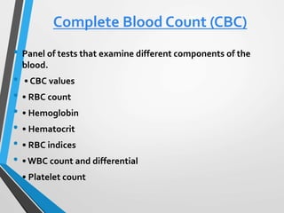
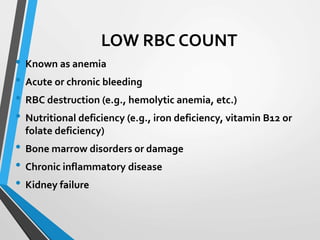
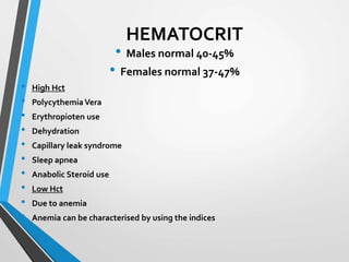
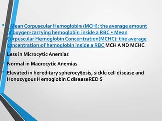
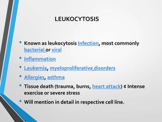
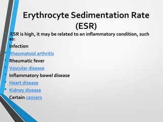
3. LOW RBC COUNT
• Known as anemia
• Acute or chronic bleeding
• RBC destruction (e.g., hemolytic anemia, etc.)
• Nutritional deficiency (e.g., iron deficiency, vitamin B12 or
folate deficiency)
• Bone marrow disorders or damage
• Chronic inflammatory disease
• Kidney failure
4. HIGH RBC COUNT
• Known as polycythemia
• Dehydration
• Pulmonary disease
• Kidney or other tumor that produces excess erythropoietin
• Smoking
• Genetic causes (altered oxygen sensing, abnormality in
hemoglobin oxygen release)
• Polycythemia vera
5. HEMOGLOBIN
• Is the protein molecule that carries
oxygen in the Red Blood Cells.
• 13.5-17.5 g/dl in males
• 12.5-15.5 g/dl in females
6. HEMATOCRIT
• Males normal 40-45%
• Females normal 37-47%
• High Hct
• PolycythemiaVera
• Erythropioten use
• Dehydration
• Capillary leak syndrome
• Sleep apnea
• Anabolic Steroid use
• Low Hct
• Due to anemia
• Anemia can be characterised by using the indices
7. • RBC indices
• Mean Corpuscular Volume (MCV ):a measurement of the average size of
RBCs Normal 80-100fL
• Low MCV indicates RBCs are smaller than normal (microcytic
• iron deficiency anemia, or thalassemias, Congenital sideroblastic
Anemia, Lead Poisoning, pyridoxine deficiency, anemia of chronic
disease
• High MCV indicates RBCs are larger than normal (macrocytic)
• MEGALOBLASTIC MACROCYTIC ANEMIAMacrocytes in bone marrow
smear
• Medications affecting folate metabolism
• Vit B12 deficiency (Pernicious Anemia)
• Folate deficiency (Alcohol related often)
• Atrophic Gastitis
8. • • Mean Corpuscular Hemoglobin (MCH): the average amount
of oxygen-carrying hemoglobin inside a RBC • Mean
Corpuscular Hemoglobin Concentration(MCHC): the average
concentration of hemoglobin inside a RBC MCH AND MCHC
• Less in Microcytic Anemias
• Normal in Macrocytic Anemias
• Elevated in hereditary spherocytosis, sickle cell disease and
Honozygous Hemoglobin C diseaseRED S
9. RBC indices
• • Red Cell Distribution Width (RDW): a variation in the size of RBCs
• Low value indicates uniformity in size of RBCs
• High value indicates mixed population of small and large
RBCs; immature RBCs tend to be larger. For example, in iron
deficiency anemia or pernicious anemia, there is high variation
(anisocytosis) in RBC size (along with variation in shape –
poikilocytosis), causing an increase in the RDW.
10. WHITE BLOOD CELL COUNT
• The normal number of WBCs in the blood is
• 4,000-10,000 white blood cells per microliter (mcL). Normal
value ranges may vary slightly among different labs.
• There are five basic white blood cell types:
• • Neutrophils 45-70%
• • Eosinophils 1-5%
• • Basophils 0-1%
• • Lymphocytes 25-45%
• • Monocytes 2-12%
Each WBC cell type has its' own unique features.
11. LEUKOPENIA
• Low white cell count may be due to acute viral infections,
such as with a cold or influenza. It can be associated with
chemotherapy, radiation
• therapy, myelofibrosis and aplastic anemia (failure of white
cell, red cell and platelet production). HIV and AIDS are also
a threat to white cells.
• Other causes of low white blood cell count include systemic
lupus erythematosus, Hodgkin's lymphoma, some types of
cancer, typhoid, malaria, tuberculosis, dengue, rickettsial
infections, enlargement of
• the spleen, folate deficiencies, psittacosis, sepsis and Lyme
disease. Many other causes exist, such as deficiency in
certain minerals, such as copperand zinc.
12. LEUKOCYTOSIS
• Known as leukocytosis Infection, most commonly
bacterial or viral
• Inflammation
• Leukemia, myeloproliferative disorders
• Allergies, asthma
• Tissue death (trauma, burns, heart attack) ¢ Intense
exercise or severe stress
• Will mention in detail in respective cell line.
13. Neutrophils
• These are the most common of the WBCs and serve as the
primary defense against infection.The typical response to
infection or serious injury is an increased production of
neutrophils.
• Bands/Stabs Early in the response to infection, immature
forms of neutrophils will be seen.These are call Stab or
Band cells.The presence of these immature cells is called a
"shift to the left" and can be the earliest sign of a WBC
response, even before the WBC becomes elevated.
14.
15.
16. Eosinophils
• These cells play a role in allergic disorders and in
combating parasitic infections. • Elevations in
eosinophil counts are associated with: • Allergic
reactions • Parasite infections • Chronic skin infections
• Some cancers • Decreases in eosinophil counts are
associated with: • Stress • Steroid exposure • Anything
that may suppressWBC production generally.
17.
18. Basophils
• These cells can digest bacteria and other foreign bodies
(phagocytosis) and also have some role in allergic reactions.
• Elevations in basophil counts are associated with: • Some
cancers • Some allergic reactions • Some infections •
Radiation exposure • Diminished basophil counts are
associated with: • Stress reactions • Some allergic reactions
• Hyperthyroidism • Prolonged steroid expo
19.
20. Monocytes
• These cells respond to inflammation, infection and foreign
bodies by ingesting and digesting the foreign material. •
Increased monocyte counts are associated with: • Recovery
from an acute infection •Viral illness • Parasitic infections •
Collagen disease • Some cancers • Decreased monocyte counts
are associated with: • HIV infection • Rheumatoid arthritis •
Steroid exposure • Some cancers
21.
22. Lymphocytes
• These cells play both an immediate and delayed role in
response to infection or inflammation. • Increased numbers of
lymphocytes are seen in: • Most viral infections • Some
bacterial infections • Some cancers • Graves' disease •
Decreased numbers of lymphocytes are seen in: • Steroid
exposure • Some cancers • Immunodeficiency • Renal failure •
Lupus
23.
24. Erythrocyte Sedimentation Rate
(ESR)
• ESR is high, it may be related to an inflammatory condition, such
as:
• Infection
• Rheumatoid arthritis
• Rheumatic fever
• Vascular disease
• Inflammatory bowel disease
• Heart disease
• Kidney disease
• Certain cancers