

Recommended
More Related Content
What's hot
What's hot (20)
Similar to airway assessment part-1-Anesthesia
Similar to airway assessment part-1-Anesthesia (20)
More from NISAR ARAIN
More from NISAR ARAIN (20)
Recently uploaded
Recently uploaded (20)
airway assessment part-1-Anesthesia
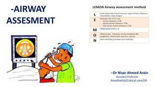
- 1. -AIRWAY ASSESMENT --Dr Nisar Ahmed Arain Assistant Professor Anesthetist/Critical care/ER
- 2. -I-PREOPERATIVE AIRWAY ASSESMENT INCIDENCE OF DIFFICULT INTUBATION --a-1 – 3 out of 100 of Anesthetized patients have Difficult Intubation. --b-1 – out of 1000 of anesthetized patients have Failed Intubation --c-1 - out of 10,000 of anesthetized patients are in the “cannot Intubate cannot ventilate” scenario. CAUSES OF DIFFICULT INTUBATION --1-Anesthesiologist is the most important cause, due to; --2-Inadequate preoperative assessment of the patient. --3-Inadequate equipment preparation as malfunction OR unavailability --4-Inexperience and or poor technique --5-Absence of a trained assistant
- 3. -A-CONGENITAL SYNDROME --1-Associated with one or more anatomical features which prevent easy intubation as -Trisomy 21 (Down syndrome) -Large tongue -small mouth -Narrow pharynx -Subglottic stenosis -Short Neck -Irregular Dentition and -Atlanto-Axial (cervical spine) instability -B-ANATOMICAL FEATURES --1-Teeth -Protruding Incisors (Buck Teeth) as the blade enters the mouth in cephalad direction -Inter incisor distance with maximal opening of the mouth:- If this is less than 3 cm (i.e less than 2 fingers breadth) the Flange of the blade cannot be easily inserted between teeth -Loose teeth --2-Palate:-long high arched palate --3-Tongue:-Large tongue -II-PREOPERATIVE AIRWAY ASSESMENT
- 4. --4-Jaw and Mandible:- --Receding of the Lower jaw (Retrognathia) --Hypoplastic Mandible (Micrognathia) where the pharyngeal space becomes limited because the tongue is positioned more posteriorly. The space in which the soft tissues are going to be displaced during direct laryngoscopy is reduced. --Obtuse Mandibular angle --Poor Mandibular Mobility:-The patients are asked to protrude their Mandible Here three possibilities are present -Class A – The lower Incisors can be protruded anterior to the upper incisors -Class B – The lower incisors can be brought “Edge to edge” with the upper incisors -Class C – The lower incisors cannot be brought “Edge to edge” with the upper incisors HERE BOTH CLASS B AND C ARE ASSOCIATED WITH DIFFICULT LARYNGOSCOPY -II-PREOPERATIVE AIRWAY ASSESMENT
- 5. RETROGNATHIA -receding of lower jaw -II-PREOPERATIVE AIRWAY ASSESMENT
- 6. --5-Neck --Short Large Muscular Neck --Inability of the tip of the chin to touch the chest --Limitation of Neck Extension due to presence of soft tissue Example:-Lipoma I the back of the Neck preventing Neck extension --Flexion Extension at the Cranio-Cervical junction -II-PREOPERATIVE AIRWAY ASSESMENT
- 7. -6-Distance --Decreased distance between the Tip of chin and Thyroid cartilage prominence (i.e Thyromental distance) and less than 6.5 cm or 3 finger breadths with fully extended neck, this is called “Patil’s test or examination --Decreased distance between the Tip of chin and sternum less than 12.5 cm with fully extended head and closed mouth. These distances indicate “Difficult Intubation” if decreased then expected or normal -II-PREOPERATIVE AIRWAY ASSESMENT
- 8. --7- Mallampati Classification ---It is assessed by voluntary tongue protrusion with maximum mouth opening without phonating While the patient is sitting and head in the neutral position There are Four Classes --Class – 1- Visualization of the soft palate, Uvula and Pillars. No anticipated difficulty --Class – 11-Visualization of the soft Palate, Uvula partly visualized. This may become difficult intubation --Class – 111 – Visualization of the soft palate and base of the Uvula. Anticipate difficult intubation --Class – 1V – Soft palate is not visible. Anticipate severe difficulty. The classification assigned may vary if the patient is in the supine position (instead or sitting) -II-PREOPERATIVE AIRWAY ASSESMENT
- 11. -II-PREOPERATIVE AIRWAY ASSESMENT -C – Acquired causes --1-Decreased Jaw Opening:-causing difficult laryngoscopy This may be because of the following --Muscle:-Reflex spasm of masseter and medial pterygoid (i.e, Trismus) by infection Abscess, Fracture or Tetanus --Tempro - Mandibular Joint Fibrosis (Post infection, Radio-Therapy, Trauma) Rheumatoid Arthritis or Ankylosing spondylitis --Bone:-Fracture Mandible or Maxilla --Soft tissue Tumors, edema or abscess
- 12. --2-Decreased mouth opening:- As in cases of Burn --3-Decreased Neck movement:- As in Ankylosing spondylitis or Burn of the Neck --4-Cervical spine instability or subluxation CAUSES --Degenerated diseases as Rheumatoid Arthritis and Osteoarthritis --Congenital atlanto-axial instability as in Down’s syndrome --Trauma or Fracture --Tumors, Tuberculosis --5-Airways:- --Airway edema:-due to abscess infection, trauma, angioedema, or Burns --Airway compression:-Due to Goiter or surgical Hemorrhage --Airway scarring:-Due to Radiotherapy, infection, or Burns --Airway Mass:-Due to Tumors, Polyps, or Foreign body --Airway collapse:-Due to Laryngo-tracheomalacia -II-PREOPERATIVE AIRWAY ASSESMENT
- 13. --6-Others --Morbid Obesity --Pregnancy --Acromegaly N.B:- Previous anesthetic records should always be consulted but a past record of normal tracheal intubation is not a guarantee for future anesthesia because acquired causes may occur leading to a change in the airway Occasionally, patients may have a written notification of previous airway problems -II-PREOPERATIVE AIRWAY ASSESMENT