Downloaded 5,348 times


![serology The branch of medical immunology concerned with antigen-antibody reactions in vitro is serology [serum and -ology]. The usefulness of serological test is dependent on its sensitivity and specificity . False Negatives/Positives High sensitivity prevents false negatives. False negatives occurs when there is no reaction when the Ag or Ab is present. High specificity prevents false positives. False positives occurs when there is cross reaction with another molecule.](https://image.slidesharecdn.com/diagnosticmicrobiology-100606060005-phpapp01/75/Diagnostic-microbiology-Traditional-and-Modern-Approach-49-2048.jpg)


![A technique used to measure the concentration of hormones, Drugs, enzymes, viruses, bacterial antigens and other organic substances of biological interest found in Blood, Tissues and other biological fluids Radio immuno assay Gamma Counter Principle: Uses an immune reaction [Antigen – Antibody reaction] to estimate a ligand Ag + Ag* + Ab AgAb + Ag*Ab + Ag + Ab* Unbound Ag* and Ag washed out Radioactivity of bound residue measured Ligand conc is inversely related to radioactivity [Ag : ligand to be measured ; Ag* radiolabelled ligand]](https://image.slidesharecdn.com/diagnosticmicrobiology-100606060005-phpapp01/75/Diagnostic-microbiology-Traditional-and-Modern-Approach-62-2048.jpg)


![Serovar [serotype] A serovar is a strain differentiated by serological means. Individual strains of Salmonella spp. are often distinguished by serological means. Biovar [biotype] Biovars are strains that are differentiated by biochemical or other non- serological means. Type strain "One strain of a species is designated as the type strain . It is usually one of the first strains studied and is often more fully characterized than other strains; however, it does not have to be the most representative member. Only those strains very similar to the type strain are included in a species." (p. 392, Prescott et al., 1996)](https://image.slidesharecdn.com/diagnosticmicrobiology-100606060005-phpapp01/75/Diagnostic-microbiology-Traditional-and-Modern-Approach-97-2048.jpg)
![Morphovar [morphotype] A morphovar is a strain which is differentiated on the basis of morphological distinctions. Isolate ('i-so-lit) An isolate is a pure culture derived from a heterogeneous, wild population of microorganisms . The term isolate is also applicable to eucaryotic microorganisms as well as to viruses . Classification Placement of an organism within a scheme relating different types of organisms, such as that presented in Woese's universal tree , is know as classification. Organisms are classified for scientific purposes.](https://image.slidesharecdn.com/diagnosticmicrobiology-100606060005-phpapp01/75/Diagnostic-microbiology-Traditional-and-Modern-Approach-98-2048.jpg)


The document discusses the identification of microbes in diagnostic microbiology, emphasizing the importance of studying microorganisms and their roles in ecology and health. It outlines various identification methods including phenotypic, immunological, and genotypic techniques, highlighting the laboratory processes involved from specimen collection to reporting results. Key methods include biochemical tests, serology, and molecular techniques like PCR, all aimed at accurately identifying pathogens for effective treatment.
Overview of the importance and methods for identifying microorganisms.
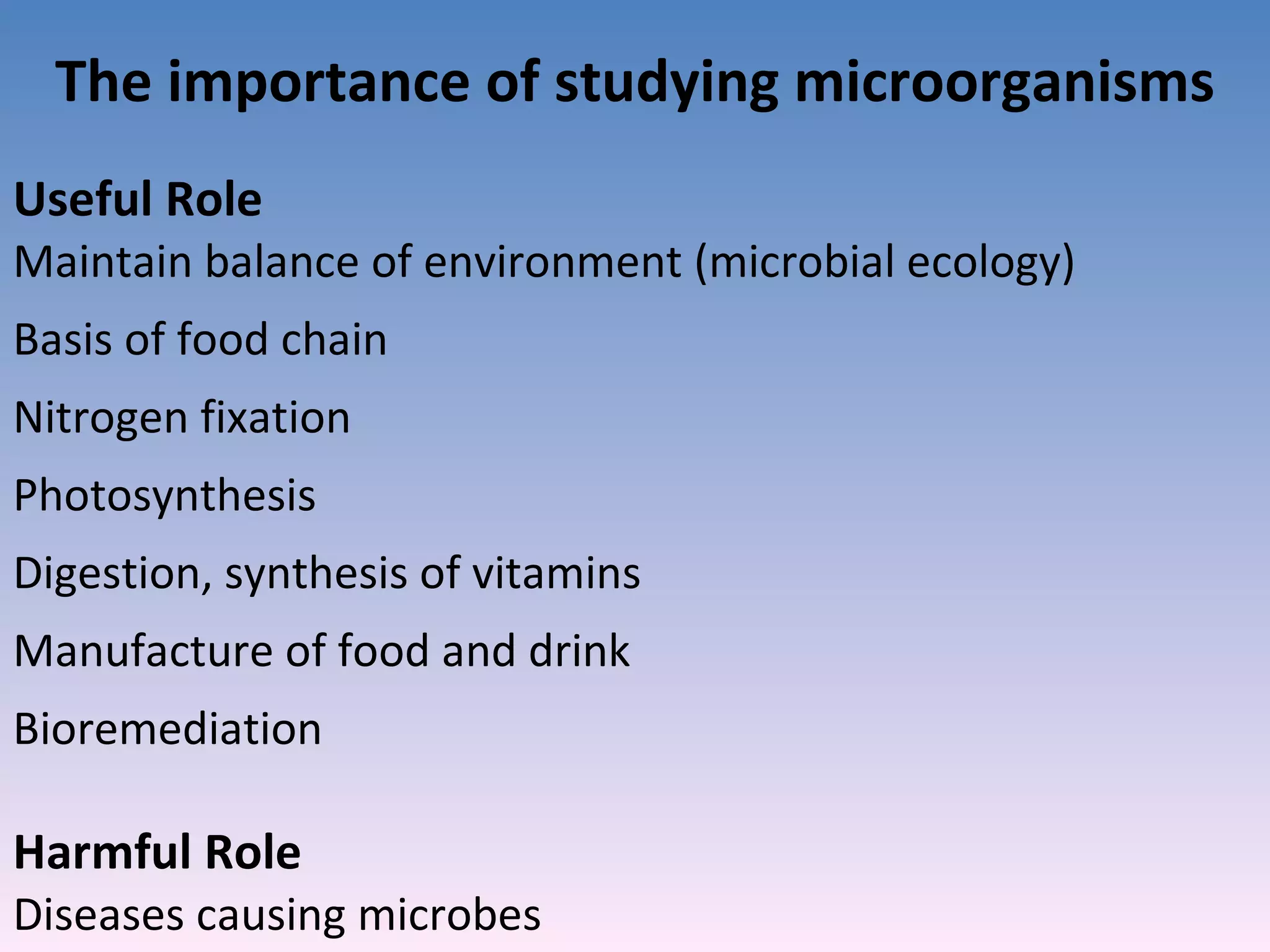
Microorganisms maintain environmental balance but can also cause diseases.
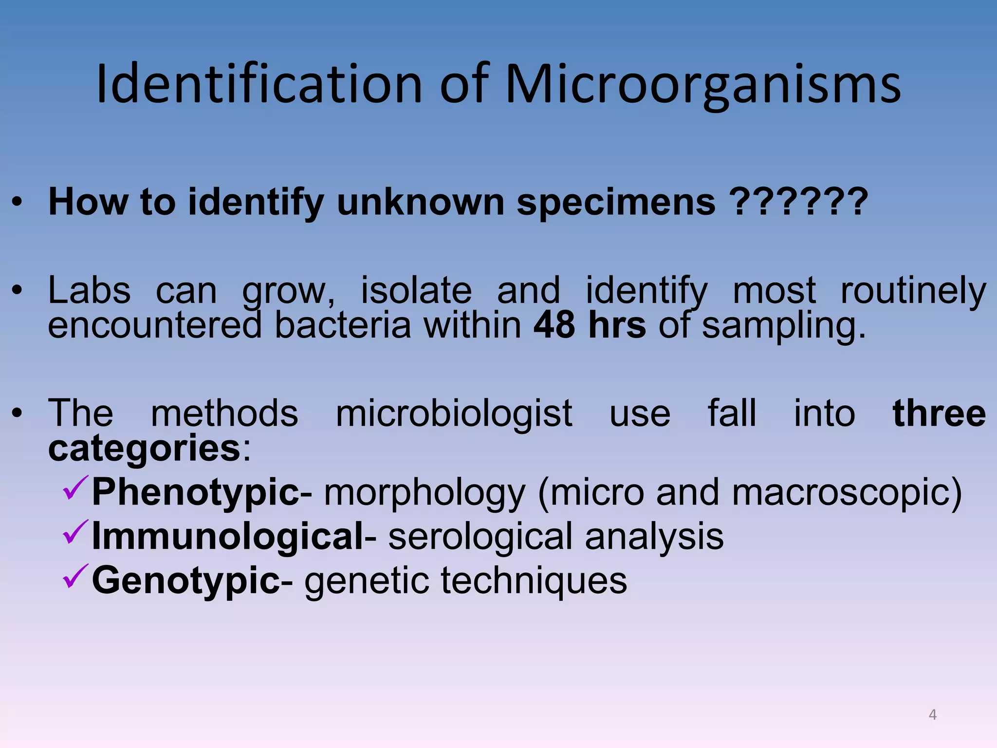
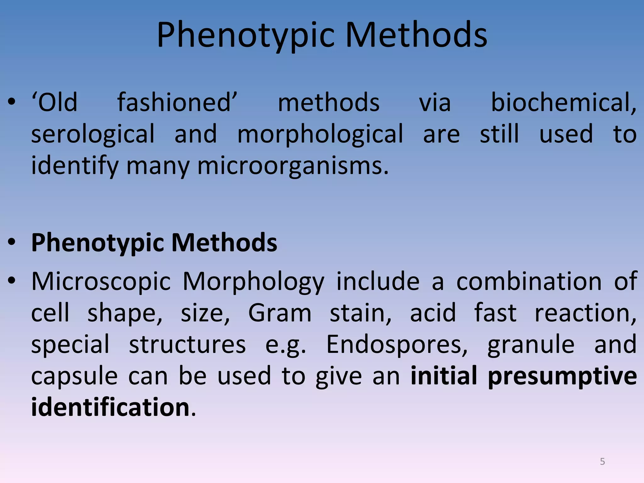
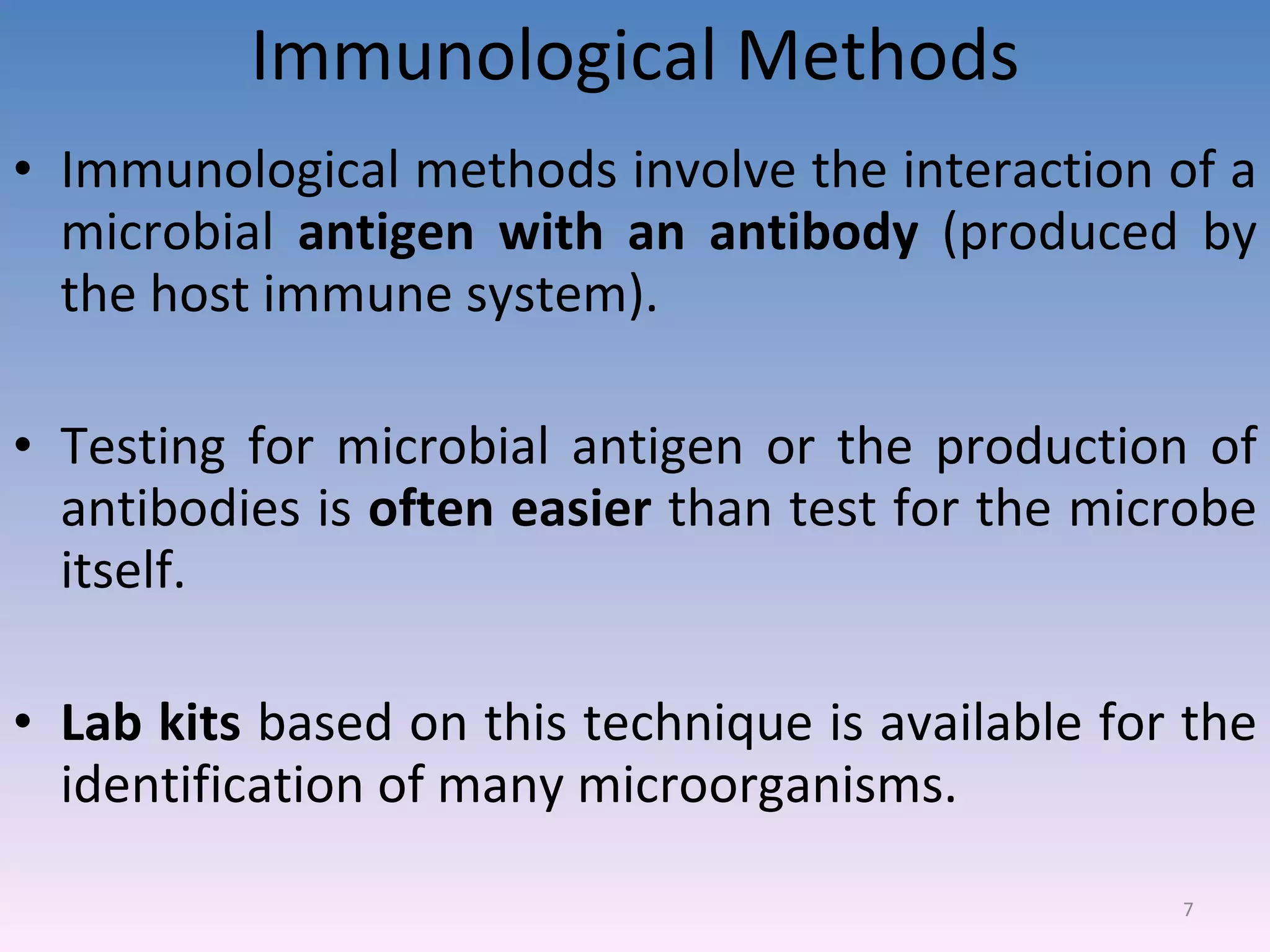
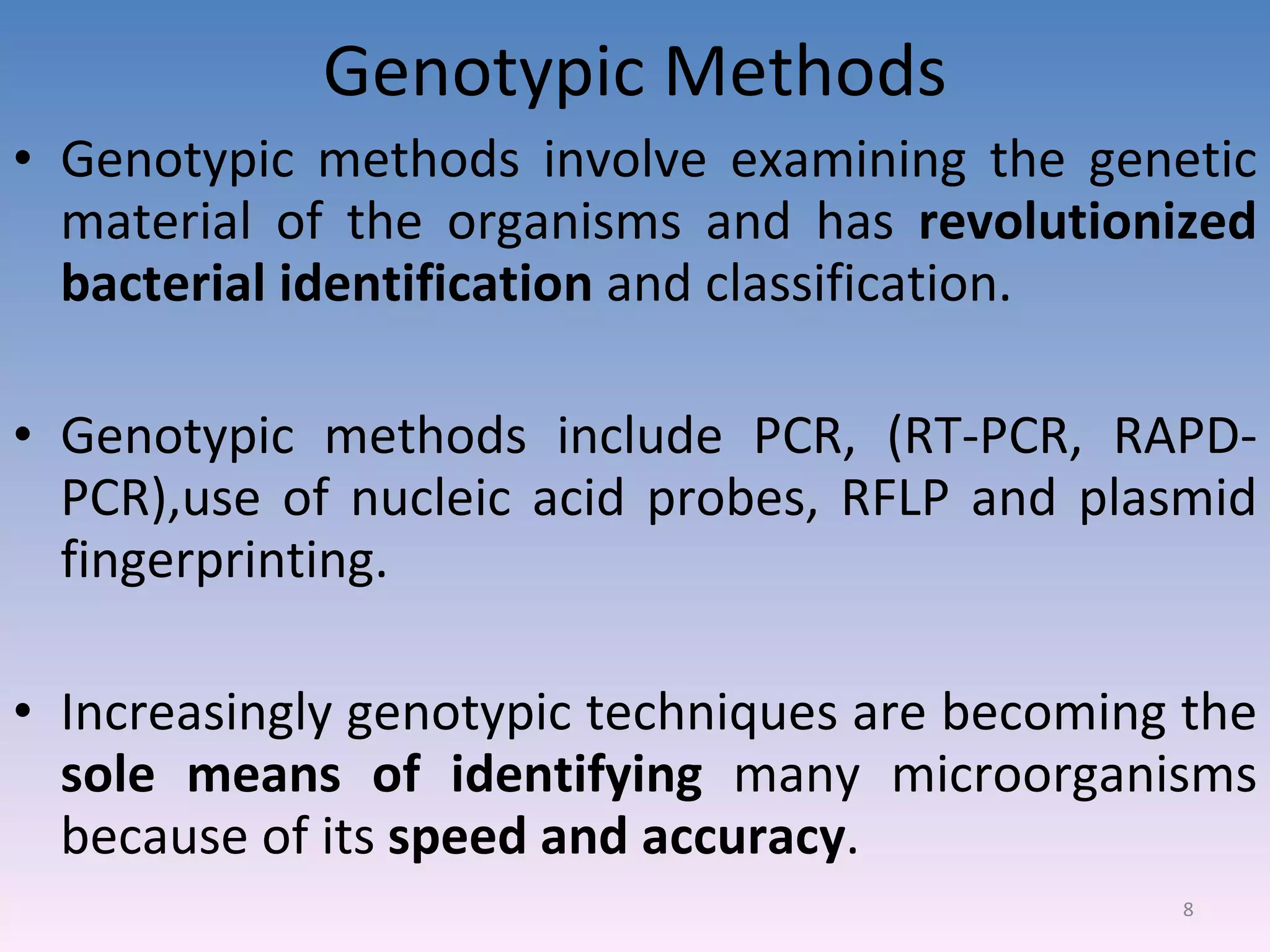
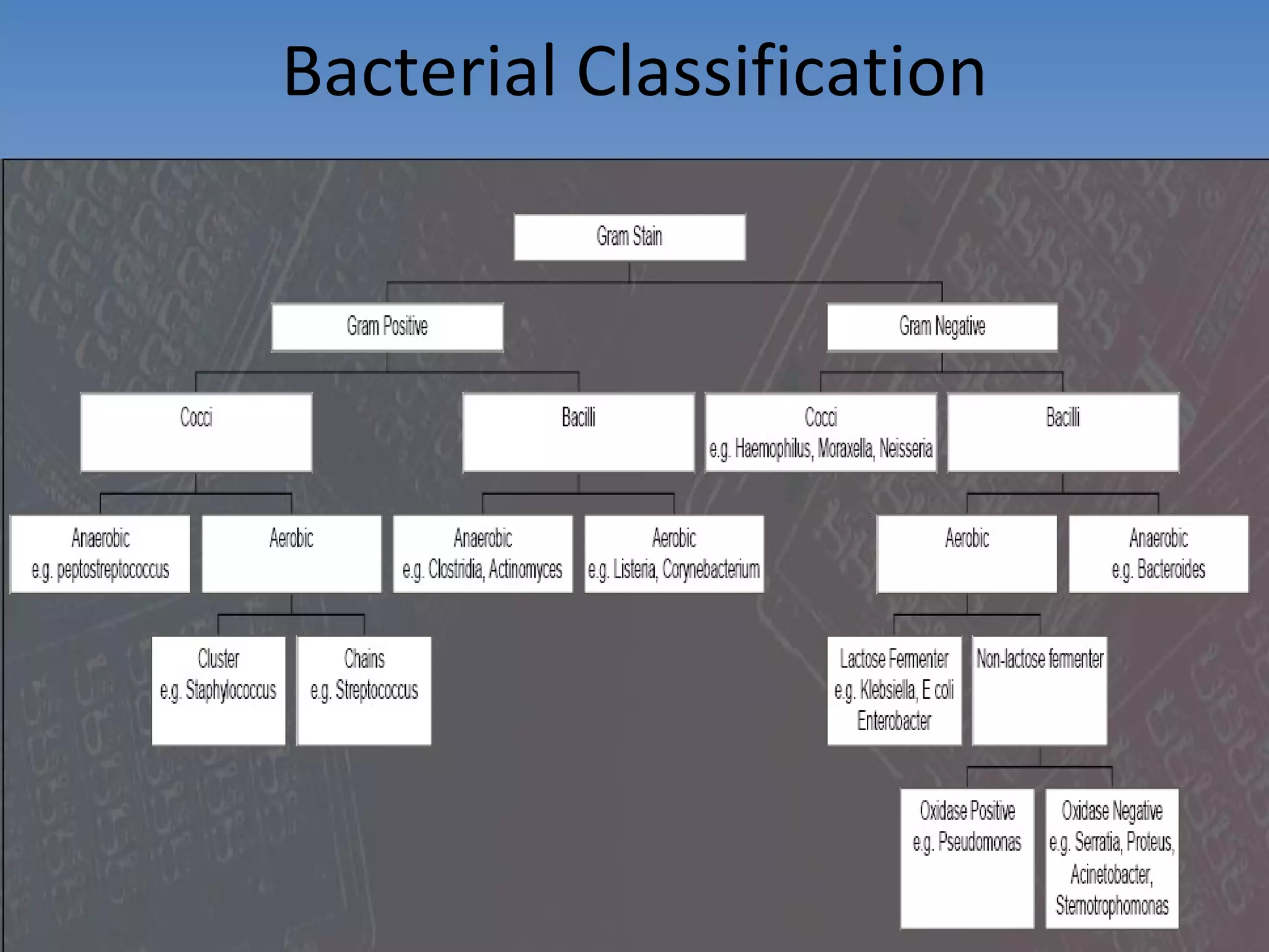
Methods: Phenotypic (morphology), Immunological (antigen-antibody), and Genotypic (genetic techniques).
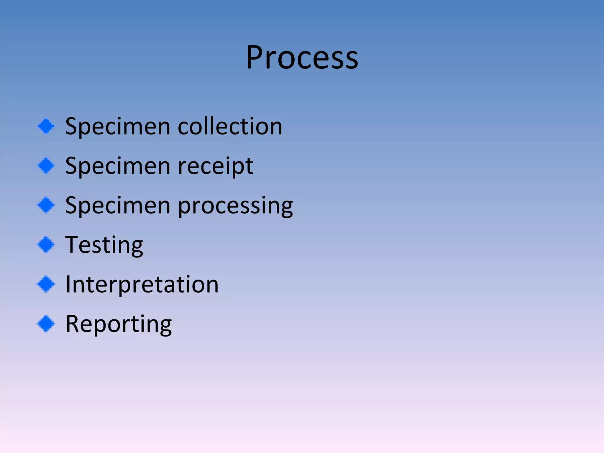
Steps from specimen collection, processing, to the importance of techniques for safety and accuracy.
Fundamental safety measures in handling specimens including hand hygiene and personal protective equipment.
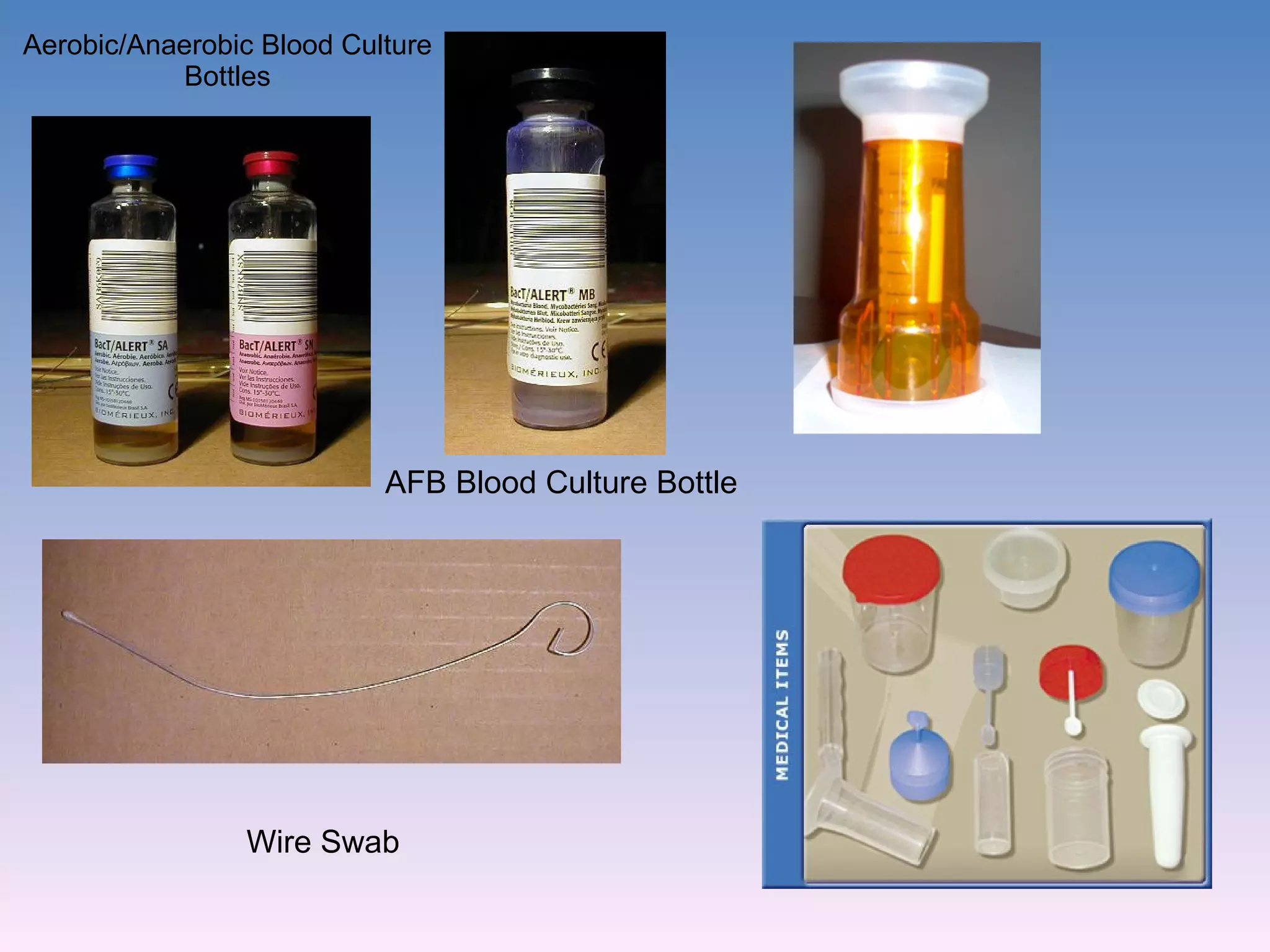
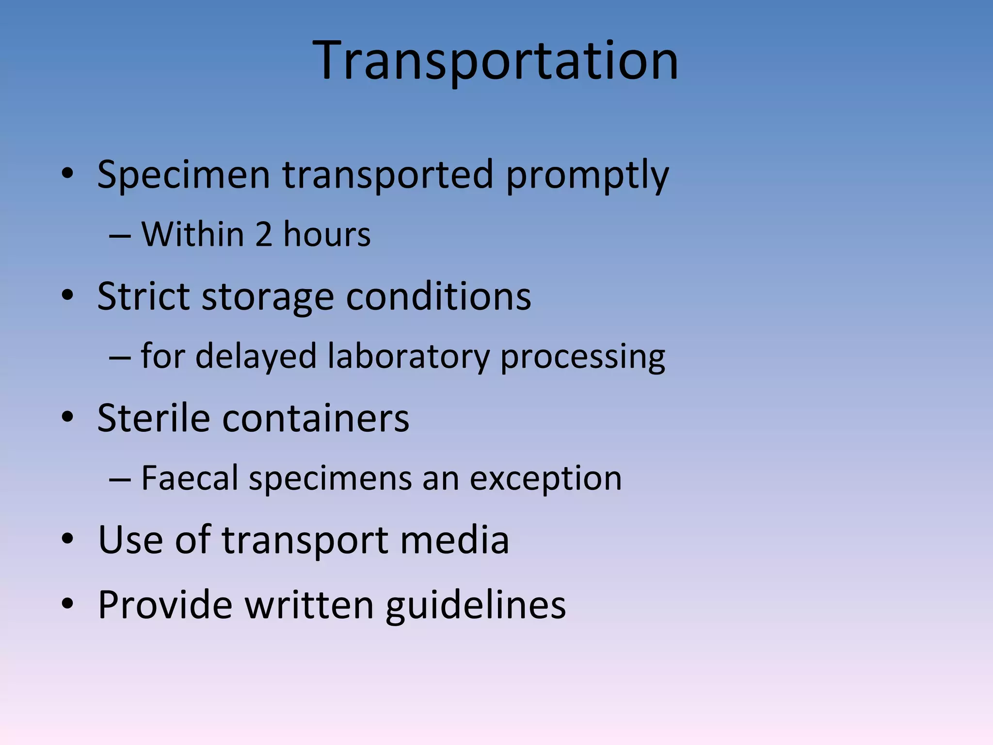
Proper techniques and materials for collecting and transporting specimens to ensure accurate results.
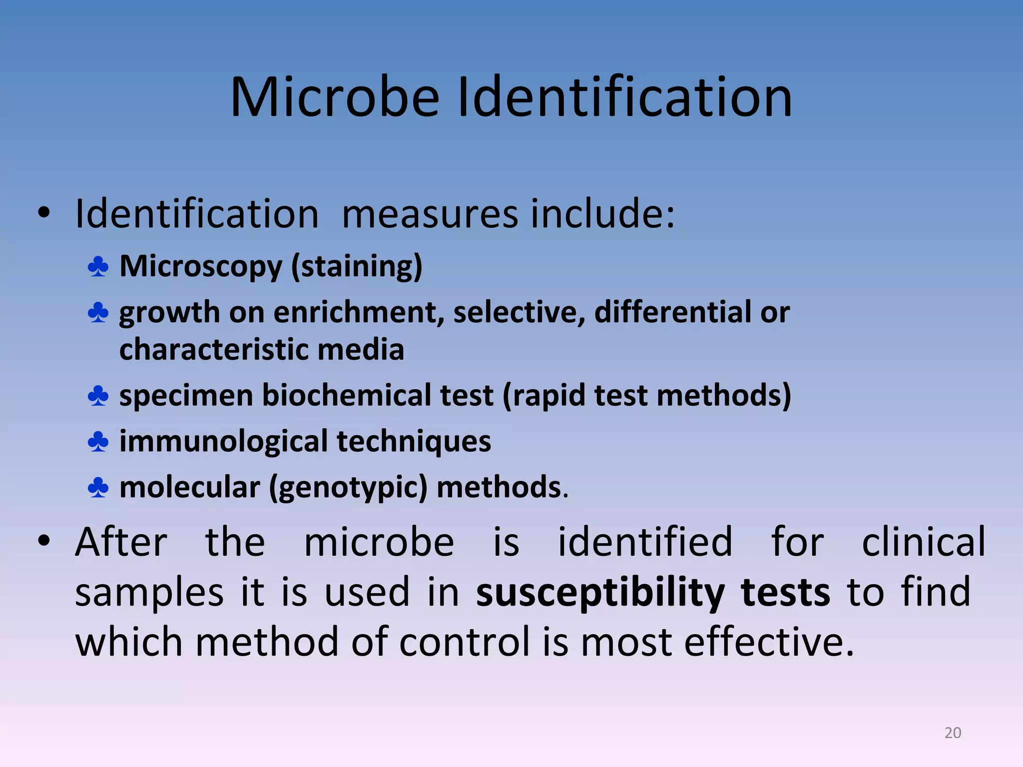
Identification measures include microscopy, culture media, biochemical testing, and susceptibility tests.
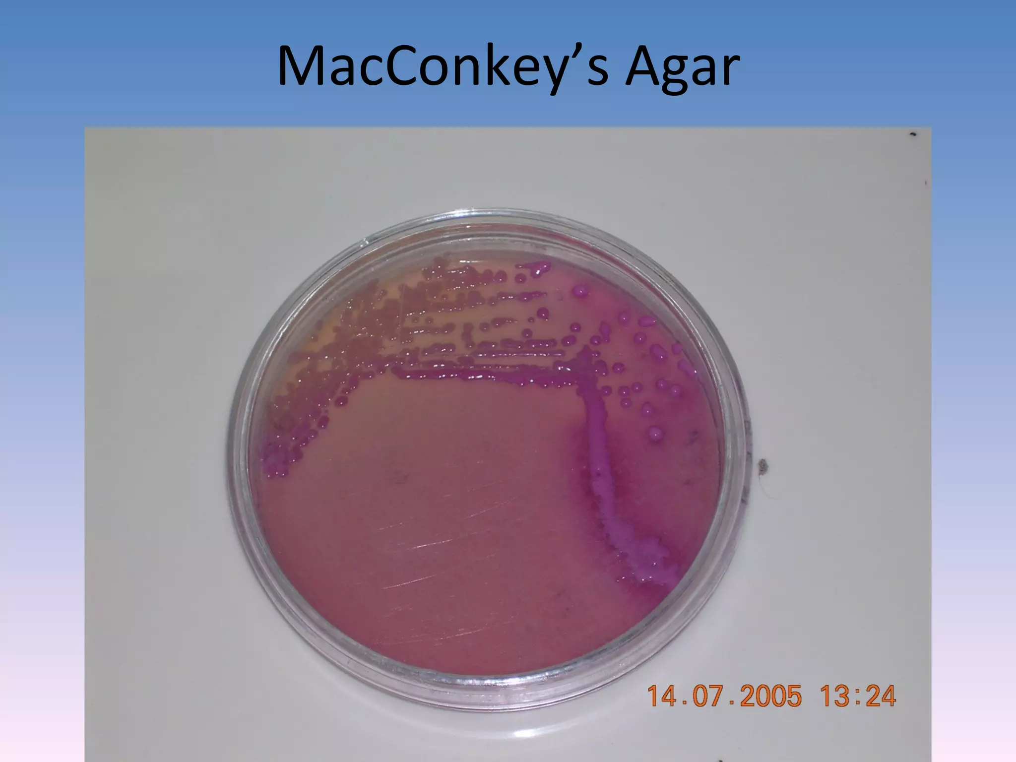
Observations of microorganisms using different media to assist in identification.
Assessing microbial resistance to various agents and understanding their metabolic capabilities.
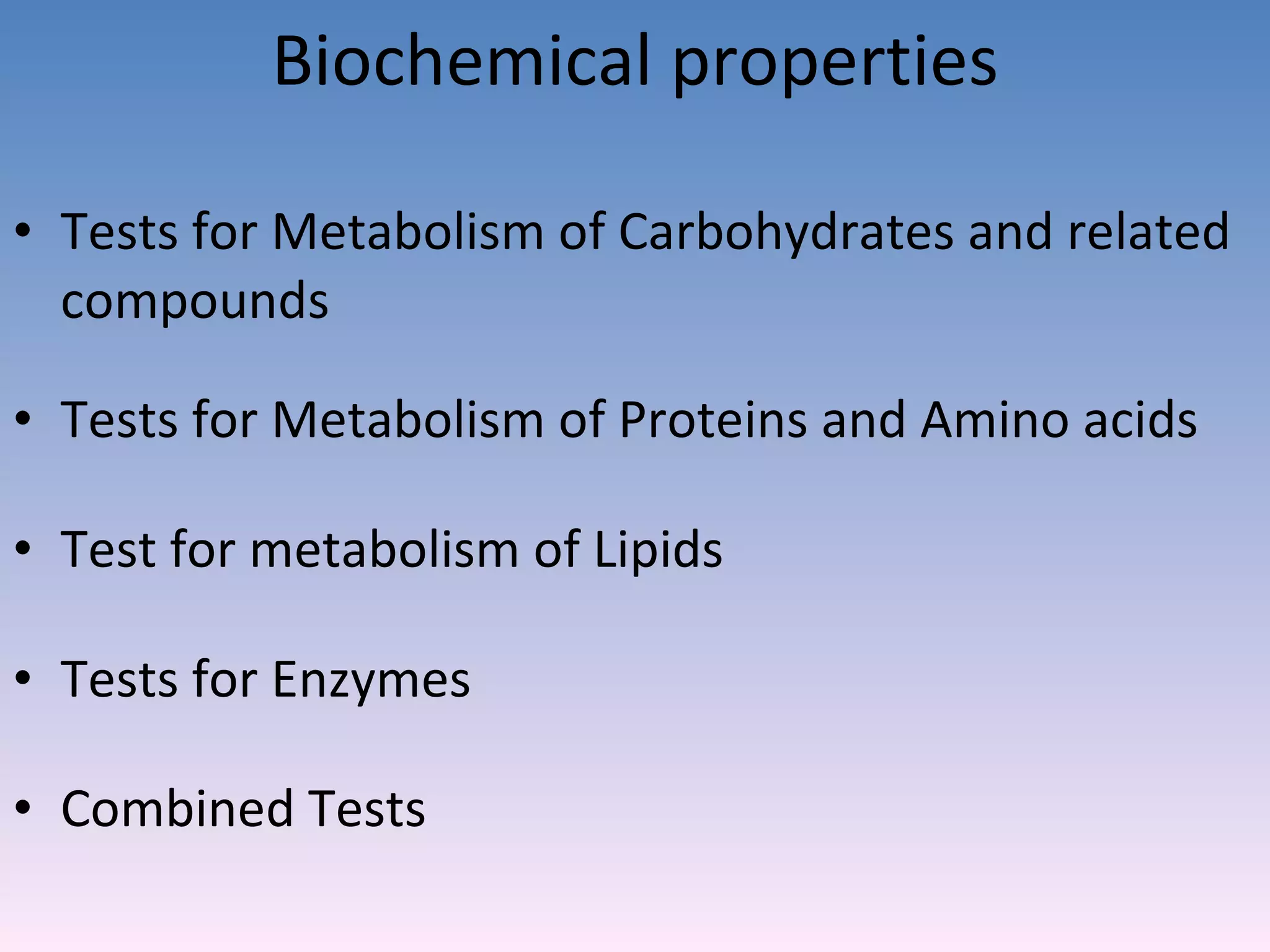
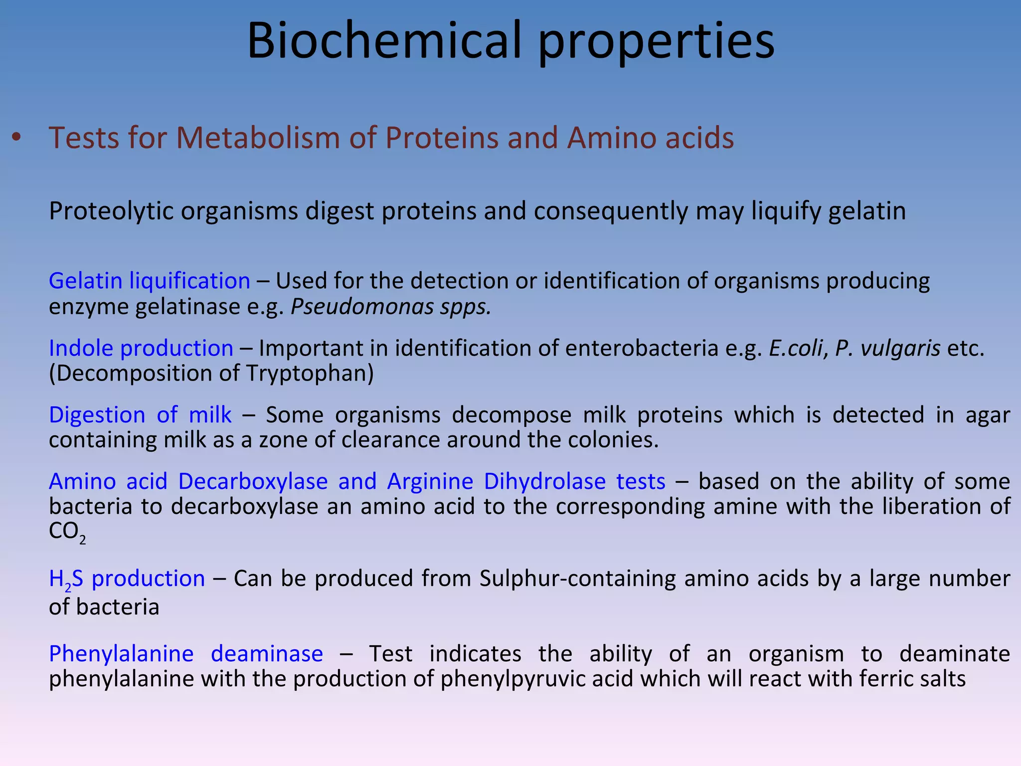
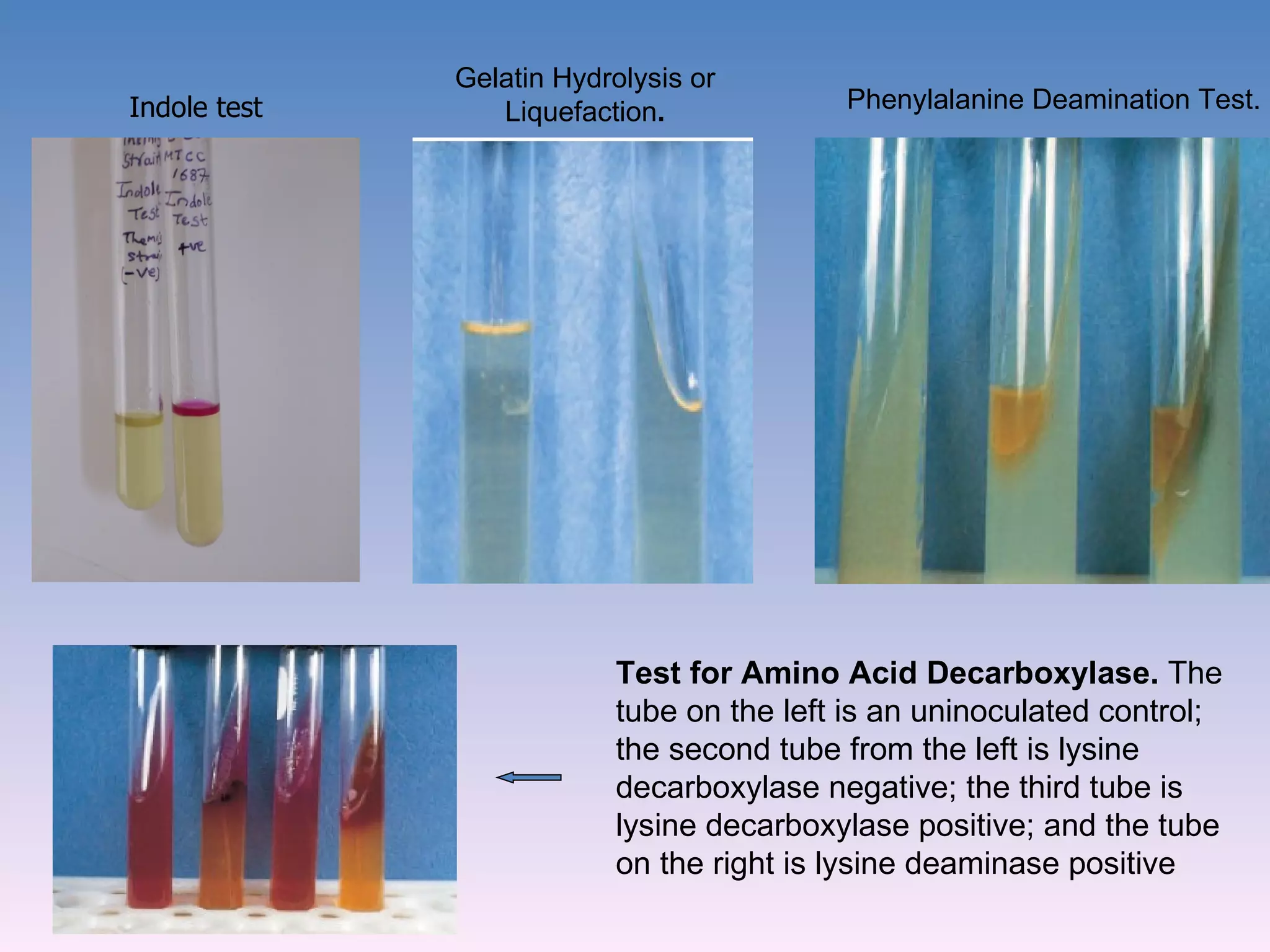
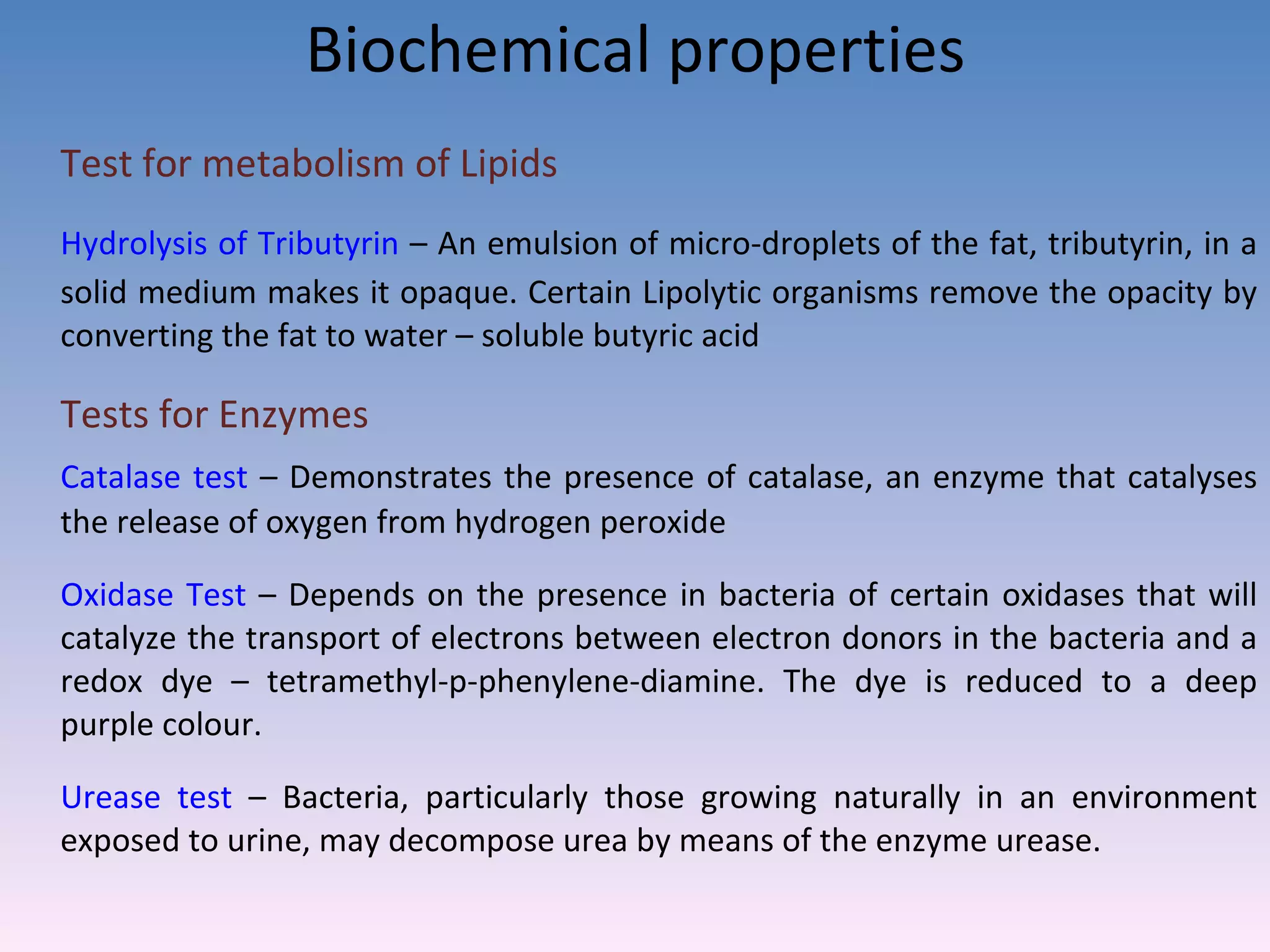
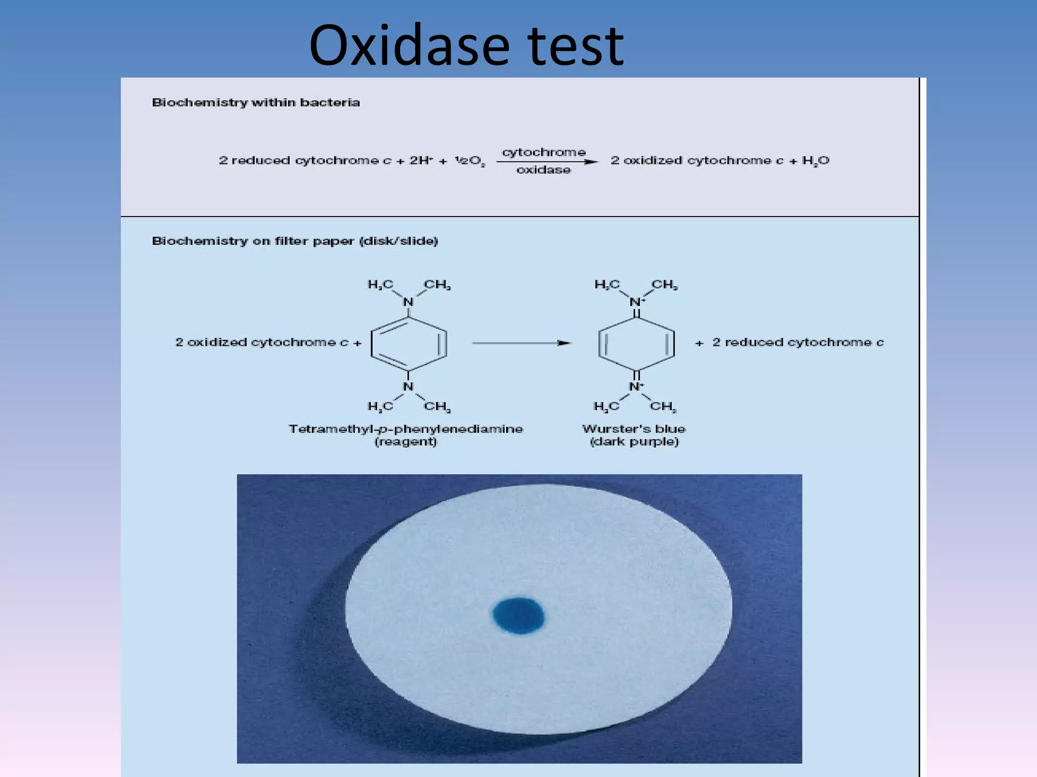
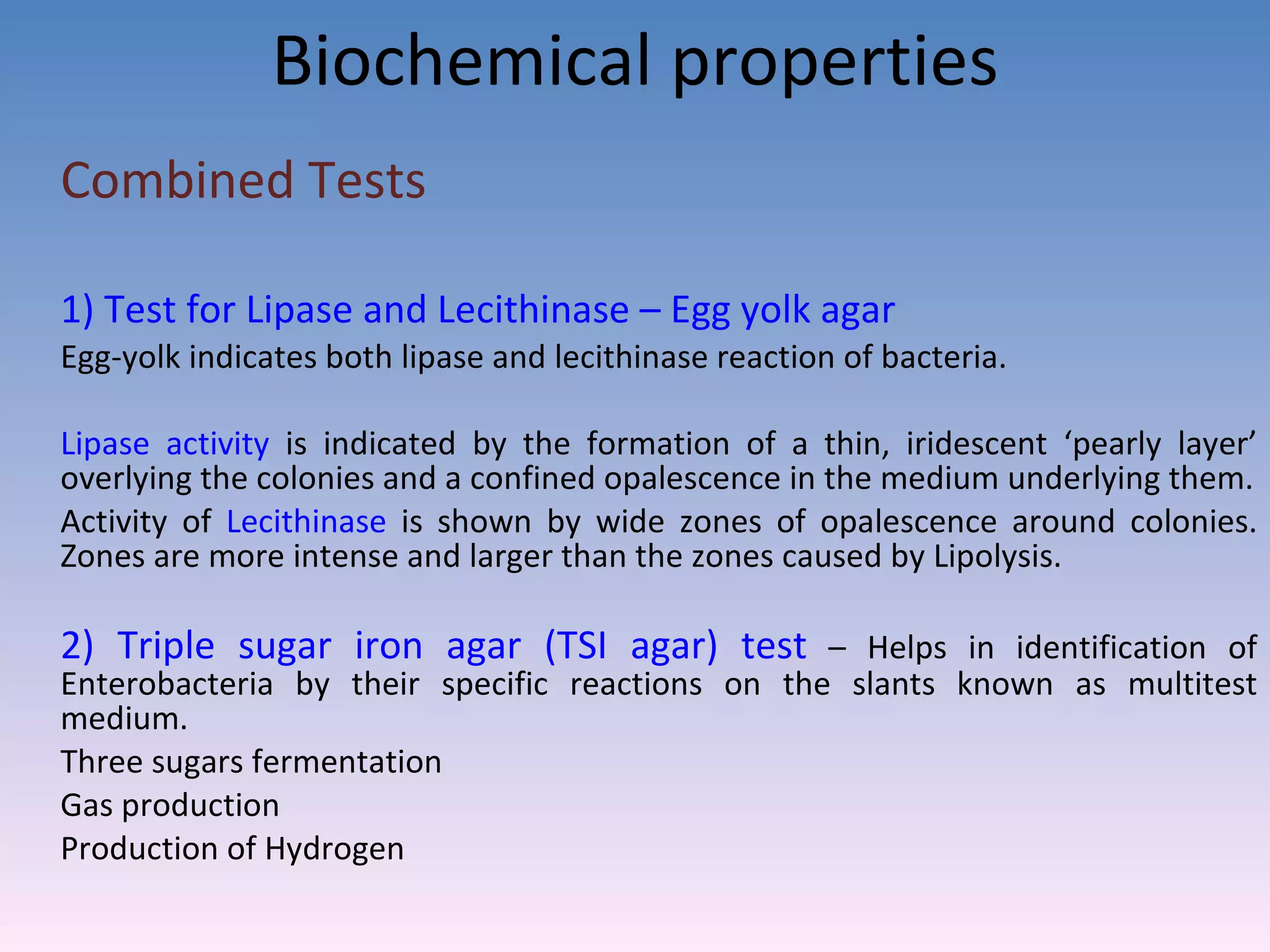
Detailed biochemical tests and reactions used to identify pathogens, e.g., fermentation, enzyme tests.
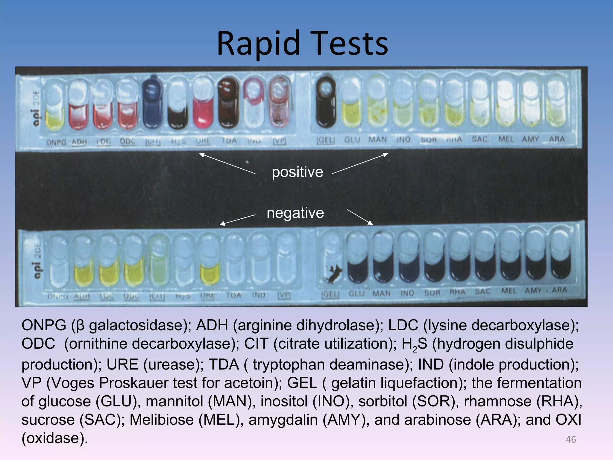
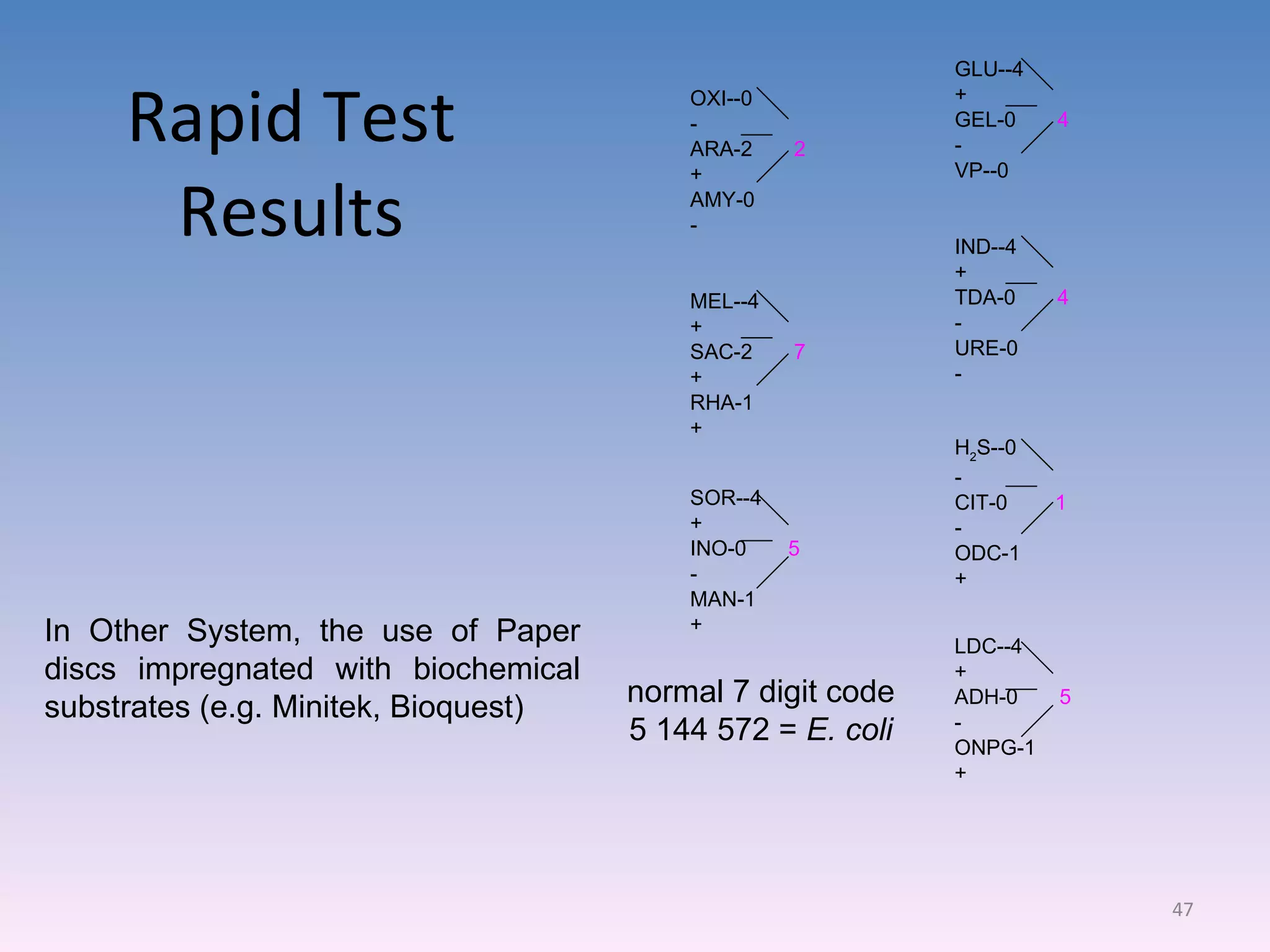
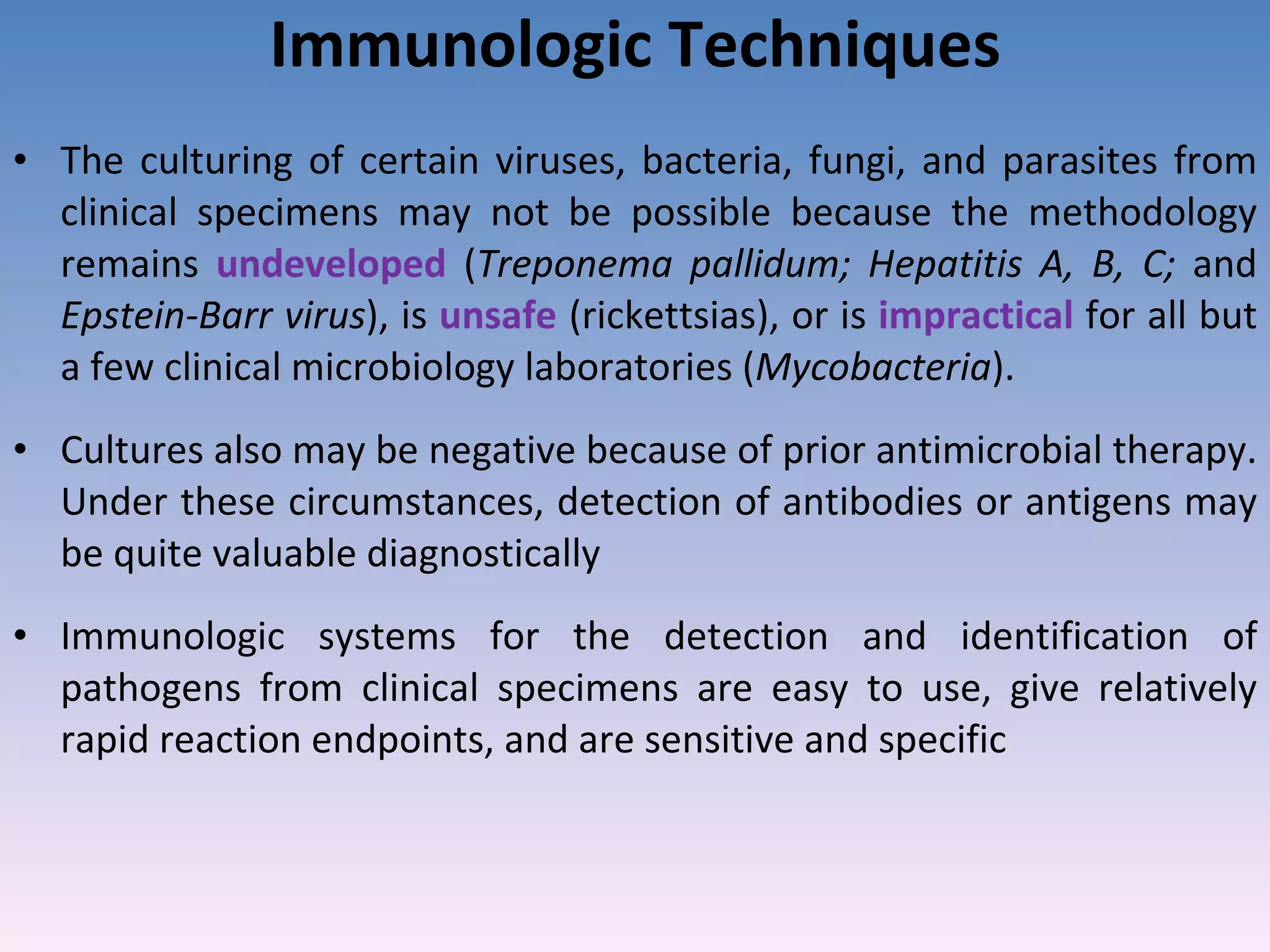
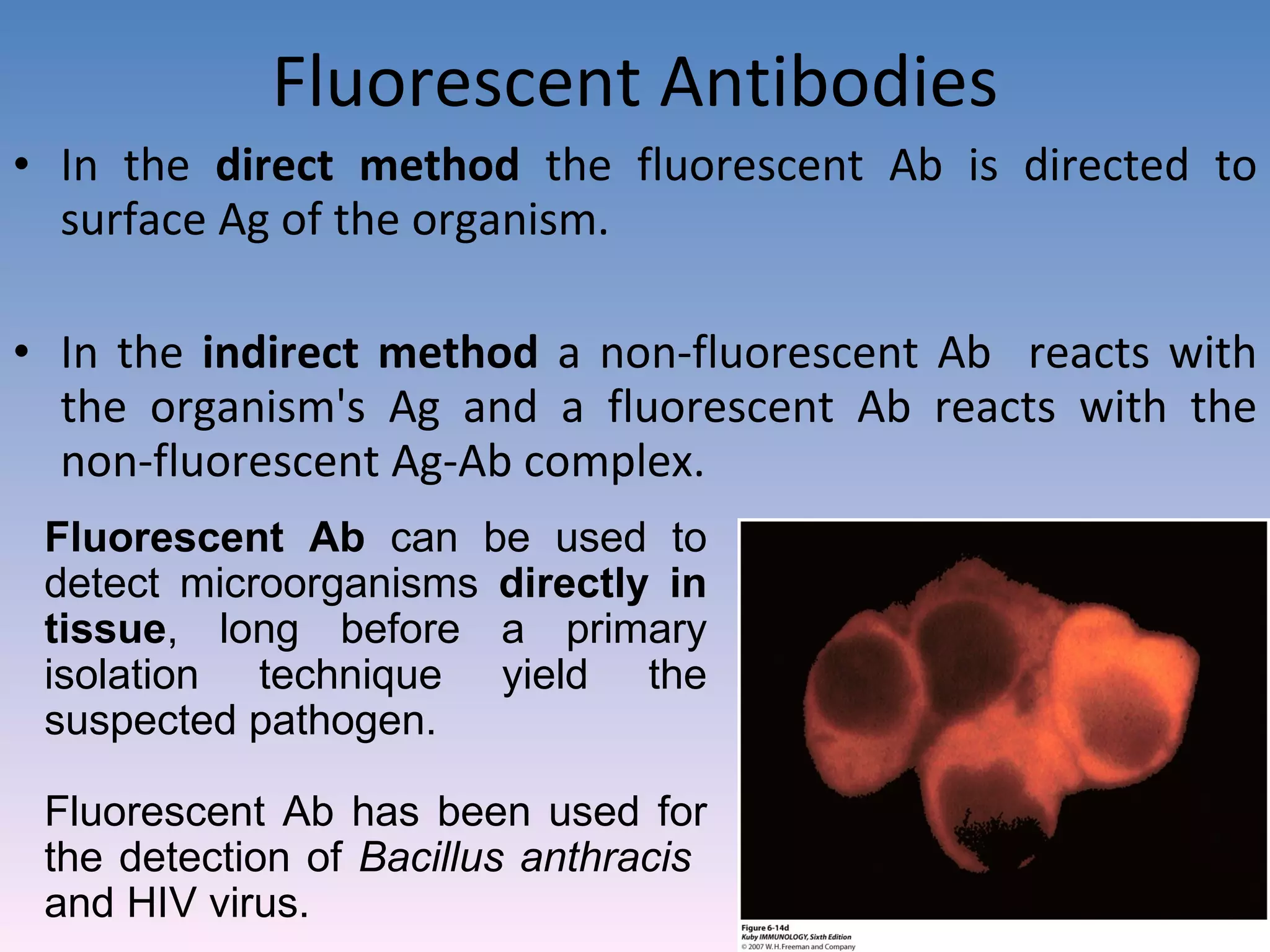
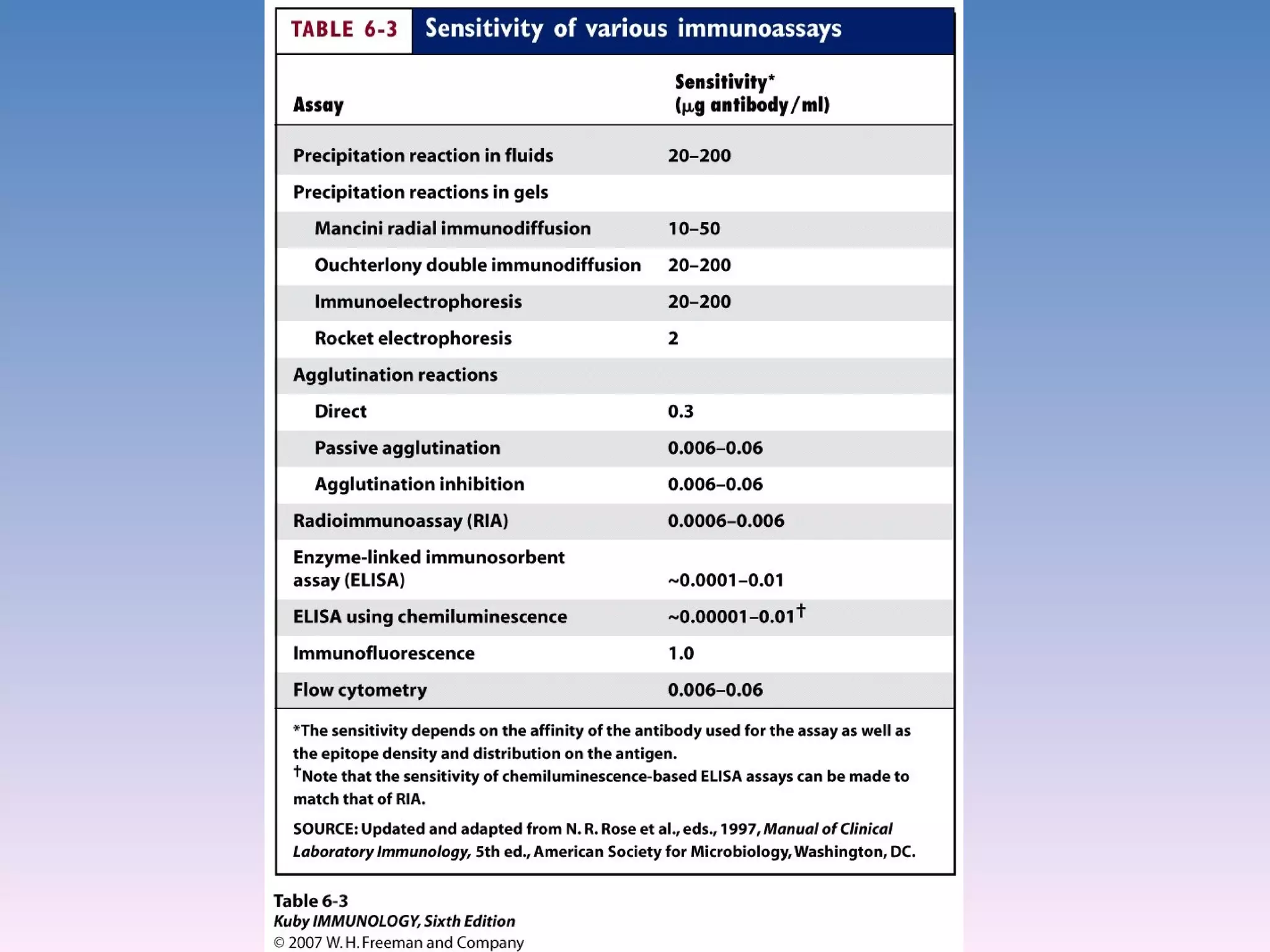
Commercial biochemical panels and techniques for quick microorganism identification.Techniques like the ELISA and immunoblot used for detecting antigens or antibodies from clinical specimens.
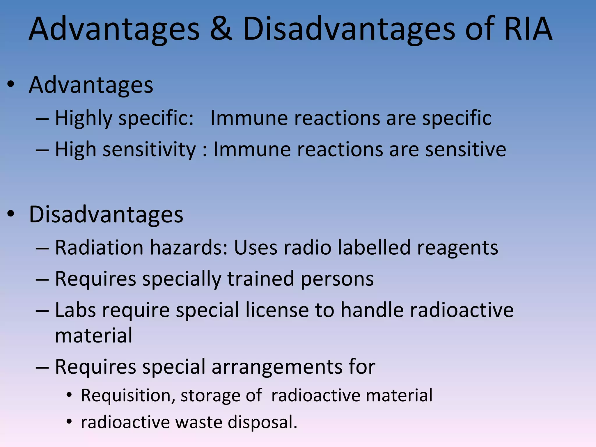
Radioimmunoassay and Immuno-electron microscopy for detecting proteins in various biological materials.
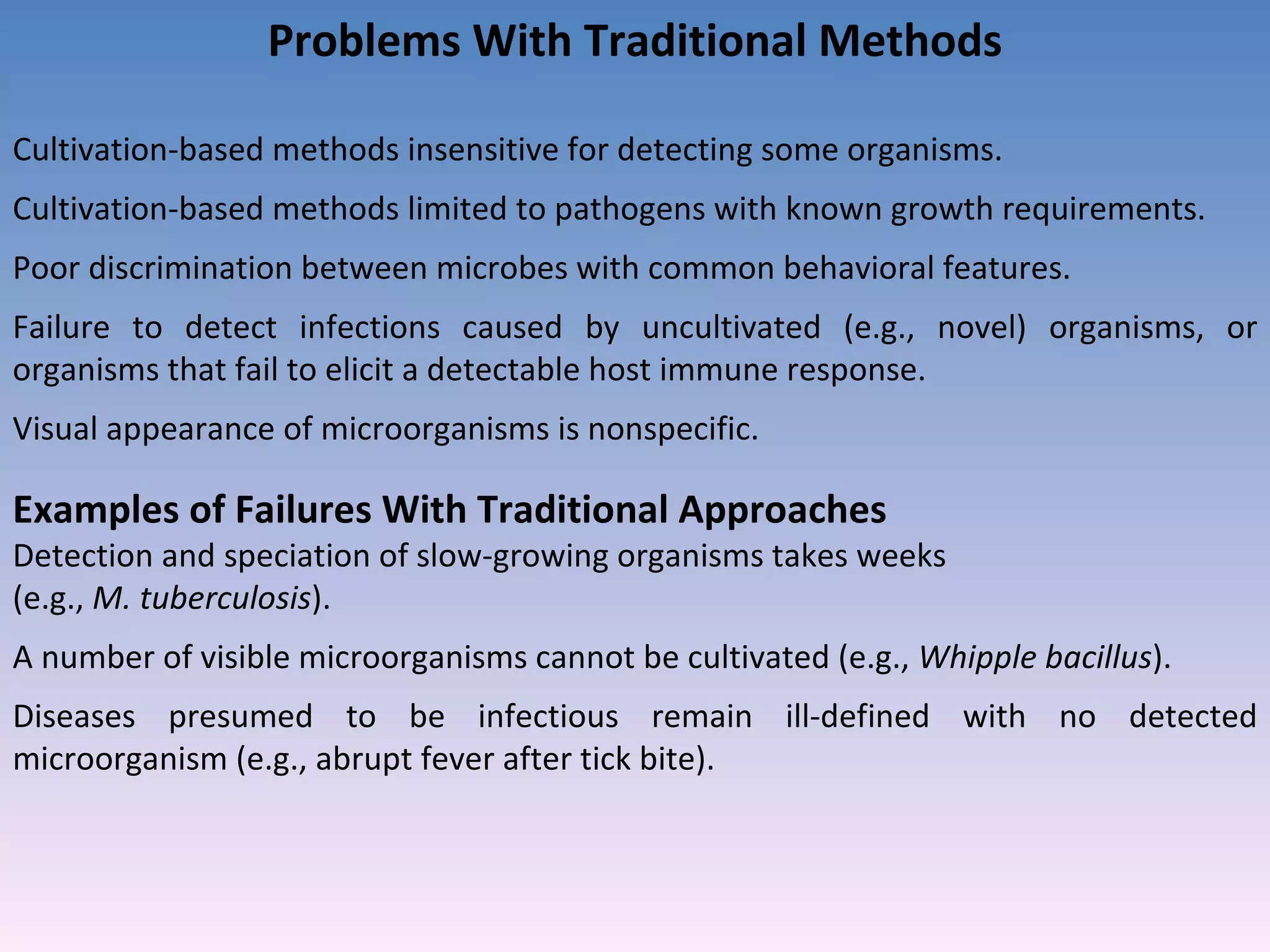
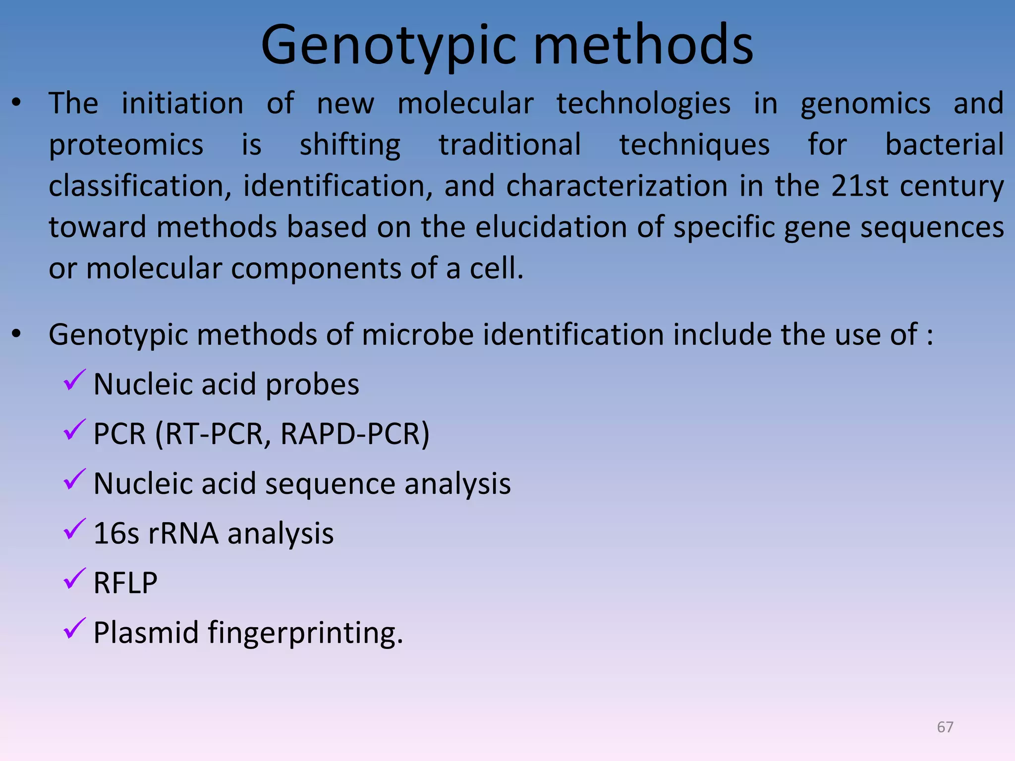
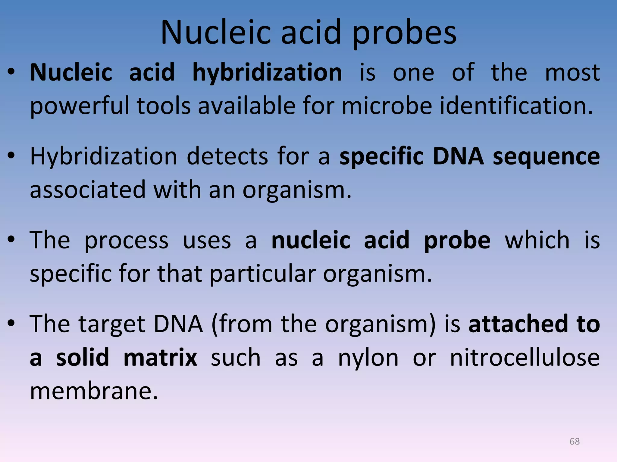
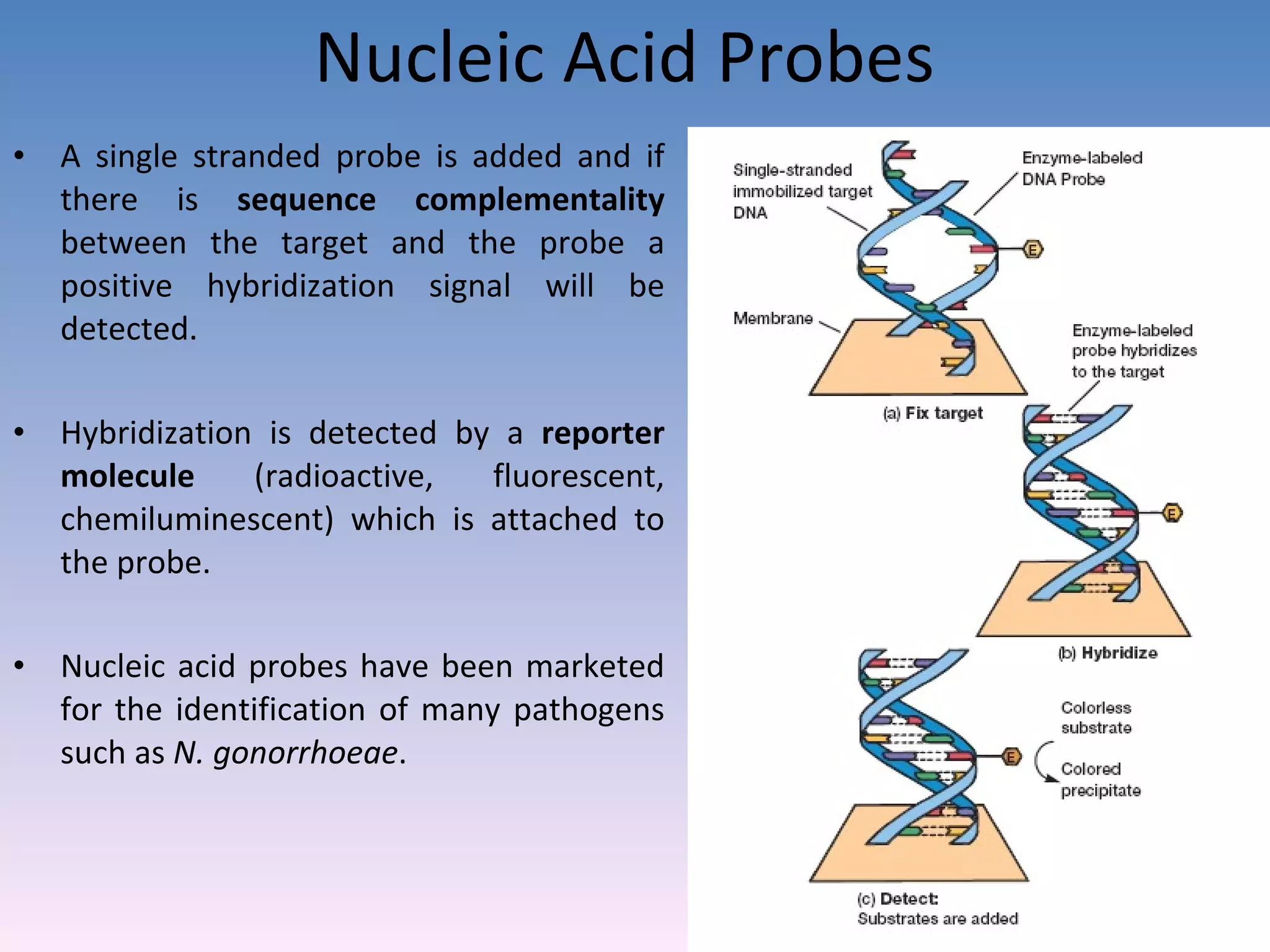
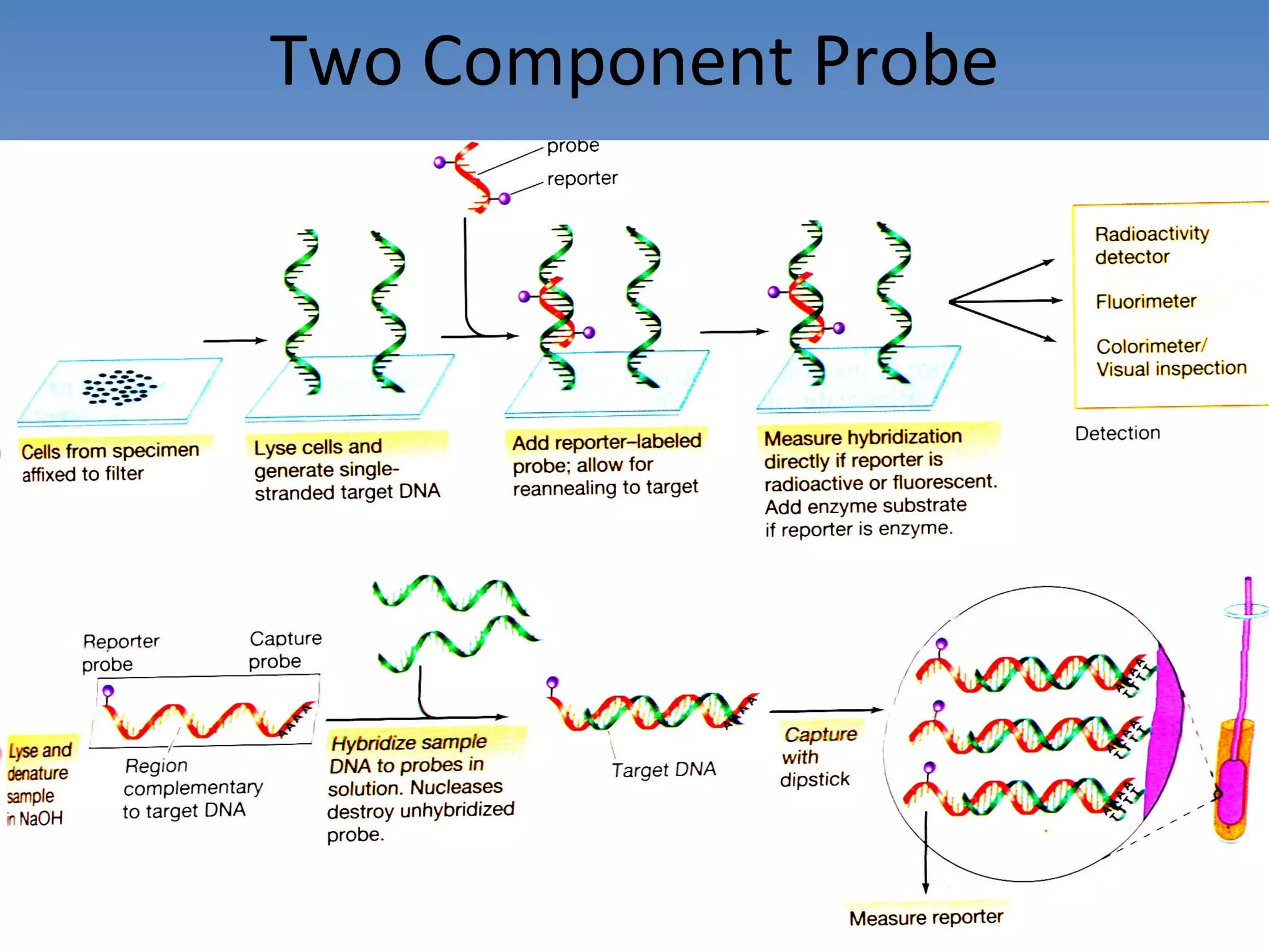
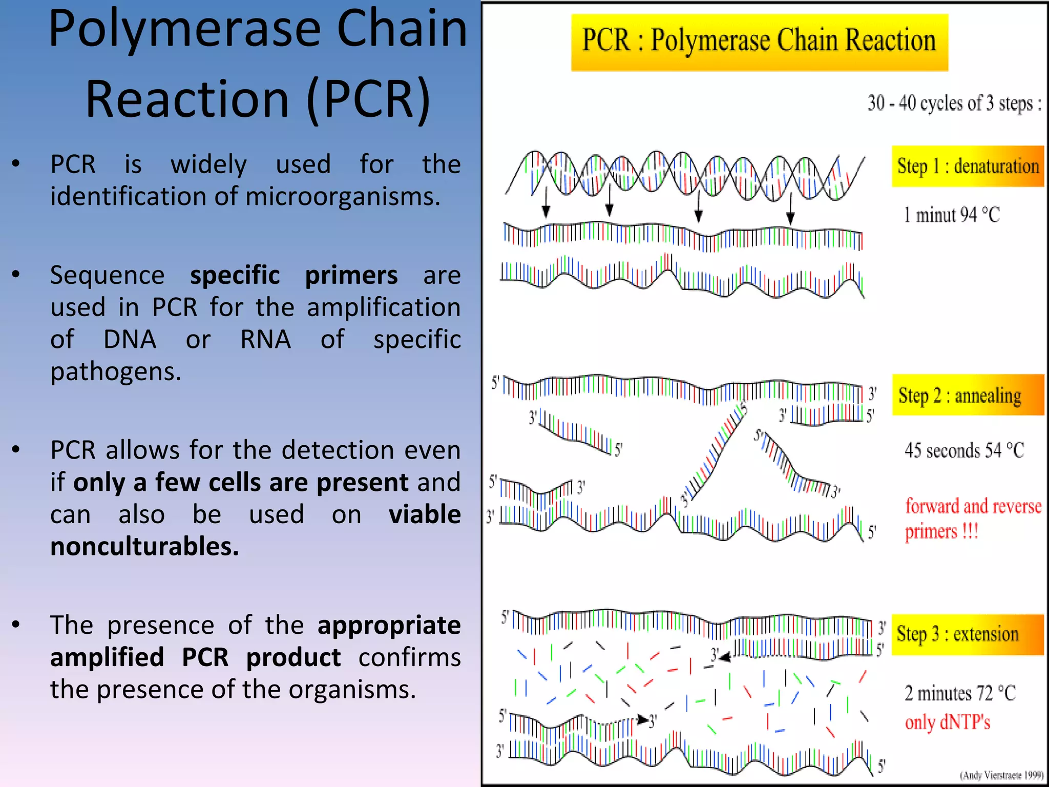
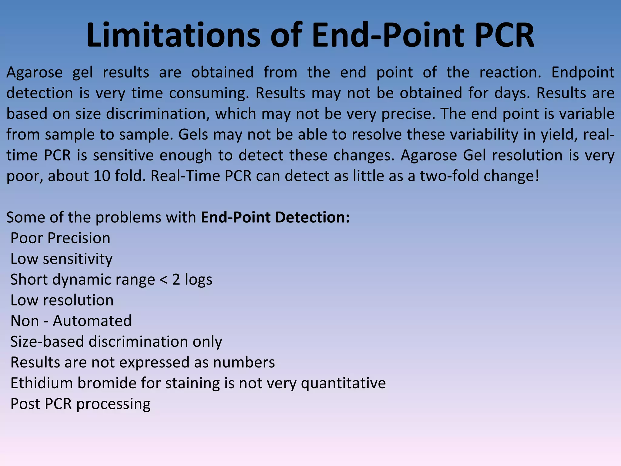
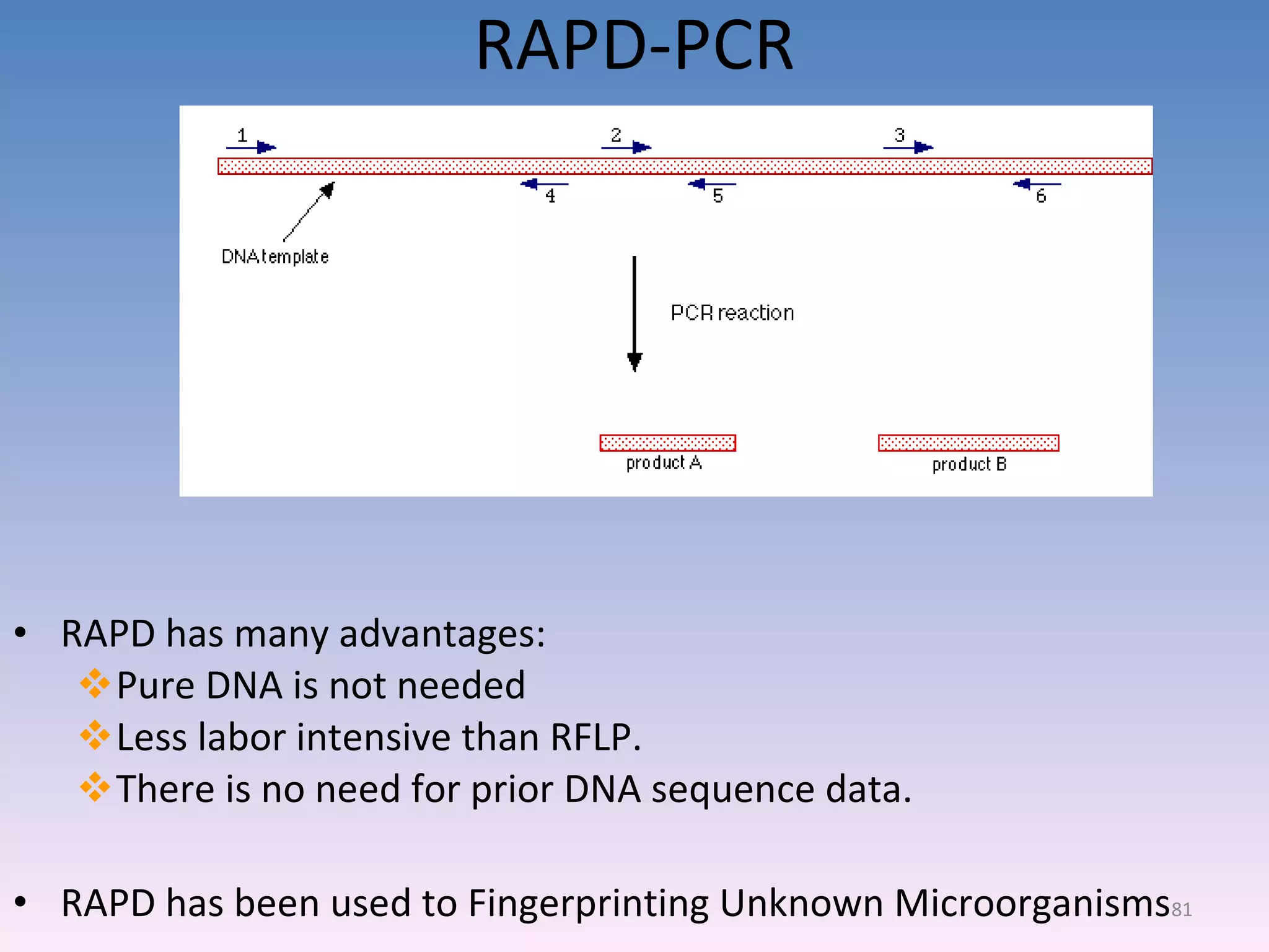
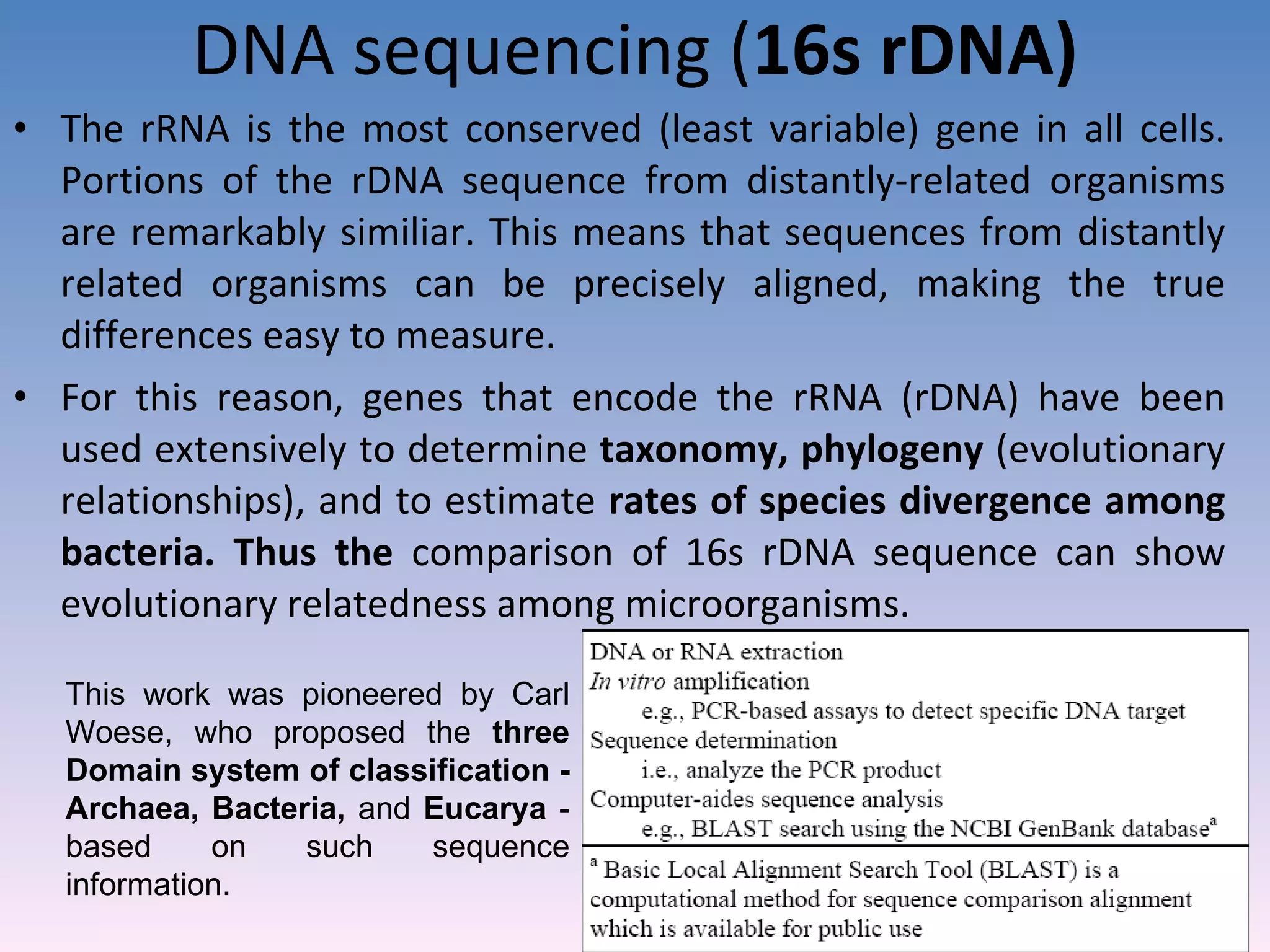
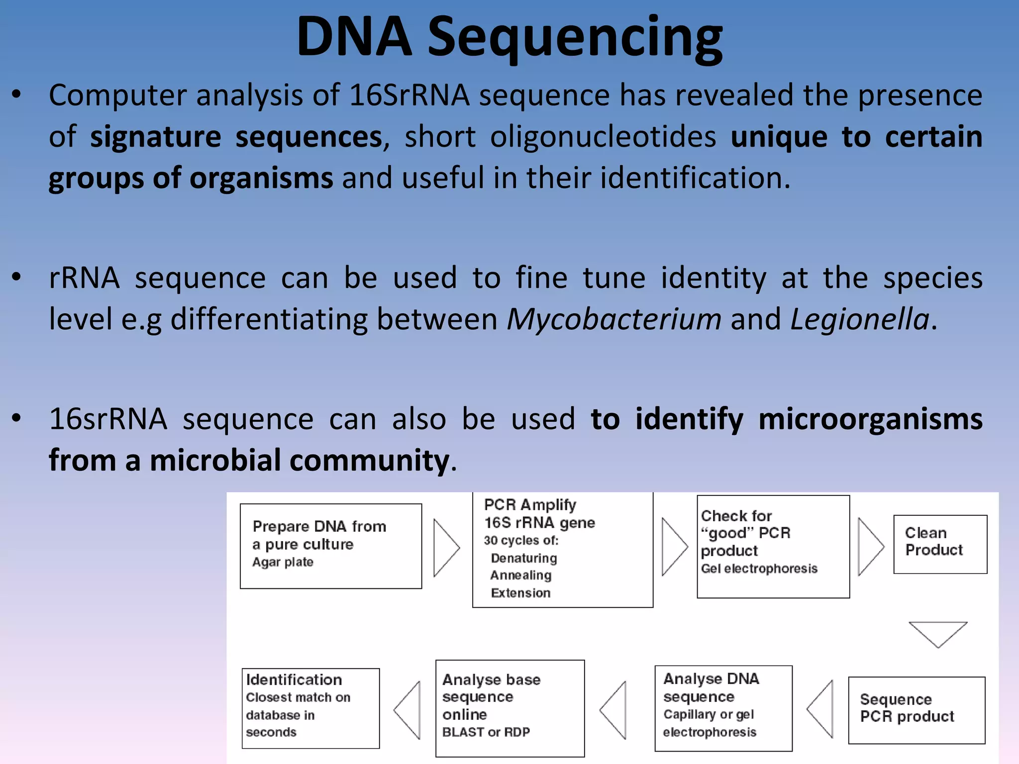
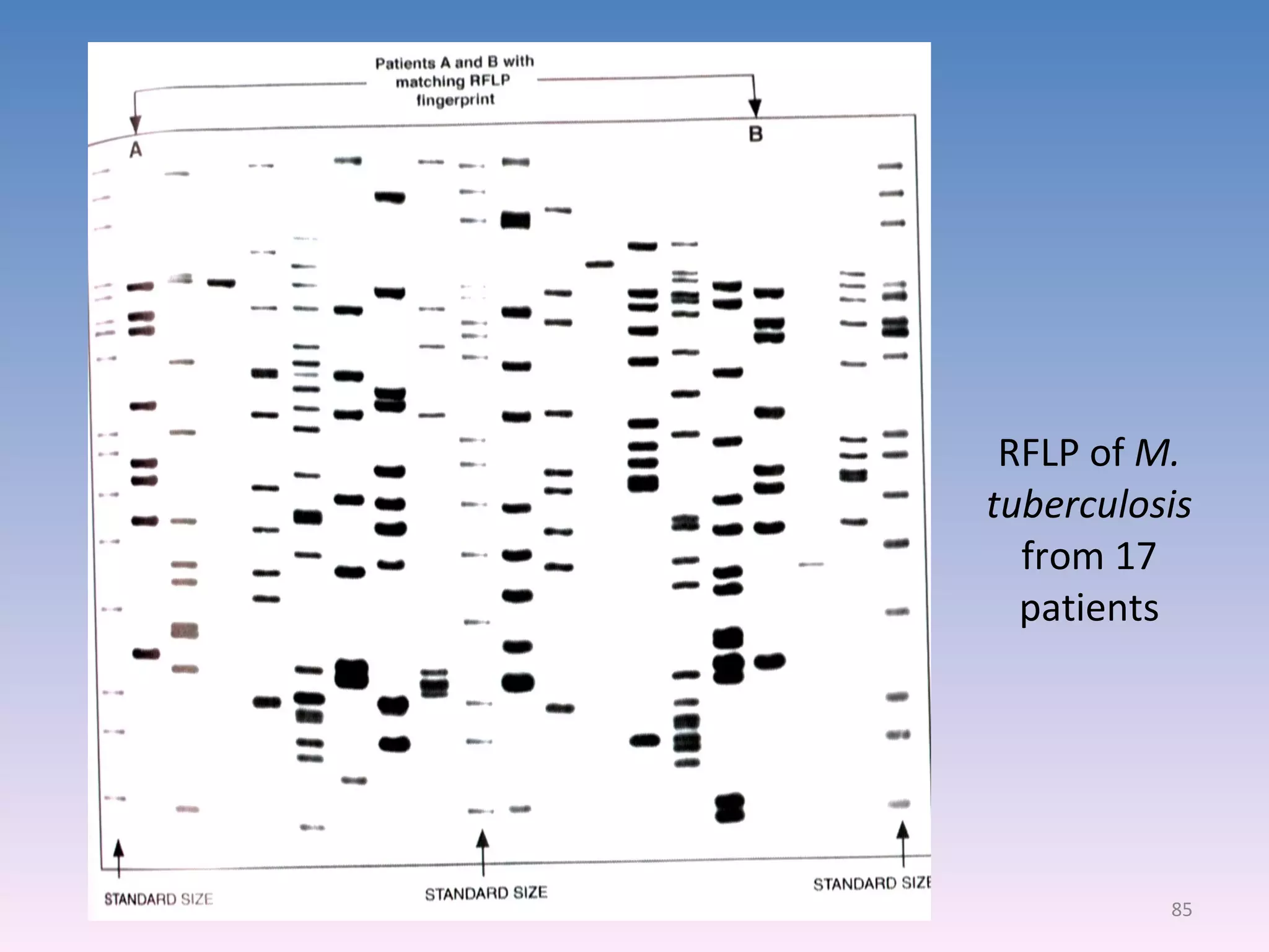
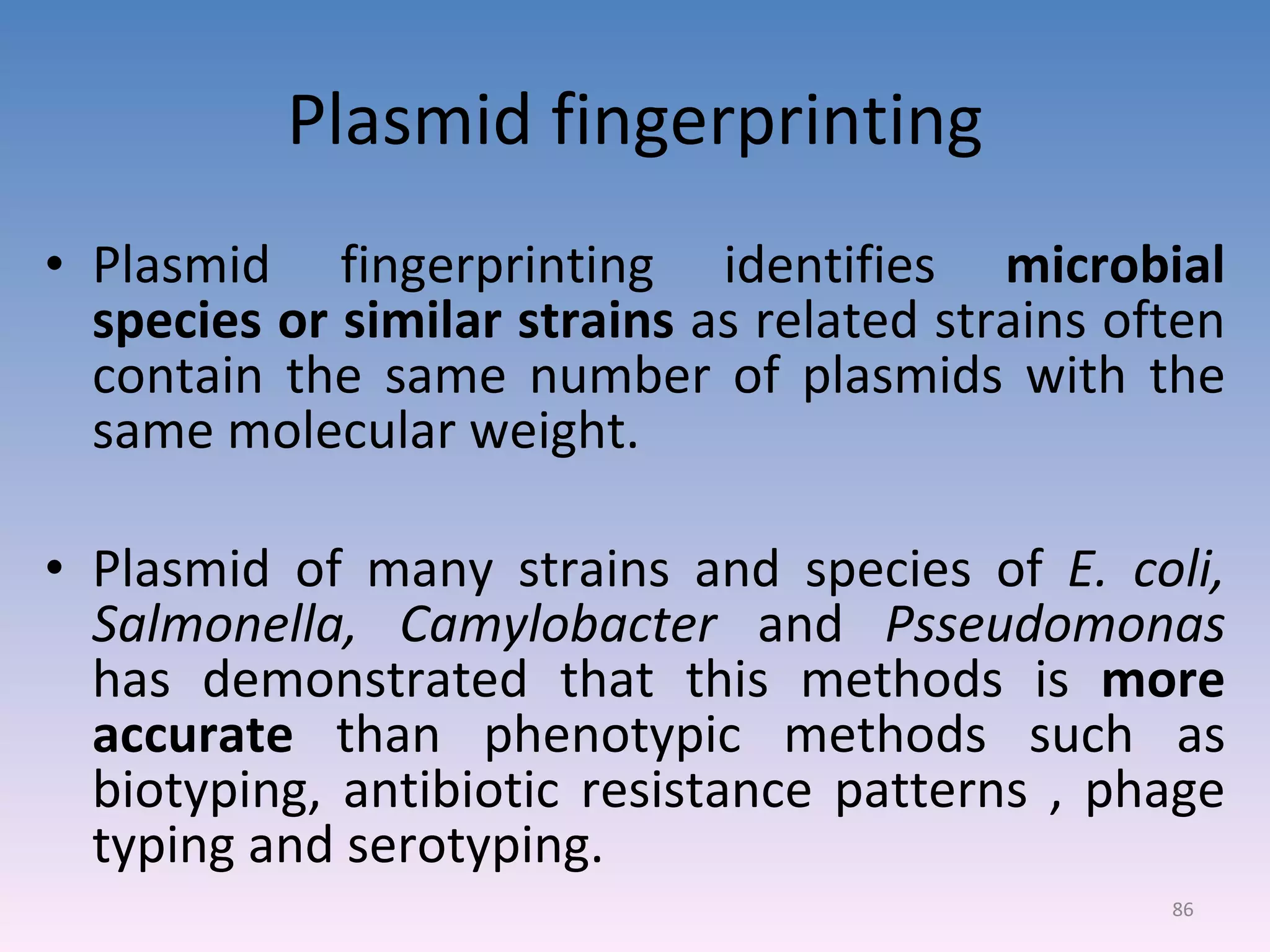
Limitations of cultivation-based microbiological methods and the rise of molecular techniques.Technologies for identification using nucleic acid probes, PCR, sequencing, and plasmid fingerprinting.
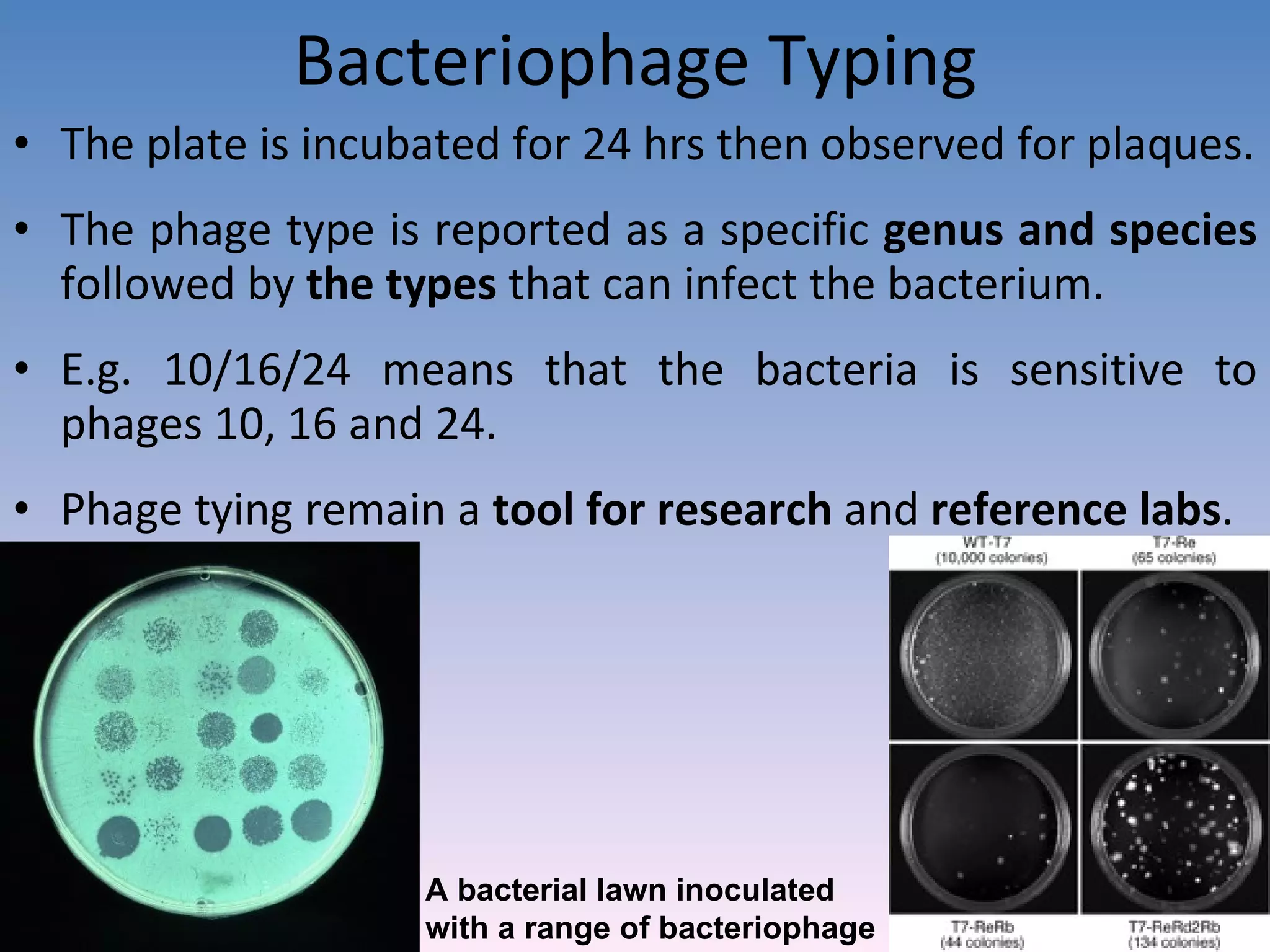
Techniques to identify microorganisms using metabolic byproducts and bacteriophage specificity.
Methods like flow cytometry for detecting unculturable microorganisms.
Definitions and classifications of microbial species, types, strains, and identification importance.
New approaches using synthetic peptides for diagnostics and the process for selecting identification technologies.
Key considerations when choosing identification systems for efficacy in laboratory settings.


