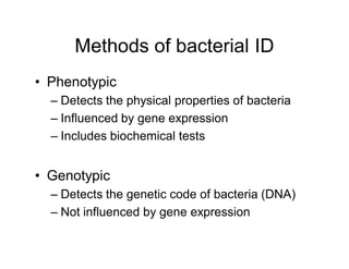
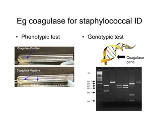
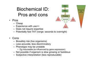
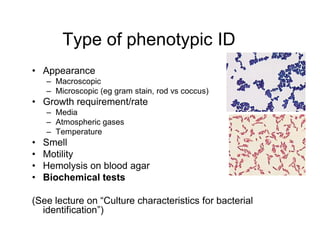
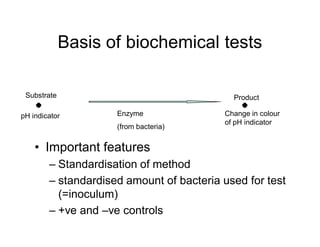
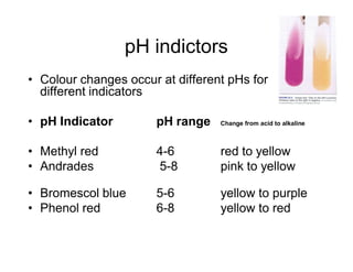
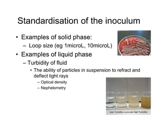
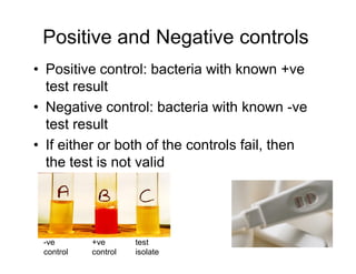
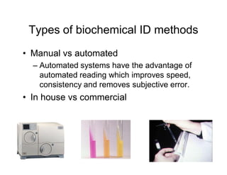
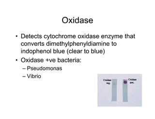
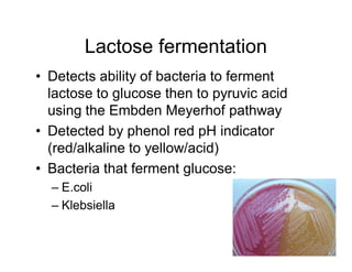
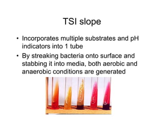
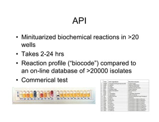
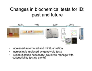
Biochemical identification of
bacteria
Dr. Paul Ingram
Infectious Diseases Physician (RPH) &
Microbiologist (PathWest Laboratories)
Outline
• Phenotypic vs genotypic tests
• Pros and cons of biochemical tests
• Basis of biochemical tests
• Examples of biochemical test
• Diagnostic algorithms
• The future of biochemical identification
tests
Methods of bacterial ID
• Phenotypic
– Detects the physical properties of bacteria
– Influenced by gene expression
– Includes biochemical tests
• Genotypic
– Detects the genetic code of bacteria (DNA)
– Not influenced by gene expression
Eg coagulase for staphylococcal ID
• Phenotypic test • Genotypic test
Coagulase
gene
Biochemical ID:
Pros and cons
• Pros
– Cheap
– Experience with use++
– Does not require expertise
– Potentially fast TAT (range: seconds to overnight)
• Cons
– Biosafety risk (live organisms)
– Less accurate, less discriminatory
– Phenotype may be unstable
• Eg induceable (ie influenced by gene expression)
– Not possible if organism is slow growing or fastidious
– Subjective interpretation (less reproducable)
Type of phenotypic ID
• Appearance
– Macroscopic
– Microscopic (eg gram stain, rod vs coccus)
• Growth requirement/rate
– Media
– Atmospheric gases
– Temperature
• Smell
• Motility
• Hemolysis on blood agar
• Biochemical tests
(See lecture on “Culture characteristics for bacterial
identification”)
Basis of biochemical tests
• Important features
– Standardisation of method
– standardised amount of bacteria used for test
(=inoculum)
– +ve and –ve controls
Substrate
pH indicator
Product
Enzyme
(from bacteria)
Change in colour
of pH indicator
pH indictors
• Colour changes occur at different pHs for
different indicators
• pH Indicator pH range Change from acid to alkaline
• Methyl red 4-6 red to yellow
• Andrades 5-8 pink to yellow
• Bromescol blue 5-6 yellow to purple
• Phenol red 6-8 yellow to red
Standardisation of the inoculum
• Examples of solid phase:
– Loop size (eg 1microL, 10microL)
• Examples of liquid phase
– Turbidity of fluid
• The ability of particles in suspension to refract and
deflect light rays
– Optical density
– Nephelometry
Positive and Negative controls
• Positive control: bacteria with known +ve
test result
• Negative control: bacteria with known -ve
test result
• If either or both of the controls fail, then
the test is not valid
-ve
control
+ve
control
test
isolate
Types of biochemical ID methods
• Manual vs automated
– Automated systems have the advantage of
automated reading which improves speed,
consistency and removes subjective error.
• In house vs commercial
Examples of common biochemical
tests used for ID of gram negative
bacteria
• Urease
• Indole
• Oxidase
• Glucose fermentation
• Lactose fermentation
• Nitrate
Urease
• Detects hydrolysis of urea to ammonia by
urease enzyme
• Ammonia causes an increase in pH which
is detected by the pH indicator (orange à
pink)
• Urease +ve bacteria:
– Proteus
– Klebsiella
Indole
• Detects indole production from tryptophan,
which produces a colour change in
combination with
dimethylaminobenzaldehyde (clear to red)
• Indole +ve bacteria:
– E.coli
– Citrobacter
Oxidase
• Detects cytochrome oxidase enzyme that
converts dimethylphenyldiamine to
indophenol blue (clear to blue)
• Oxidase +ve bacteria:
– Pseudomonas
– Vibrio
Glucose fermentation
• Detects ability of bacteria to ferment
glucose to pyruvic acid using the Embden
Meyerhof pathway
• Detected by phenol red pH indicator
(red/alkaline to yellow/acid)
• Bacteria that ferment glucose:
– E.coli
– Proteus
Lactose fermentation
• Detects ability of bacteria to ferment
lactose to glucose then to pyruvic acid
using the Embden Meyerhof pathway
• Detected by phenol red pH indicator
(red/alkaline to yellow/acid)
• Bacteria that ferment glucose:
– E.coli
– Klebsiella
Nitrate
• Detects nitrate reductase enzyme which
converts nitrate to nitrite.
• Nitrite then revealed by addition of
naphthylamine and sulfinic acid to form
diazonium dye (clear to red)
• Nitrate +ve bacteria:
– E.coli
– Klebsiella
TSI slope
• Incorporates multiple substrates and pH
indicators into 1 tube
• By streaking bacteria onto surface and
stabbing it into media, both aerobic and
anaerobic conditions are generated
API
• Minituarized biochemical reactions in >20
wells
• Takes 2-24 hrs
• Reaction profile (“biocode”) compared to
an on-line database of >20000 isolates
• Commerical test
Automated Biochemical ID systems
• Examples:
– Vitek
– Biolog
– Pheonix
– Autoscan Walkaway
• Varying capacity for:
– Number of specimens they can handle
– Size/extent of comparative database
– Interfacing with lab data program
– Turn around time
– Capacity for ID to species level
Diagnostic algorithms for bacterial
ID
• Primary tests allow genus level ID
(enterobacteriacae, “non-glucose fermenters”,
HACEK, etc)
– Gram stain
– Culture morphology
– Basic biochemical tests
• Eg Oxidase, indole, urease tests, etc
• Species level identification requires more
complex, second line tests
Example 1 of diagnostic algorithm
Indole Methyl
red
Voges
Proskauer
Citrate Urease
E.coli + + - - -
Enterobacter - - + + -
Klebsiella
pneumoniae
- - + + +
Salmonella - + - + -
Shigella - + - - -
Proteus
mirabilis
- + - +/- +
Example 2 of diagnostic algorithm
Changes in biochemical tests for ID:
past and future
• Increased automated and minituarisation
• Increasingly replaced by genotypic tests
• Is identification necessary: could we manage with
susceptibility testing alone?
1970 2000
1980 2010
Conclusions
• Biochemical tests remain critical to
bacterial identification
• Need to understand the principles of the
common/primary tests
• Biochemical tests have limitations
• In the future they will increasingly be
replaced by genotypic tests

biochemical_identifcation_of_bacteria_slides.pdf

  • 1.
    Biochemical identification of bacteria Dr.Paul Ingram Infectious Diseases Physician (RPH) & Microbiologist (PathWest Laboratories)
  • 2.
    Outline • Phenotypic vsgenotypic tests • Pros and cons of biochemical tests • Basis of biochemical tests • Examples of biochemical test • Diagnostic algorithms • The future of biochemical identification tests
  • 3.
    Methods of bacterialID • Phenotypic – Detects the physical properties of bacteria – Influenced by gene expression – Includes biochemical tests • Genotypic – Detects the genetic code of bacteria (DNA) – Not influenced by gene expression
  • 4.
    Eg coagulase forstaphylococcal ID • Phenotypic test • Genotypic test Coagulase gene
  • 5.
    Biochemical ID: Pros andcons • Pros – Cheap – Experience with use++ – Does not require expertise – Potentially fast TAT (range: seconds to overnight) • Cons – Biosafety risk (live organisms) – Less accurate, less discriminatory – Phenotype may be unstable • Eg induceable (ie influenced by gene expression) – Not possible if organism is slow growing or fastidious – Subjective interpretation (less reproducable)
  • 6.
    Type of phenotypicID • Appearance – Macroscopic – Microscopic (eg gram stain, rod vs coccus) • Growth requirement/rate – Media – Atmospheric gases – Temperature • Smell • Motility • Hemolysis on blood agar • Biochemical tests (See lecture on “Culture characteristics for bacterial identification”)
  • 7.
    Basis of biochemicaltests • Important features – Standardisation of method – standardised amount of bacteria used for test (=inoculum) – +ve and –ve controls Substrate pH indicator Product Enzyme (from bacteria) Change in colour of pH indicator
  • 8.
    pH indictors • Colourchanges occur at different pHs for different indicators • pH Indicator pH range Change from acid to alkaline • Methyl red 4-6 red to yellow • Andrades 5-8 pink to yellow • Bromescol blue 5-6 yellow to purple • Phenol red 6-8 yellow to red
  • 9.
    Standardisation of theinoculum • Examples of solid phase: – Loop size (eg 1microL, 10microL) • Examples of liquid phase – Turbidity of fluid • The ability of particles in suspension to refract and deflect light rays – Optical density – Nephelometry
  • 10.
    Positive and Negativecontrols • Positive control: bacteria with known +ve test result • Negative control: bacteria with known -ve test result • If either or both of the controls fail, then the test is not valid -ve control +ve control test isolate
  • 11.
    Types of biochemicalID methods • Manual vs automated – Automated systems have the advantage of automated reading which improves speed, consistency and removes subjective error. • In house vs commercial
  • 12.
    Examples of commonbiochemical tests used for ID of gram negative bacteria • Urease • Indole • Oxidase • Glucose fermentation • Lactose fermentation • Nitrate
  • 13.
    Urease • Detects hydrolysisof urea to ammonia by urease enzyme • Ammonia causes an increase in pH which is detected by the pH indicator (orange à pink) • Urease +ve bacteria: – Proteus – Klebsiella
  • 14.
    Indole • Detects indoleproduction from tryptophan, which produces a colour change in combination with dimethylaminobenzaldehyde (clear to red) • Indole +ve bacteria: – E.coli – Citrobacter
  • 15.
    Oxidase • Detects cytochromeoxidase enzyme that converts dimethylphenyldiamine to indophenol blue (clear to blue) • Oxidase +ve bacteria: – Pseudomonas – Vibrio
  • 16.
    Glucose fermentation • Detectsability of bacteria to ferment glucose to pyruvic acid using the Embden Meyerhof pathway • Detected by phenol red pH indicator (red/alkaline to yellow/acid) • Bacteria that ferment glucose: – E.coli – Proteus
  • 17.
    Lactose fermentation • Detectsability of bacteria to ferment lactose to glucose then to pyruvic acid using the Embden Meyerhof pathway • Detected by phenol red pH indicator (red/alkaline to yellow/acid) • Bacteria that ferment glucose: – E.coli – Klebsiella
  • 18.
    Nitrate • Detects nitratereductase enzyme which converts nitrate to nitrite. • Nitrite then revealed by addition of naphthylamine and sulfinic acid to form diazonium dye (clear to red) • Nitrate +ve bacteria: – E.coli – Klebsiella
  • 19.
    TSI slope • Incorporatesmultiple substrates and pH indicators into 1 tube • By streaking bacteria onto surface and stabbing it into media, both aerobic and anaerobic conditions are generated
  • 20.
    API • Minituarized biochemicalreactions in >20 wells • Takes 2-24 hrs • Reaction profile (“biocode”) compared to an on-line database of >20000 isolates • Commerical test
  • 21.
    Automated Biochemical IDsystems • Examples: – Vitek – Biolog – Pheonix – Autoscan Walkaway • Varying capacity for: – Number of specimens they can handle – Size/extent of comparative database – Interfacing with lab data program – Turn around time – Capacity for ID to species level
  • 22.
    Diagnostic algorithms forbacterial ID • Primary tests allow genus level ID (enterobacteriacae, “non-glucose fermenters”, HACEK, etc) – Gram stain – Culture morphology – Basic biochemical tests • Eg Oxidase, indole, urease tests, etc • Species level identification requires more complex, second line tests
  • 23.
    Example 1 ofdiagnostic algorithm Indole Methyl red Voges Proskauer Citrate Urease E.coli + + - - - Enterobacter - - + + - Klebsiella pneumoniae - - + + + Salmonella - + - + - Shigella - + - - - Proteus mirabilis - + - +/- +
  • 24.
    Example 2 ofdiagnostic algorithm
  • 25.
    Changes in biochemicaltests for ID: past and future • Increased automated and minituarisation • Increasingly replaced by genotypic tests • Is identification necessary: could we manage with susceptibility testing alone? 1970 2000 1980 2010
  • 26.
    Conclusions • Biochemical testsremain critical to bacterial identification • Need to understand the principles of the common/primary tests • Biochemical tests have limitations • In the future they will increasingly be replaced by genotypic tests