Young & Hot Surat ℂall Girls Dindoli 8527049040 WhatsApp AnyTime Best Surat ℂ...
Optical Coherence Tomography : A Tool for Assessment of Conduits Used in Coronary Artery Bypass Surgery
1. Optical Coherence Tomography : A Tool for Assessment
of Conduits Used in Coronary Artery Bypass Surgery
Alex Brown
Department of Cardiothoracic Surgery, Boston University School of Medicine
Study is Supported by NIH grant (R01 HL84080) awarded to Principal Investigator Dr. Robert S Poston
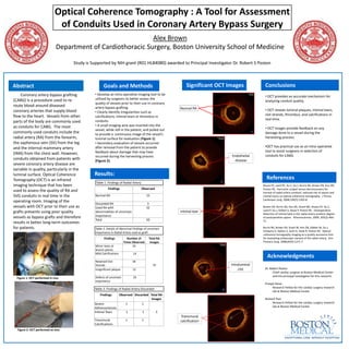
Abstract Goals and Methods Significant OCT Images Conclusions
Coronary artery bypass grafting • Develop an intra-operative imaging tool to be
• OCT provides an accurate mechanism for
(CABG) is a procedure used to re- utilized by surgeons to better assess the
analyzing conduit quality.
quality of vessels prior to their use in coronary
route blood around diseased artery bypass grafting. Normal RA • OCT reveals luminal plaques, intimal tears,
coronary arteries that supply blood • Clearly identify irregularities such as
flow to the heart. Vessels from other clot strands, thrombus, and calcifications in
calcifications, intimal tears or thrombus in
conduits. real-time.
parts of the body are commonly used
as conduits for CABG. The most • A small imaging wire was inserted into the
vessel, while still in the patient, and pulled out • OCT Images provide feedback on any
commonly used conduits include the to provide a continuous image of the vessel’s damage done to a vessel during the
radial artery (RA) from the forearm, luminal surface for evaluation (Figure 1). harvesting process.
the saphenous vein (SV) from the leg • Secondary evaluation of vessels occurred
and the internal mammary artery after removal from the patient to provide •OCT has practical use as an intra-operative
feedback about damage that may have tool to assist surgeons in selection of
(IMA) from the chest wall. However, conduits for CABG.
occurred during the harvesting process Endothelial
conduits obtained from patients with (Figure 2). disease
severe coronary artery disease are
variable in quality, particularly in the
luminal surface. Optical Coherence Results:
Tomography (OCT) is an infrared References
Table 1: Findings of Radial Artery
imaging technique that has been Brazio PS, Laird PC, Xu C, Gu J, Burris NS, Brown EN, Kon ZN,
Findings Observed Poston RS. Harmonic scalpel versus electrocautery for
used to assess the quality of RA and harvest of radial artery conduits: reduced risk of spasm and
SVG conduits in real time in the Normal RA 15 intimal injury on optical coherence tomography. J Thorac
Cardiovasc Surg. 2008;136(5):1302-8.
operating room. Imaging of the
Discarded RA 3
vessels with OCT prior to their use as Used RA with 32 Brown EN, Burris NS, Kon ZN, Grant MC, Brazio PS, Xu C,
Laird P, Gu J, Kallam S, Desai P, Poston RS. Intraoperative
grafts prevents using poor quality abnormalities of uncertain Intimal tear
detection of intimal lipid in the radial artery predicts degree
vessels as bypass grafts and therefore importance of postoperative spasm. Atherosclerosis. 2009; 205(2):466-
Total 50 71.
results in better long-term outcomes
for patients. Table 2: Details of Abnormal Findings of uncertain Burris NS, Brown EN, Grant M, Kon ZN, Gibber M, Gu J,
importance in Radial Artery used as graft Schwartz K, Kallam S, Joshi A, Vitali R, Poston RS. Optical
coherence tomography imaging as a quality assurance tool
Findings Number of Total RA for evaluating endoscopic harvest of the radial artery. Ann
Times Observed Images Thoracic Surg. 2008;85(4):1271-7.
Minor tears at 10
branch points
Mild Calcifications 14
Acknowledgments
Retained Clot 18
Strands 32 Intraluminal
Insignificant plaque 12 clot Dr. Robert Poston
Chief cardiac surgeon at Boston Medical Center
Defects of uncertain 29 and the principal investigator for this research.
Figure 1: OCT performed in vivo
importance
Pranjal Desai
Table 3: Findings of Radial Artery Discarded Research Fellow for the cardiac surgery research
lab at Boston Medical Center
Findings Observed Discarded Total RA
Images Richard Tran
Severe 1 1 Research Fellow for the cardiac surgery research
Altherosclerosis lab at Boston Medical Center
Intimal Tears 1 1 3
Transmural
Transmural 1 1 calcification
Calcifications
Figure 2: OCT performed ex vivo