1. CT Clinical
Engines
2013
Edition
www.siemens.com/ct-acute-care
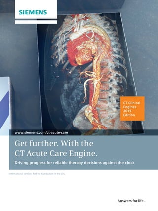
Get further. With the
CT Acute Care Engine.
Driving progress for reliable therapy decisions against the clock
International version. Not for distribution in the U.S.
Answers for life.
2. What is a CT Clinical Engine?
• powerful combination of software applications and
A
scanner features – tailored to meet your clinical challenges
• solution that helps you get the most from your CT scanner
A
With a CT Clinical Engine, you can continually enhance
speed, workflow efficiency, and diagnostic information.
2
3. 2013 Edition: Overview
How far can you get
with your CT?
Rising patient expectations, increasing efficiency needs: The best way to meet these
challenges is to get detailed diagnostic information faster.
Whether it’s for triple rule-out of acute chest pain, for stroke assessment, poly-trauma,
or acute abdominal pain – the CT Acute Care Engine provides clinical functionality
that delivers decisive results for all of these challenging indications. Automatic Case
Preparation has your case ready for reading as soon as it is opened. Above all, speed and
dependability add confidence for critical decisions made against the clock.
Driving progress for reliable therapy decisions against the clock.
3
4. Rapid
generation of images
for comprehensive
surgical planning
in thorax and
spine trauma.
4
6. Rapid generation of images
for surgical planning
Rapid Results Technology and Bone Vessel Technology produce the decision basis in the acute care
Isolation Mode scenario, whether in severe trauma cases or rule-out of
aortic dissections. Save time in the “golden hour” by auto-
In trauma cases requiring surgical intervention for the spine,
matically creating just the right amount of information –
the orthopedic surgeon usually requires several standard-
for standardized and reproducible surgical planning.
ized views of the affected region to aid in a comprehensive
pre-procedural workup. Generating these different views Bone Vessel Isolation Mode enables you to selectively
can be time-consuming, which is particularly challenging in highlight vessels, soft tissue, and bone fragments caused
an emergency. Furthermore, conventional single-volume by fractures. It assists you in alignment assessment when
rendered techniques can make it challenging to selectively there is a suspicion of acute trauma to the spine. Bone
highlight bone fragments attributed to a fracture. Vessel Isolation Mode facilitates identification of spinal
fractures, injuries to the spinal cord, and damage to the
With Rapid Results Technology you can automatically gen-
vascular system, thus providing a sound basis for surgical
erate visualizations of the spine and general vessels in var-
planning.
ious types and orientations. Be creative and design your
own personal protocols that suit your institution’s standards
best. Define your workflow once and let Rapid Results
6
7. 2013 Edition: What‘s the top innovation?
Unparalleled speed whenever time is of the essence fractures or lesions. Additionally, with the STRATON tube
The SOMATOM Definition Edge and Rapid Results and the newly designed gantry using Siemens’ most ad-
Technology vanced patient table, this high spatial resolution is
achieved even at an acquisition speed of up to 230 mm/s.
In acute care scenarios unconscious or severely injured
This takes motion out of the equation, increasing the di-
patients have to be scanned quickly. Here the “golden
agnostic reliability in crucial cases like acute care patients.
hour” to diagnosis mandates precise localization and iden-
tification of critical injuries. Therefore one of the most By combining the strengths of the SOMATOM Definition
challenging demands is providing high acquisition speed Edge and Rapid Results Technology Siemens offers a fast
without compromising spatial resolution. and reliable basis for tackling the challenges in the acute
care setting.
The SOMATOM Definition Edge with the Stellar Detector
is in a class of its own. The new level of image detail with
a routine spatial resolution of up to 0.30 mm provided
by the Stellar Detector allows visualizations of very fine
7
8. syngo.CT Neuro Perfusion* syngo.CT Cardiac Function – Enhancement
Lifesaving decisions, when
every second counts
Quantitative evaluation of dynamic CT data Localize the perfusion defect – assess the
syngo.CT Neuro Perfusion* hemodynamic relevance
syngo.CT Cardiac Function – Enhancement**
In acute stroke it may be challenging to differentiate the
core infarct from tissue at risk for infarction (penumbra). A coronary CTA might yield an intermediate coronary ste-
This is important, however, as the latter is potentially nosis with unclear hemodynamic relevance. The evaluation
salvageable with further therapy. in standard cardiac planes may be challenging, since
the correlation to the coronary vessel in question can be
With a range of unique features syngo.CT Neuro Perfusion
difficult.
helps you easily assess the potential benefit of treatment.
It directly visualizes tissue at risk in 3-D color maps, based syngo.CT Cardiac Function – Enhancement solves this
on the mismatch between blood volume and flow. Alter- issue. It features AHA-compliant 17-segment polar maps
natively, feel free to select individual mismatch parame- for first pass myocardial perfusion data. Perfusion defects
ters such as Siemens’ Time-To-Drain. Refined algorithms are easily localized and the overlay with the VRT in the
offer automated gray matter segmentation so you can Hybrid View helps to correlate a defect with the supplying
immediately focus on the relevant tissues. coronary artery.
Rule out coronary artery disease in less than a minute
syngo.CT Coronary Analysis
When suspecting an acute coronary syndrome, it is essen-
tial to assess the entire coronary tree. Severe stenoses may
degrade a detailed visualization of the coronary vessels.
8
9. 2013 Edition: What else is new?
syngo.CT Coronary Analysis syngo.CT Vascular Analysis
Nevertheless, you may need to make a confident decision In syngo.CT Vascular Analysis, reference markers are now
in a very short period of time. displayed in the VRT, enabling an easy placement at, e.g.
ostia or the iliac bifurcation. The exact position can now
syngo.CT Coronary Analysis now features a robust seg-
be fine-tuned through direct scrolling in cross sections
mentation of the coronary vessels and provides a compre-
along the curved centerline. Also, the system automati-
hensive visualization of the coronary tree, despite high-
cally provides effective vessel diameters, based on cross-
grade stenoses. You can reliably assess the case and make
sectional area and perimeter.
a sound decision – even when time is close.
* Not commercially available in the U.S.
Comprehensive length and diameter measurements ** Optional
for therapy planning
syngo.CT Vascular Analysis
Your benefits at a glance
Accurate measurement is key to reliable AAA and TAA stent
planning. Inexact placement of start and end points of a
• ore comprehensive – differentiate between the
M
distance measurement compromises the optimal choice of
core infarct and penumbra
the implant device. The calculation of the effective vessel
diameter can be cumbersome, since vessel cross sections • asier – detect perfusion defects and correlate
E
are usually noncircular. them with the supplying coronary artery
• ounder – experience robust coronary
S
segmentation despite high-grade stenoses
9
10. Quick risk assessment and coronary Comprehensive global and local left Right ventricular analysis –
age calculation ventricular analysis even with MinDose data
syngo.CT CaScoring syngo.CT Cardiac Function syngo.CT Cardiac Function –
Right Ventricular Analysis*
Automatic detection of pulmonary Dynamic quantitative myocardial Volumetric quantification and differentiation
filling defects perfusion assessment of lipid, fibrous, and calcified plaques
syngo.CT PE CAD** syngo Volume Perfusion CT Body – syngo Circulation Plaque Analysis*
Myocardium*
Get further – with our
CT Acute Care Engine
10
11. Do you know the whole picture in acute care CT?
Dynamic vessel evaluation Zero-click tracing of the main Accurate bone removal with Dual Energy
syngo.CT Dynamic Angio* general vessels syngo.CT DE Direct Angio*
syngo.CT Vascular Analysis –
Autotracer*
Detailed visualization of the cerebral Zero-delay quantitative aortic annulus Quantification of myocardial iodine uptake
vasculature assessment with Dual Energy
syngo.CT Neuro DSA syngo.CT Cardiac Function – Valve Pilot** syngo.CT DE Heart PBV*
*
Optional
**
Optional and not commercially
available in the U.S.
and optional applications
11