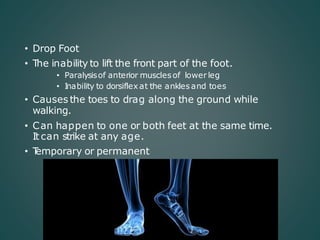
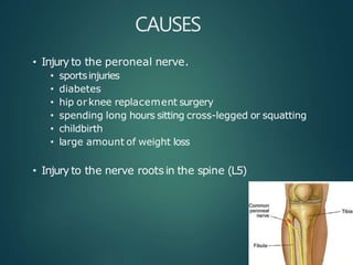
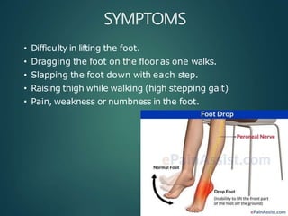
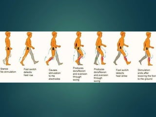
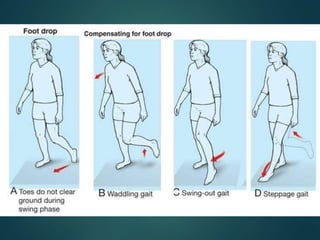
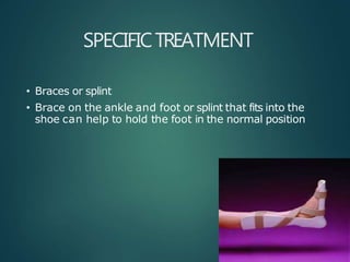
FOOT DROP
• Drop Foot
• T
he inability to lift the front part of the foot.
• Paralysisof anterior muscles of lower leg
• Inability to dorsiflex at the ankles and toes
• Causes the toes to drag along the ground while
walking.
• Can happen to one or both feet at the same time.
It can strike at any age.
• T
emporary or permanent
CAUSES
• Injury to the peroneal nerve.
• sportsinjuries
• diabetes
• hip orknee replacement surgery
• spending long hours sitting cross-legged or squatting
• childbirth
• large amount of weight loss
• Injury to the nerve roots in the spine (L5)
• Neurological conditions that can contribute to foot
drop include:
• stroke
• multiple sclerosis(MS)
• cerebral palsy
• Charcot-Marie-T
ooth disease
• Conditions that cause the muscles to progressively
weaken or deteriorate may cause foot drop:
• muscular dystrophy
• amyotrophic lateral sclerosis (Lou Gehrig’s disease)
• polio
• Rupture of Anterior Tibialis
• Fracture of fibula
• Compartment Syndrome
• Diabetes
• Alcohol Abuse
VULNERABILITYOF PERONEAL NERVE
• Funiculi of the peroneal nerve - larger and less
connective tissue
• Fewer autonomic fibers, so in any injury, motor and
sensory fibers bear the brunt of the trauma.
• More superficial course, especially at the fibular
neck
• Adheres closely to the periosteum of the proximal
fibula
MUSCLES
• DORSIFLEXORS
TIBIALISANTERIOR
EXTENSOR HALLUCIS LONGUS
EXTENSOR DIGITORUM LONGUS
PERONEUS TERTIUS
• EVERTORS
PERONEUS LONGUS
PERONEUS BREVIS
SYMPTOMS
• Difficulty in lifting the foot.
• Dragging the foot on the floor as one walks.
• Slapping the foot down with each step.
• Raising thigh while walking (high stepping gait)
• Pain, weakness or numbness in the foot.
GAITCYCLE
• Swing phase (SW): The period of time when the foot
isnot in contact with the ground. Inthose cases
where the foot never leaves the ground (foot drag)
- phase when all portions of the foot are in forward
motion.
• Initial contact (IC): when the foot initially makes
contact with the ground; representsbeginning of
the stance phase - foot strike.
• T
erminal contact (T
C): when the foot leaves the
ground -end of the stance phase or beginning of
the swing phase - foot off. .
FOOTDROP
• Drop foot SW: Greater flexion at the knee to
accommodate the inability to dorsiflex - stair
climbing movement.
• Drop foot IC: Instead of normal heel-toe foot strike,
foot may either slap the ground or the entire foot
may be planted on the ground all at once.
• Drop foot TC: T
erminal contact isquite different -
inability to support theirbody weight –walker can
be used
IMAGING
• X-Ray
Post-T
raumatic - tibia/fibula and ankle - any
bony injury.
Anatomic dysfunction (eg. Charcot joint)
• Ultrasonography
If bleeding issuspected in a patient with a hip
or knee prosthesis
• Magnetic Resonance Neurography
T
umor or a compressive mass lesion to the
peroneal nerve
ELECTROMYELOGRAM
• Thisstudy can confirm the type of neuropathy,
establish the site of the lesion, estimate extent of
injury
, and provide a prognosis.
• Sequential studies are useful to monitor recovery
of acute lesions.
TREATMENT
• Depends on the underlying cause.
• If cause is successfully treated foot drop may
improve or even disappear.
• Medical treatment - Painful Paresthesia
• Sympathetic block
• Amitriptyline
• Nortriptyline
• Pregabalin
• Laproscopic Synovectomy
SPECIFICTREATMENT
• Braces or splint
• Brace on the ankle and foot or splint that fits into the
shoe can help to hold the foot in the normal position
PHYSICAL THERAPY
• Exercises that strengthen the leg muscles
• Maintain the range of motion in knee and ankle
• Improve gait problems associated with foot drop.
NERVE STIMULATION
Stimulating the nerve (peroneal nerve) improves foot
drop especially if it caused by a stroke.
SURGICAL REPAIR
• Foot drop due to direct trauma to the dorsiflexors
generally requires surgical repair
.
• When nerve insult isthe cause - restore the nerve
continuity - nerve grafting or repair.
• If there isno significant neuronal recovery at one
year - tendon transfer maybe considered.
• Bridal procedure
• Neurotendinous transpositon
BRIDALSPROCEDURE
• Tendon to bone attachment - posterior tibial tendon is
attatched to the second cuneiform bone.
• T
endon to tendon attachment
NEUROTENDINOUS
TRANSPOSITION
• Lateral head of gastronemius istransposed to the
tendons of the anterior muscle group with simultaneous
transposition of the proximal end of deep peroneal
nerve.
• T
he nerve issutured to the motor nerve of the
gartronemius
• Active voluntary dorsiflexion of foot
• AFTER TENDON TRANSFER
Cast and Non-Weight Bearing ambulation for 6
weeks
• PHYSIOTHERAPY
T
o correct gait abnormalities
• CHRONIC AND CONTRACTURE CASES
Achilles tendon lengthening
• Inpatientswhom foot drop isdue to neurologic and
anatomic factors (polio, charcot joint ) - Arthodesis
• Subtalar Stabilising procedure or Triple Arthodesis can be
done.
COMPLICATIONS
• Surgical procedure-wound infection may occur.
• Nerve graft failure
• Intendon transferprocedures- recurrent deformity
• Inarthrodeses or fusion procedures-pseudoarthrosis,
delayed union,or nonunion.
THANKYOU

Foot drop

  • 1.
  • 2.
    • Drop Foot •T he inability to lift the front part of the foot. • Paralysisof anterior muscles of lower leg • Inability to dorsiflex at the ankles and toes • Causes the toes to drag along the ground while walking. • Can happen to one or both feet at the same time. It can strike at any age. • T emporary or permanent
  • 3.
    CAUSES • Injury tothe peroneal nerve. • sportsinjuries • diabetes • hip orknee replacement surgery • spending long hours sitting cross-legged or squatting • childbirth • large amount of weight loss • Injury to the nerve roots in the spine (L5)
  • 4.
    • Neurological conditionsthat can contribute to foot drop include: • stroke • multiple sclerosis(MS) • cerebral palsy • Charcot-Marie-T ooth disease • Conditions that cause the muscles to progressively weaken or deteriorate may cause foot drop: • muscular dystrophy • amyotrophic lateral sclerosis (Lou Gehrig’s disease) • polio
  • 5.
    • Rupture ofAnterior Tibialis • Fracture of fibula • Compartment Syndrome • Diabetes • Alcohol Abuse
  • 6.
    VULNERABILITYOF PERONEAL NERVE •Funiculi of the peroneal nerve - larger and less connective tissue • Fewer autonomic fibers, so in any injury, motor and sensory fibers bear the brunt of the trauma. • More superficial course, especially at the fibular neck • Adheres closely to the periosteum of the proximal fibula
  • 7.
    MUSCLES • DORSIFLEXORS TIBIALISANTERIOR EXTENSOR HALLUCISLONGUS EXTENSOR DIGITORUM LONGUS PERONEUS TERTIUS • EVERTORS PERONEUS LONGUS PERONEUS BREVIS
  • 8.
    SYMPTOMS • Difficulty inlifting the foot. • Dragging the foot on the floor as one walks. • Slapping the foot down with each step. • Raising thigh while walking (high stepping gait) • Pain, weakness or numbness in the foot.
  • 9.
    GAITCYCLE • Swing phase(SW): The period of time when the foot isnot in contact with the ground. Inthose cases where the foot never leaves the ground (foot drag) - phase when all portions of the foot are in forward motion. • Initial contact (IC): when the foot initially makes contact with the ground; representsbeginning of the stance phase - foot strike. • T erminal contact (T C): when the foot leaves the ground -end of the stance phase or beginning of the swing phase - foot off. .
  • 12.
    FOOTDROP • Drop footSW: Greater flexion at the knee to accommodate the inability to dorsiflex - stair climbing movement. • Drop foot IC: Instead of normal heel-toe foot strike, foot may either slap the ground or the entire foot may be planted on the ground all at once. • Drop foot TC: T erminal contact isquite different - inability to support theirbody weight –walker can be used
  • 15.
    IMAGING • X-Ray Post-T raumatic -tibia/fibula and ankle - any bony injury. Anatomic dysfunction (eg. Charcot joint) • Ultrasonography If bleeding issuspected in a patient with a hip or knee prosthesis • Magnetic Resonance Neurography T umor or a compressive mass lesion to the peroneal nerve
  • 16.
    ELECTROMYELOGRAM • Thisstudy canconfirm the type of neuropathy, establish the site of the lesion, estimate extent of injury , and provide a prognosis. • Sequential studies are useful to monitor recovery of acute lesions.
  • 17.
    TREATMENT • Depends onthe underlying cause. • If cause is successfully treated foot drop may improve or even disappear. • Medical treatment - Painful Paresthesia • Sympathetic block • Amitriptyline • Nortriptyline • Pregabalin • Laproscopic Synovectomy
  • 18.
    SPECIFICTREATMENT • Braces orsplint • Brace on the ankle and foot or splint that fits into the shoe can help to hold the foot in the normal position
  • 19.
    PHYSICAL THERAPY • Exercisesthat strengthen the leg muscles • Maintain the range of motion in knee and ankle • Improve gait problems associated with foot drop.
  • 20.
    NERVE STIMULATION Stimulating thenerve (peroneal nerve) improves foot drop especially if it caused by a stroke.
  • 21.
    SURGICAL REPAIR • Footdrop due to direct trauma to the dorsiflexors generally requires surgical repair . • When nerve insult isthe cause - restore the nerve continuity - nerve grafting or repair. • If there isno significant neuronal recovery at one year - tendon transfer maybe considered. • Bridal procedure • Neurotendinous transpositon
  • 22.
    BRIDALSPROCEDURE • Tendon tobone attachment - posterior tibial tendon is attatched to the second cuneiform bone. • T endon to tendon attachment
  • 23.
    NEUROTENDINOUS TRANSPOSITION • Lateral headof gastronemius istransposed to the tendons of the anterior muscle group with simultaneous transposition of the proximal end of deep peroneal nerve. • T he nerve issutured to the motor nerve of the gartronemius • Active voluntary dorsiflexion of foot
  • 24.
    • AFTER TENDONTRANSFER Cast and Non-Weight Bearing ambulation for 6 weeks • PHYSIOTHERAPY T o correct gait abnormalities • CHRONIC AND CONTRACTURE CASES Achilles tendon lengthening • Inpatientswhom foot drop isdue to neurologic and anatomic factors (polio, charcot joint ) - Arthodesis • Subtalar Stabilising procedure or Triple Arthodesis can be done.
  • 25.
    COMPLICATIONS • Surgical procedure-woundinfection may occur. • Nerve graft failure • Intendon transferprocedures- recurrent deformity • Inarthrodeses or fusion procedures-pseudoarthrosis, delayed union,or nonunion.
  • 26.