Oe.om,mastoiditis
•Download as PPTX, PDF•
1 like•71 views

otitis externa, otitis media ,furuncle, mastoiditis

Recommended
More Related Content
What's hot
What's hot (20)
Similar to Oe.om,mastoiditis
Similar to Oe.om,mastoiditis (20)
Recently uploaded
Recently uploaded (20)
Oe.om,mastoiditis
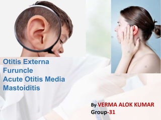
- 1. Otitis Externa Furuncle Acute Otitis Media Mastoiditis By-VERMA ALOK KUMAR Group-31
- 4. Anatomy and Physiology • Consists of the auricle and EAM • Skin-lined • Approximately 2.5 cm in length • Ends at tympanic membrane • Auricle is mostly skin-lined cartilage • External auditory meatus – Cartilage: ~40%, Bony: ~60% – S-shaped, Narrowest portion at bony-cartilage junction
- 5. Anatomy and Physiology • EAC is related to various contiguous structures – Tympanic membrane – Mastoid – Glenoid fossa – Cranial fossa – Infra-temporal fossa
- 6. • Innervation: cranial nerves V ,VII, IX, X, and greater auricular nerve • Arterial supply: superficial temporal, posterior and deep auricular branches • Venous drainage: superficial temporal and posterior auricular veins • Lymphatics
- 7. OTITIS EXTERNA • An acute or chronic infection of the whole or a part of the skin of the external ear canal • Otitis externa is often referred to as "swimmer’s ear" because repeated exposure to water can make the ear canal more vulnerable to inflammation
- 8. Etiology
- 9. Speculum findings: • the canal may be so swollen that a view into the ear is impossible • In swimmers, divers and surfers, chronic water exposure can lead to the growth of bony swellings in the canal known as exostoses. These can interfere with the drainage of wax and predispose to infection.
- 10. Organisms 1. Pseudomonas species 2. Staphylococci 3. Streptococci/Gram negative rods 4. Fungi (Aspergillus/Candida species)
- 11. Acute Otitis Externa (AOE) • “swimmer’sear” • Pre-inflammatory stage • Acute inflammatory stage • Mild • Moderate • Severe
- 12. Factors contributing to AOE • High humidity • Water exposure • Maceration of canal skin • High environmental temperature • Local trauma • Perspiration • Allergy • Stress • Removal of normal skin lipids • Absence of cerumen • Alkaline pH of canal
- 13. AOE: Pre-inflammatory Stage • Oedema of stratum corneum and plugging of apopilosebaceous unit • Symptoms: pruritus and sense of fullness • Signs: mild edema • Starts the itch/scratch cycle
- 14. AOE: Mild to Moderate Stage • Progressive infection • Symptoms • Pain • Increased pruritus • Signs • Erythema • Increasing edema • Canal debris, discharge
- 15. AOE: Severe Stage • Severe pain, worse with ear movement • Signs • Lumen obliteration • Purulent otorrhoea • Involvement of peri- auricular soft tissue
- 16. AOE: Treatment • Most common pathogens: P. aeruginosa and S. aureus, E.coli and proteus. • Four principles • Frequent canal cleaning; swab or suction • With sever EO, placement of a wick made of sponge or gauze provides a pathway for drops to be delivered to the EAC wall skin for 48-72 hours! • Topical antibiotics, and if severe>> Systemic or PO-ABT • Pain control • Instructions for prevention
- 17. AT A GLANCE. . . • Otalgia • Tenderness on palpation or manipulation (Tragus sign) • Ear fullness • Conductive hearing loss. • Erythema of meatus and canal • Swelling and obstruction of canal • Crusting and discharge • Odor!
- 18. Necrotizing (malignant) External Otitis(NEO) • Potentially lethal infection of EAC and surrounding structures • Pseudomonas aeruginosa is the usual culprit • Risk Factors: - Diabetes Mellitus - Elderly - Immunocompromised state - Human Immunodeficiency Virus (HIV) • Typically seen in diabetics and immunocompromised patients
- 19. NEO: Signs & Symptoms • Similar to Otitis Externa except • Severe, unrelenting Ear Pain and Headache • Persistent discharge • Does not respond to topical medications • Commonly associated with Diabetes Mellitus • Granulation tissue in posterior and inferior canal • Pathognomonic for necrotizing otitis • Occurs at bone-cartilage junction • Extra-auricular findings • Cervical Lymphadenopathy • Trismus (TMJ involvement) • Facial Nerve Palsy or paralysis • (Bell's Palsy) • Associated with poor prognosis
- 20. NEO: Diagnosis, Prevention and Treatment: • Prognosis; Reportedly mortality 20-53% • Diagnosis : History, Physical Examn, Labs and Imaging: - Labs; CBC, Culture of discharge, ESR, Serum glucose, Serum creatinine. - Radiology; CT, or MRI (ear) • Prevention: - Avoid use of cotton swabs in ear and other canal trauma. - Use caution when irrigating ear of high risk patients. - Treat eczema of ear canal and other pruritic dermatitis
- 21. NEO: Treatment • Intravenous antibiotics for at least 4 weeks – with serial gallium scans monthly • Local canal debridement until healed • Pain control • Use of topical agents controversial • Hyperbaric oxygen experimental • Surgical debridement for refractory cases
- 22. NEO: Mortality • Death rate essentially unchanged despite newer antibiotics (37% to 23%) • Higher with multiple cranial neuropathies (60%) • Recurrence not uncommon (9% to 27%) • May recur up to 12 months after treatment
- 23. Chronic Otitis Externa • Acute otitis externa occurs in 4 of every 1000 people per year • Otitis externa is defined as chronic when the duration of the infection exceeds 4 weeks or when more than 4 episodes occur in 1 year • Bacterial, fungal, dermatological aetiologies COE: Symptoms • Unrelenting pruritus • Mild discomfort • Dryness, Crusting, and flaking of canal skin
- 24. COE: Signs • Asteatosis • Dry, flaky skin • Hypertrophied skin • Muco-purulent otorrhoea (occasional)
- 25. COE: Treatment • Similar to that of AOE • Topical antibiotics, frequent cleanings • Topical Steroids • Surgical intervention • Failure of medical treatment • Goal is to enlarge and resurface the EAC
- 26. Radiation-Induced Otitis Externa • OE occurring after radiotherapy • Often difficult to treat • Limited infection treated like COE • Involvement of bone requires surgical debridement and skin coverage
- 27. Furunculosis • Acute localized infection • Lateral 1/3 of posterosuperior canal • Obstructed apopilosebaceous unit • Pathogen: S. aureus
- 28. Furunculosis: Symptoms • Localized pain • Pruritus • Hearing loss (if lesion occludes canal)
- 29. Furunculosis: Signs • Edema • Erythema • Tenderness • Occasional fluctuance
- 30. Furunculosis: Treatment • Local heat • Analgesics • Oral anti-staphylococcal antibiotics • Incision and drainage reserved for localized abscess • IV antibiotics for soft tissue extension
- 32. Acute inflammation in middle ear < 3 weeks (1 month) Often associated with a viral upper respiratory infection Most common reason for medical therapy for children younger than 5 years Recurrent otitis media: At least 4 episodes/ year At least 3 episodes/ 6 months (with adequate therapy) Acute otitis media
- 33. Most children have at least one episode of AOM (by age 3, 50-85%) Peak incidence age 6-15 months Increased incidence in the fall and winter Only 20% are adults >700 milion cases/year Epidemiology
- 34. Eustachian tube is lined with respiratory mucosa Responds together with nasopharynx mucosa Edema > narrowed lumen negative middle ear pressure Influx of pathogens from nasopharynx is possible Causes >
- 35. Causes Inflammatory response in middle ear worsens the obstruction Trigger: Allergies Upper respiratory tract infections GER (especially children) Adenoid hypertrophy Other
- 36. Viral (30-70%) RSV Rhinovirus Coronavirus Influenza, parainfluenza Bacterial (55%) Streptococcus pneumoniae (44%) Haemophilus influenzae (41%) Moraxella catarrhalis (14%) Gram negative enteric bacteria S. Aureus • Combined (15%) Causes
- 37. Age: <7 Their Eustachian tubes are short, floppy, horizontal and poorly functioning Risk factors
- 38. Handbook of Pediatric Otolaryngology : A Practical Guide for Evaluation and Management of Pediatric Ear, Nose, and Throat Disorders
- 39. Risk factors Genetic predisposition Eustachian tube dysfunction Allergic tendencies Bottle feeding (first 3 months) (breast milk contains lactoferrin, oligosaccharide and surface immunoglobulin A that inhibit bacterial colonization) (sucking generates negative pressure) Incorrect posture while breastfeeding
- 40. Risk factors Underlying pathology Unrepaired cleft palate Parental smoking Large familys/attending daycare Immunocompromised states
- 41. Otalgia (not always) Fever Hearing loss (speech delay for children) Headache Nausea Cough Rhinitis Conjunctivitis Signs and symptoms
- 42. Pneumatic otoscopy/otoscopy: Red or opaque eardrum Retracted eardrum Immobile or hypo-mobile eardrum Presence of fluid behind eardrum (purulent, serous, mucoid) Retraction pockets Bullous myringitis Physical Examination
- 45. Otorrhea (in case of tympanostomy tube, perforation) Mastoid tenderness Anteriorly rotated pinna Tympanometry Audiometry Inspection or pharynx and nasal cavity Physical Examination
- 46. Diagnosis Acute onset of signs and syptoms The presence of middle ear effusion (hypomobile eardrum, air-fluid level) Signs and symptoms of middle ear inflamation (erythema, otalgia)
- 47. Acute mastoiditis Abscess formation Facial paralysis Otitis media with effusion PersistentAOM RecurrentAOM Hearing loss Perforation of eardrum Complications
- 48. Complications (rare) Lateral sinus thrombosis Otitic hydrocephalus Septic shock Meningitis Encephalitis Extradural abscess Labyrinthitis
- 49. Antibacterial therapy for: Children of age <6months 6 months to 2 years with severe illness Recurrent or billateral AOM Immunocompromised patients Patients with a perforated tympanic membrane Pain management (Ibuprofen, Diclofenac, paracetamol) Decongestants and/or antihistamines, nasal steroids Treatment
- 50. Antibacterial therapy Amoxicilin 750-1500mg/day 50-100 mg/kg/day (has not recived amoxicilin in past 30 days and has no allergy to penicilin) Amoxicillin-clavulanate 875/125mg/day 90/6.4 mg/kg/day (alternative for amoxicilin) Ceftriaxone 1-2g/day 50mg/kg/day or Cefuroxim 500mg/day 30mg/kg/day Azithromycin, clarithromycin, erythromycin in case of allergy to penicilin 5-7-10 days
- 52. Non-drug Treatment Myringotomy in case of sevare pain Tympanocentesis in case of severe pain andas a diagnostic procedure if there is no improvement with • 2nd line of antibiotics (local anesthesia) (narcosis)
- 53. Avoiding risk factors if possible Vaccination: ? S. Pneumonia (PCV-7) Influenza • Adenoidectomy • Polipectomy Preventive measures
- 54. Differential diagnosis Otitis externa Impacted cerumen or foreign body in ear Tympanosclerosis Otitis media with effusion Injury of the ear
- 55. MASTOIDITIS
- 56. • INTRODUCTION • The mastoid process is the portion of the temporal bone of the skull that is behind the ear which contains open, air-containing spaces. • DEFINITION • It is an inflammation of the mastoid process behind the ear and of the air space connecting it to the cavity of the middle ear.
- 57. • CLASSIFICATION 1.Acute mastoiditis: It is a rare complication of acute otitis media. 2.Chronic mastoiditis: It is most commonly associated with CSOM or with cholesteatoma formations. • CAUSES & RISK FACTORS 1.Infection of the middle ear. 2.Injury of the mastoid bones and cells. 3. Cholesteatoma. 4.Upper respiratory infection.
- 58. SIGNS AND SYMPTOMS 1.Otalgia. 2.Swelling on the mastoid bone. 3.Perforation of the ear drum. 4.Loss of hearing. 5.Severe pain at eating time. 6.Increased cranial pressure. 7.Painless discharge from the affected ear. 8.Otorrhoea(purulent discharge) may be odorless or foul smelling. 9.Nausea, vomiting.
- 59. PATHOPHYSIOLOGY
- 60. DIAGNOASTIC EVALAUTION 1.History collection. 2.Physical examination. 3.Mastoid bone x ray. 4. CT scan. 5.Lab: CBC, DLC, Blood culture, tympanocentesis. 6.Audiography.
- 61. Treatment • MEDICAL MANAGEMENT 1. Antibiotic and steroid eardrop for infection and inflammation, e.g. Ciplox-D. 2. Ear-irrigation: For removing purulentdischarge. 3. Analgesics drugs: Aspirin, Nimuslide. • SURGICAL MANAGEMENT 1. Mastoidectomy: It is a surgical procedure that removes diseases mastoid air cells. 2. Myringotomy: It is a surgical procedure in which a tiny incision is created in the eardrum relieves pressure caused by excessive buildup of fluid orpus. 3. Tympanoplasty: also called eardrum repair. It is the surgical reconstruction of the perforated eardrum or the small bones of the middle ear.