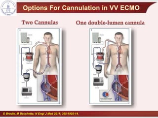
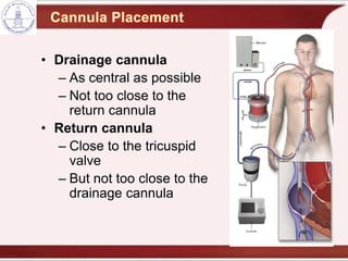
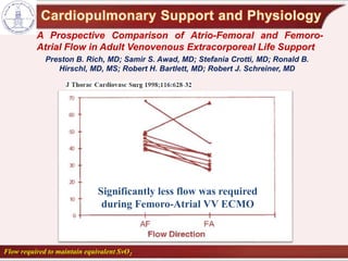
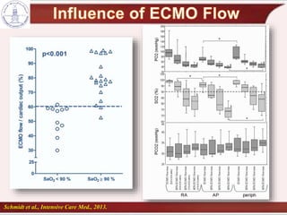
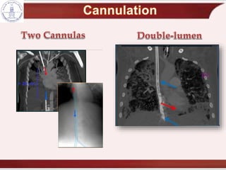
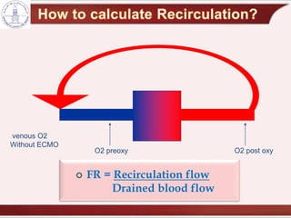
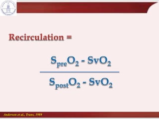
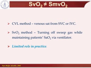
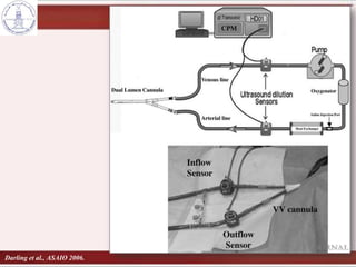
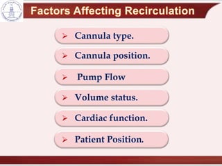
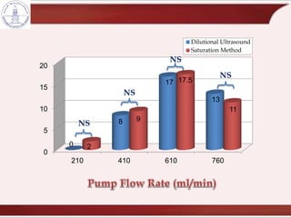
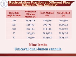
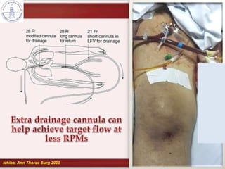
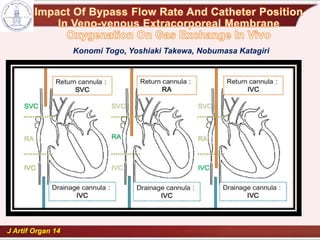
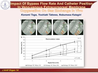
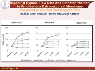
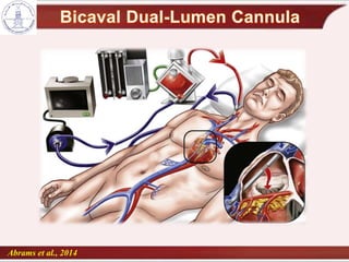
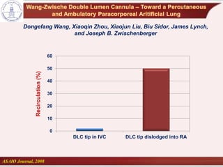
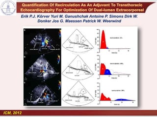
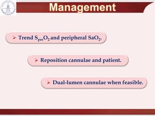
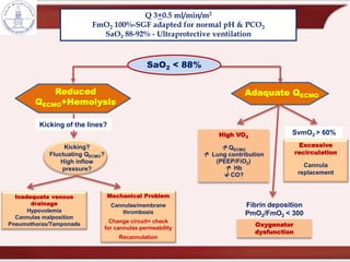
Assistant Professor
Critical Care Medicine
Cairo University
D Brodie, M Bacchetta; N Engl J Med 2011; 365:1905-14.
• Drainage cannula
– As central as possible
– Not too close to the
return cannula
• Return cannula
– Close to the tricuspid
valve
– But not too close to the
drainage cannula
Significantly less flow was required
during Femoro-Atrial VV ECMO
A Prospective Comparison of Atrio-Femoral and Femoro-
Atrial Flow in Adult Venovenous Extracorporeal Life Support
Preston B. Rich, MD; Samir S. Awad, MD; Stefania Crotti, MD; Ronald B.
Hirschl, MD, MS; Robert H. Bartlett, MD; Robert J. Schreiner, MD
Flow required to maintain equivalent SvO2
Cannulation in
jugular vein
not possible.
Higher risk for
femoral
vein/caval
thrombosis(?)
Schmidt et al., Intensive Care Med., 2013.
Pedersen et al., Ann Thorac Surg 1997
Q =
DP p r4
8 h L
Flow is proportional to the power of 4 of radius
inversely proportional to tubing length and viscosity
1797-1869
Joseph-Frédéric-Benoît Charrière 1803-1876
1 Charrière = 1 Fr = 1/3 mm
29 Fr.
25 Fr.
23 Fr.
21 Fr.
19 Fr.Drainage cannula
23 Fr.
21 Fr.
19 Fr.
17 Fr.15 Fr.
Return cannula
Admission
Return
– Less Recirculation.
– Single access.
– Possible ambulation.
– Bigger cannula and
smaller lumen.
– Image guidance is
mandatory.
Mobilization is possible .
It probably reduces critical
illness polyneuropathy,
delirium and muscle
atrophy.
It may reduce time on
ventilation and improve
outcome post lung
transplantation.
 Size: 24 Fr.
 Bloodflow approx. 2.0 L/min.
 Recirculation.
 Indication: CO2 removal.
 Also available: 18 Fr, 22 Fr jugular.
 188 cannulation attempts.
 11 cannulation failures.
 3 arterial punctures.
• One leading to distal necrosis.
 1 SVC laceration VCS.
 1 fatal hemothorax.
• SVC perforation by Reinfusion Cannula.
Thomas Pranikoff, MD; Ronald B. Hirschl, MD’; ‘Robert Remenapp, RRT;
Fresca Swaniker, MD and Robert H. Bartlett, MD, FCCP
Chest 1999; 115:818-822.
Transesophageal Echocardiographic Guided Placement of a
Right Internal Jugular Dual-Lumen Venovenous Extracorporeal
Membrane Oxygenation (ECMO) Catheter
Mazzeffi M J Cardiothorac Vasc Anesth, 2013
Mid-esophageal four-chamber TEE view
with white arrow showing improperly
positioned cannula in the right ventricle.
Modified mid-esophageal bicaval TEE
view using color Doppler compare mode
showing return blood flow in the center of
the right atrium directed towards the
tricuspid valve. (Color version of figure is
available online).
Dolch et al, ASAIO, 2011.
Daniel Hind, Neill Calvert, Richard McWilliams, Andrew Davidson,
Suzy Paisley, Catherine Beverley, Steven Thomas
 Venous thrombosis in 25 patients.
 Fatal PE in 2 patients.
 “The true incidence of thromboembolic
events is highly underestimated by clinical
evaluation”.
Rastan AJ et al., Int J Artif Organs 2006; 29:1121-31.
infection
Air
Blood flow / drainage.
Fraction of oxygen delivered through membrane.
Membrane function / efficiency.
Recirculation.
Abrams et al, ASAIO 2014
 Proportion of oxygenated blood captured by the
ECMO system and reinjected into the ECMO circuit,
instead of being pumped
 Does not share in patient oxygenation
O2 post oxy
venous O2
Without ECMO
O2 preoxy
 FR = Recirculation flow
Drained blood flow
Anderson et al., Trans, 1989
 CVL method – venous sat from SVC or IVC.
 SvO2 method – Turning off sweep gas while
maintaining patients’ SaO2 via ventilator.
 Limited role in practice.
Van Heijst ASAIO, 2001
Darling et al., ASAIO 2006.
 Cannula type.
 Cannula position.
 Pump Flow
 Volume status.
 Cardiac function.
 Patient Position.
0
5
10
15
20
210 410 610 760
0
8
17
13
2
9
17.5
11
Dilutional Ultrasound
Saturation Method
NS
NS
NS
NS
CVL Method
(%)
SvO2 Method
(%)
Ultrasound
Dilution
Method (%)
Flow Rate
(ml/kd – min)
62.5+6.945.0+6.936.0+12.8150
54.8+13.539.3+9.532.8+9.3125
39.1+17.631.7+8.224.6+9.4100
29.5+15.724.5+12.620.2+6.575
29.6+15.712.4+10.013.0+4.050
Ichiba, Ann Thorac Surg 2000
J Artif Organ 14
Konomi Togo, Yoshiaki Takewa, Nobumasa Katagiri
J Artif Organ 14
Konomi Togo, Yoshiaki Takewa, Nobumasa Katagiri
J Artif Organ 14
Konomi Togo, Yoshiaki Takewa, Nobumasa Katagiri
Massimo Bonacchi, MD; Guy Harmelin, MD; Adriano Peris, MD
and Guido Sani, MD
The Journal of Thoracic and Cardiovascular Surgery, November 2011
Massimo Bonacchi, MD; Guy Harmelin, MD; Adriano Peris, MD
and Guido Sani, MD
The Journal of Thoracic and Cardiovascular Surgery, November 2011
RCT of 30 patients with severe ARDS
X-configuration (n = 16)
Vs
Standard 2-site setup (n = 14)
Higher SaO2
Facilitated less ventilator support
Recirc at 4L/min: 8.4% vs 37.9%
∆ Recirc per L/min: 2.9% vs 11.1%
Lindstrom, perfusion 2012
Oxygenated
return blood
Tricuspid valve
prevents
recirculation
Venous drainage
(to circuit)
Mixed drainage
(to circuit)
Recirculation
Effective flow
Oxygenated
return blood
Abrams et al., 2014
Dongefang Wang, Xiaoqin Zhou, Xiaojun Liu, Biu Sidor, James Lynch,
and Joseph B. Zwischenberger
ASAIO Journal, 2008
0
10
20
30
40
50
60
DLC tip in IVC DLC tip dislodged into RA
Recirculation(%)
ICM, 2012
Erik P.J. Körver Yuri M. Ganushchak Antoine P. Simons Dirk W.
Donker Jos G. Maessen Patrick W. Weerwind
 Trend SpreO2 and peripheral SaO2.
 Reposition cannulae and patient.
 Dual-lumen cannulae when feasible.
Q 3+0.5 ml/min/m2
FmO2 100%-SGF adapted for normal pH & PCO2
SaO2 88-92% - Ultraprotective ventilation
Reduced
QECMO+Hemolysis
Adaquate QECMO
SaO2 < 88%
SvmO2 > 60%
Excessive
recirculation
Cannula
replacement
Fibrin deposition
PmO2/FmO2 < 300
Oxygenator
dysfunction
High VO2
 QECMO
 Lung contribution
(PEEP/FiO2)
 Hb
 CO?
Kicking?
Fluctuating QECMO?
High inflow
pressure?
Kicking of the lines?
Mechanical Problem
Cannulas/membrane
thrombosis
Change circuit+ check
for cannulas permeability
Recannulation
Inadequate venous
drainage
Hypovolemia
Cannulas malposition
Pneumothorax/Tamponade
 Cannula Choice:
 The bigger the better.
 Careful positioning preferably
ultrasound gided.
 Transesophageal echocardiography or
fluoroscopic guidance is advisable (esp.
Avalon).
 Good monitoring to avoid complications.
 Recirculation compromises ECMO efficiency
 Ultrasound dilution may help quantify
recirculation.
 Development of bicaval dual-lumen cannulae
have helped minimize recirculation.
 Efforts to decrease recirculation are helpful in
maximizing oxygen delivery.
cannulation and recirculation in vv ecmo

cannulation and recirculation in vv ecmo

  • 1.
    Assistant Professor Critical CareMedicine Cairo University
  • 3.
    D Brodie, MBacchetta; N Engl J Med 2011; 365:1905-14.
  • 4.
    • Drainage cannula –As central as possible – Not too close to the return cannula • Return cannula – Close to the tricuspid valve – But not too close to the drainage cannula
  • 6.
    Significantly less flowwas required during Femoro-Atrial VV ECMO A Prospective Comparison of Atrio-Femoral and Femoro- Atrial Flow in Adult Venovenous Extracorporeal Life Support Preston B. Rich, MD; Samir S. Awad, MD; Stefania Crotti, MD; Ronald B. Hirschl, MD, MS; Robert H. Bartlett, MD; Robert J. Schreiner, MD Flow required to maintain equivalent SvO2
  • 8.
    Cannulation in jugular vein notpossible. Higher risk for femoral vein/caval thrombosis(?)
  • 10.
    Schmidt et al.,Intensive Care Med., 2013.
  • 11.
    Pedersen et al.,Ann Thorac Surg 1997
  • 12.
    Q = DP pr4 8 h L Flow is proportional to the power of 4 of radius inversely proportional to tubing length and viscosity 1797-1869
  • 13.
  • 14.
    29 Fr. 25 Fr. 23Fr. 21 Fr. 19 Fr.Drainage cannula
  • 15.
    23 Fr. 21 Fr. 19Fr. 17 Fr.15 Fr. Return cannula
  • 18.
  • 19.
    – Less Recirculation. –Single access. – Possible ambulation. – Bigger cannula and smaller lumen. – Image guidance is mandatory.
  • 22.
    Mobilization is possible. It probably reduces critical illness polyneuropathy, delirium and muscle atrophy. It may reduce time on ventilation and improve outcome post lung transplantation.
  • 23.
     Size: 24Fr.  Bloodflow approx. 2.0 L/min.  Recirculation.  Indication: CO2 removal.  Also available: 18 Fr, 22 Fr jugular.
  • 26.
     188 cannulationattempts.  11 cannulation failures.  3 arterial punctures. • One leading to distal necrosis.  1 SVC laceration VCS.  1 fatal hemothorax. • SVC perforation by Reinfusion Cannula. Thomas Pranikoff, MD; Ronald B. Hirschl, MD’; ‘Robert Remenapp, RRT; Fresca Swaniker, MD and Robert H. Bartlett, MD, FCCP Chest 1999; 115:818-822.
  • 27.
    Transesophageal Echocardiographic GuidedPlacement of a Right Internal Jugular Dual-Lumen Venovenous Extracorporeal Membrane Oxygenation (ECMO) Catheter Mazzeffi M J Cardiothorac Vasc Anesth, 2013 Mid-esophageal four-chamber TEE view with white arrow showing improperly positioned cannula in the right ventricle. Modified mid-esophageal bicaval TEE view using color Doppler compare mode showing return blood flow in the center of the right atrium directed towards the tricuspid valve. (Color version of figure is available online).
  • 28.
    Dolch et al,ASAIO, 2011.
  • 29.
    Daniel Hind, NeillCalvert, Richard McWilliams, Andrew Davidson, Suzy Paisley, Catherine Beverley, Steven Thomas
  • 34.
     Venous thrombosisin 25 patients.  Fatal PE in 2 patients.  “The true incidence of thromboembolic events is highly underestimated by clinical evaluation”. Rastan AJ et al., Int J Artif Organs 2006; 29:1121-31.
  • 35.
  • 37.
    Blood flow /drainage. Fraction of oxygen delivered through membrane. Membrane function / efficiency. Recirculation.
  • 38.
    Abrams et al,ASAIO 2014  Proportion of oxygenated blood captured by the ECMO system and reinjected into the ECMO circuit, instead of being pumped  Does not share in patient oxygenation
  • 40.
    O2 post oxy venousO2 Without ECMO O2 preoxy  FR = Recirculation flow Drained blood flow
  • 41.
    Anderson et al.,Trans, 1989
  • 42.
     CVL method– venous sat from SVC or IVC.  SvO2 method – Turning off sweep gas while maintaining patients’ SaO2 via ventilator.  Limited role in practice. Van Heijst ASAIO, 2001
  • 44.
    Darling et al.,ASAIO 2006.
  • 45.
     Cannula type. Cannula position.  Pump Flow  Volume status.  Cardiac function.  Patient Position.
  • 46.
    0 5 10 15 20 210 410 610760 0 8 17 13 2 9 17.5 11 Dilutional Ultrasound Saturation Method NS NS NS NS
  • 47.
    CVL Method (%) SvO2 Method (%) Ultrasound Dilution Method(%) Flow Rate (ml/kd – min) 62.5+6.945.0+6.936.0+12.8150 54.8+13.539.3+9.532.8+9.3125 39.1+17.631.7+8.224.6+9.4100 29.5+15.724.5+12.620.2+6.575 29.6+15.712.4+10.013.0+4.050
  • 48.
  • 49.
    J Artif Organ14 Konomi Togo, Yoshiaki Takewa, Nobumasa Katagiri
  • 50.
    J Artif Organ14 Konomi Togo, Yoshiaki Takewa, Nobumasa Katagiri
  • 51.
    J Artif Organ14 Konomi Togo, Yoshiaki Takewa, Nobumasa Katagiri
  • 52.
    Massimo Bonacchi, MD;Guy Harmelin, MD; Adriano Peris, MD and Guido Sani, MD The Journal of Thoracic and Cardiovascular Surgery, November 2011
  • 53.
    Massimo Bonacchi, MD;Guy Harmelin, MD; Adriano Peris, MD and Guido Sani, MD The Journal of Thoracic and Cardiovascular Surgery, November 2011
  • 54.
    RCT of 30patients with severe ARDS X-configuration (n = 16) Vs Standard 2-site setup (n = 14) Higher SaO2 Facilitated less ventilator support
  • 55.
    Recirc at 4L/min:8.4% vs 37.9% ∆ Recirc per L/min: 2.9% vs 11.1% Lindstrom, perfusion 2012 Oxygenated return blood Tricuspid valve prevents recirculation Venous drainage (to circuit) Mixed drainage (to circuit) Recirculation Effective flow Oxygenated return blood
  • 56.
  • 57.
    Dongefang Wang, XiaoqinZhou, Xiaojun Liu, Biu Sidor, James Lynch, and Joseph B. Zwischenberger ASAIO Journal, 2008 0 10 20 30 40 50 60 DLC tip in IVC DLC tip dislodged into RA Recirculation(%)
  • 58.
    ICM, 2012 Erik P.J.Körver Yuri M. Ganushchak Antoine P. Simons Dirk W. Donker Jos G. Maessen Patrick W. Weerwind
  • 59.
     Trend SpreO2and peripheral SaO2.  Reposition cannulae and patient.  Dual-lumen cannulae when feasible.
  • 60.
    Q 3+0.5 ml/min/m2 FmO2100%-SGF adapted for normal pH & PCO2 SaO2 88-92% - Ultraprotective ventilation Reduced QECMO+Hemolysis Adaquate QECMO SaO2 < 88% SvmO2 > 60% Excessive recirculation Cannula replacement Fibrin deposition PmO2/FmO2 < 300 Oxygenator dysfunction High VO2  QECMO  Lung contribution (PEEP/FiO2)  Hb  CO? Kicking? Fluctuating QECMO? High inflow pressure? Kicking of the lines? Mechanical Problem Cannulas/membrane thrombosis Change circuit+ check for cannulas permeability Recannulation Inadequate venous drainage Hypovolemia Cannulas malposition Pneumothorax/Tamponade
  • 61.
     Cannula Choice: The bigger the better.  Careful positioning preferably ultrasound gided.  Transesophageal echocardiography or fluoroscopic guidance is advisable (esp. Avalon).  Good monitoring to avoid complications.
  • 62.
     Recirculation compromisesECMO efficiency  Ultrasound dilution may help quantify recirculation.  Development of bicaval dual-lumen cannulae have helped minimize recirculation.  Efforts to decrease recirculation are helpful in maximizing oxygen delivery.