презентация Glossofaryngeas
•Download as PPT, PDF•
0 likes•109 views

презентация Glossofaryngeas

Recommended
More Related Content
What's hot
What's hot (20)
Viewers also liked
Viewers also liked (18)
Similar to презентация Glossofaryngeas
Similar to презентация Glossofaryngeas (20)
More from Ajaindu Shrivastava
More from Ajaindu Shrivastava (20)
Recently uploaded
Recently uploaded (20)
презентация Glossofaryngeas
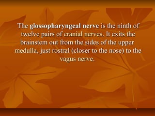
- 1. TheThe glossopharyngeal nerveglossopharyngeal nerve is the ninth ofis the ninth of twelve pairs oftwelve pairs of cranial nervescranial nerves. It exits the. It exits the brainstem out from the sides of the upperbrainstem out from the sides of the upper medullamedulla, just rostral (closer to the nose) to the, just rostral (closer to the nose) to the vagusvagus nervenerve..
- 2. FunctionsFunctions There are a number of functions of the glossopharyngeal nerve:There are a number of functions of the glossopharyngeal nerve: It receivesIt receives sensory fibressensory fibres from the posterior one-thirdfrom the posterior one-third of theof the tonguetongue, the, the tonsilstonsils, the, the pharynxpharynx, the, the middle earmiddle ear and theand the carotid bodycarotid body.. It suppliesIt supplies parasympatheticparasympathetic fibres to thefibres to the parotid glandparotid gland via thevia the otic ganglionotic ganglion.. It suppliesIt supplies motor fibresmotor fibres toto stylopharyngeus musclestylopharyngeus muscle It contributes to theIt contributes to the pharyngeal plexuspharyngeal plexus..
- 3. nucleusnucleus The glossopharyngeal nerve, being mostly sensory, does not have aThe glossopharyngeal nerve, being mostly sensory, does not have a cranial nerve nucleuscranial nerve nucleus of its own. Instead it must project into manyof its own. Instead it must project into many different structures in the brainstem:different structures in the brainstem: Solitary nucleusSolitary nucleus: Taste from the posterior one-third of the tongue and: Taste from the posterior one-third of the tongue and information frominformation from carotid baroreceptorscarotid baroreceptors andand carotid body chemoreceptorscarotid body chemoreceptors Spinal nucleus of the trigeminal nerveSpinal nucleus of the trigeminal nerve: Visceral pain as well as somatic: Visceral pain as well as somatic sensory fibers from the skin of the outer ear.sensory fibers from the skin of the outer ear. Nucleus ambiguusNucleus ambiguus: The: The lower motor neuronslower motor neurons for thefor the stylopharyngeusstylopharyngeus muscle.muscle. Inferior salivatory nucleusInferior salivatory nucleus:: ParasympatheticParasympathetic input to theinput to the parotidparotid andand mucous glandsmucous glands
- 4. PathPath From theFrom the medulla oblongatamedulla oblongata, the glossopharyngeal nerve passes laterally, the glossopharyngeal nerve passes laterally across theacross the flocculusflocculus, and leaves the skull through the central part of the, and leaves the skull through the central part of the jugular foramenjugular foramen, in a separate sheath of the, in a separate sheath of the duradura matermater, lateral to and in, lateral to and in front of the vagus and accessory nerves. Within the jugular foramen, thefront of the vagus and accessory nerves. Within the jugular foramen, the glossopharyngeal nerve forms the superior ganglion (the glossopharyngealglossopharyngeal nerve forms the superior ganglion (the glossopharyngeal neve is also associated with an inferior ganglion).neve is also associated with an inferior ganglion). In its passage through the jugular foramen, it grooves the lower border of theIn its passage through the jugular foramen, it grooves the lower border of the petrouspetrous partpart of theof the temporal bonetemporal bone; and, at its exit from the; and, at its exit from the skullskull, passes forward, passes forward between thebetween the internal jugular veininternal jugular vein andand internal carotid arteryinternal carotid artery. It descends in front of. It descends in front of the latter vessel, and beneath thethe latter vessel, and beneath the styloidstyloid processprocess and the muscles connected with it,and the muscles connected with it, to the lower border of theto the lower border of the stylopharyngeusstylopharyngeus. It then curves forward, forming an arch. It then curves forward, forming an arch on the side of the neck and lying upon theon the side of the neck and lying upon the stylopharyngeusstylopharyngeus andand middle pharyngeal constrictormiddle pharyngeal constrictor muscle. From there it passes under cover of themuscle. From there it passes under cover of the hyoglossushyoglossus muscle, and is finally distributed to themuscle, and is finally distributed to the palatine tonsilpalatine tonsil, the mucous, the mucous membrane of the fauces and base of the tongue, and the mucous glands of themembrane of the fauces and base of the tongue, and the mucous glands of the mouth.mouth.
- 5. BranchesBranches 1. Tympanic1. Tympanic 2. Stylopharyngeal2. Stylopharyngeal 3. Tonsillar3. Tonsillar 4. Nerve to carotid sinus4. Nerve to carotid sinus 5. Branches to the posterior third of tongue5. Branches to the posterior third of tongue 6. Lingual branches6. Lingual branches 7. A communicating branch to the Vagus nerve7. A communicating branch to the Vagus nerve Note: The glossopharyneal nerve contributes in theNote: The glossopharyneal nerve contributes in the formation of the pharyngeal plexus along with the vagusformation of the pharyngeal plexus along with the vagus nerve.nerve.
- 6. Testing the glossopharyngeal nerveTesting the glossopharyngeal nerve The gag reflex is absent in patients withThe gag reflex is absent in patients with damage to the glossopharyngeal nerve as it isdamage to the glossopharyngeal nerve as it is responsible for the afferent limb of the reflexresponsible for the afferent limb of the reflex
- 7. Testing the glossopharyngeal nerveTesting the glossopharyngeal nerve The gag reflex is absent in patients withThe gag reflex is absent in patients with damage to the glossopharyngeal nerve as it isdamage to the glossopharyngeal nerve as it is responsible for the afferent limb of the reflexresponsible for the afferent limb of the reflex