Download to read offline

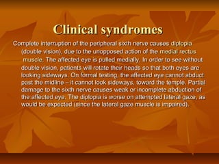


The abducens nerve controls the lateral rectus muscle of the eye and allows for sideways movement of the eye. Damage to the peripheral nerve causes double vision due to the unopposed medial rectus muscle. Damage to the abducens nucleus in the brainstem causes internuclear ophthalmoplegia, affecting both eyes simultaneously with impaired lateral movement. Cortical lesions affecting conjugate gaze are discussed elsewhere.


