
Recommended
More Related Content
What's hot
What's hot (20)
Similar to Clinical examination of the equine respiratory system
Similar to Clinical examination of the equine respiratory system (20)
Recently uploaded
Recently uploaded (20)
Clinical examination of the equine respiratory system
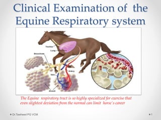
- 1. Clinical Examination of the Equine Respiratory system Dr.Tawheed PG VCM 1 The Equine respiratory tract is so highly specialized for exercise that even slightest deviation from the normal can limit horse`s career
- 2. Anatomy and Physiology Nostrils, nasal cavity ,pharynx, larynx, trachea and lungs Provides oxygen to blood, Removes waste gases CO2 Dr.Tawheed PG VCM 2 The nose is for breathing, the mouth is for eating
- 3. What can we learn Location of pathology for example :- upper v/s lower Pathophysiology for example:- obstructive v/s restrictive Etiology for example:- infectious v/s allergic v/s parasitic Dr.Tawheed PG VCM 3
- 4. What do we need to know Signalment:- (Age, Breed, sex) In examining the equine respiratory system, the only one of these that really matters is AGE Rhodococcus equi infection occurs in foals between 3 weeks to 5 month of age COPD is disease of adult horse Dr.Tawheed PG VCM 4
- 5. Chief complaints • Abnormal breathing pattern • Cough • Sneeze • Nasal discharge • Nasal rubbing • Epistaxis • Abnormal lung sounds • Lost or increase chest resonance( with percussion) • Tachypnea(increased rate) • Dyspnea(labored breathing) Dr.Tawheed PG VCM 5
- 6. Chief complaints • Noisy Breathing • Voice change • Malodorous breath • Subcutaneous emphysema • Exercise intolerance • Weight loss Dr.Tawheed PG VCM 6
- 7. History History is exceptionally important. We should make sure to note the following: • Involvement of individual or multiple animals • Onset ( slow progressive to per acute) • Duration(hrs, days, weeks, months years) • Seasonality • Association with time of feeding • Out door environment( dust, pollutants. toxins) • Hygiene of the indoor environment( tobacco smoke, ventilation, dust exposure mold,) • Anthelmintics • Recent travel/transport • Previous medication and response to treatment Dr.Tawheed PG VCM 7
- 8. Physical examination The respiratory physical exam should begin by observing the horse from a distance. This should be done in a quiet area, noting breathing pattern, stance, sneezing, coughing, respiratory rate and respiratory effort. This is ideal time to take history Dr.Tawheed PG VCM 8
- 9. First Examine the Nostrils Dr.Tawheed PG VCM 9 Flared nostrils can indicate pain or increased effort to bring in air Check both nostrils for presence of airflow and whether or not it is equal on both sides
- 10. Nostrils Dr.Tawheed PG VCM 10 Any nasal exudates should be noted. Important considerations are: unilateral/bilateral, +/- blood, color, consistency. Examine the alar fold for any abnormalities or masses
- 11. Examine the mouth Dr.Tawheed PG VCM 11 It is important to check the nostrils and mouth for odor since this is often associated with the presence of anaerobic bacteria which may be contributing to an infection. This is also a good time to listen to the horse’s breathing, noting any abnormal masses Also check the oral and nasal mucous membrane color.
- 12. Examine the Inter mandibular lymph Nodes Dr.Tawheed PG VCM 12 Palpate the inter mandibular lymph nodes, noting any enlargement or painful response to palpation.
- 13. Examine the Sinuses Dr.Tawheed PG VCM 13
- 14. Percuss the Sinuses Dr.Tawheed PG VCM 14 Percuss the sinuses. Resonance will be greatly increased if the tongue is held out of the mouth while percussing. Compare the resonance on both sides while also noting if the horse seems painful or objects to this procedure. Also check for facial symmetry, general attitude and expression
- 15. Palpate the larynx Dr.Tawheed PG VCM 15 Palpate the dorsal aspect of the larynx on both sides noting any asymmetry or muscle atrophy. If laryngeal hemiplegia is present the muscular process of the arytenoid cartilage may feel more pronounced with muscle atrophy
- 16. Auscultate and palpate the trachea Dr.Tawheed PG VCM 16 Palpate the trachea for any abnormalities such as irregular cartilage rings or fractures. The trachea can also be ausculted for any abnormal sounds.
- 17. Examine the jugular vein Dr.Tawheed PG VCM 17 Examine the jugular veins for patency and filling. This is a good estimation of hydration status along with checking skin turgor. Any distention or pulsation of the jugular veins may indicate a pleural effusion or cranial thoracic mass obstructing the return of blood flow to the heart
- 18. Examine the Ventral Thorax and Abdomen Dr.Tawheed PG VCM 18 Check for any edema along the ventral thorax and abdomen. A pleural effusion can inhibit blood flow from the ventrum of the thorax, leading to ventral edema.
- 19. Auscultate the Lungs Dr.Tawheed PG VCM 19
- 20. Auscultate the Lungs Dr.Tawheed PG VCM 20 A rebreathing bag may be used to help cause the horse to breathe more deeply when lung sounds are quiet. Make sure to hold the bag away from the nose so that it does not block the nostrils and thereby inhibit inspiration. Also note how well the horse tolerates this procedure and whether or not any coughing is elicited, as well as how quickly the horse recovers after the bag is removed.
- 21. Normal lung sounds Bronchial sound- generated in the large air ways Vesicular sounds- generated in the large airways, but heard peripherally after attenuation through aerated parenchyma Dr.Tawheed PG VCM 21
- 22. Abnormal lung sounds Changes in sound transmission Consolidated areas- lung sounds will be louder, because sounds carried more efficiently Pleural effusion- lung sounds will be quieter, but heart sounds will be louder Pneumothorax- Both lung and heart sounds will sound quieter Increased inspiratory sounds- extra thoracic or large airways obstruction Increased expiratory sounds- lower air way obstruction Other abnormal lung sounds- Crackles, wheezes Dr.Tawheed PG VCM 22
- 23. Ancillary Diagnostic test Arterial blood gas analysis Arterial blood gas determinations are the most sensitive indicator of respiratory function readily available to the clinician. Dr.Tawheed PG VCM 23
- 24. Arterial blood gas analysis • Ventilation is best assessed using PaCO2 as a guide • Decreased alveolar ventilation results in hypercapnia(respiratory acidosis, ) • Cause of low PaO2 include hypo ventilation, right to left intra pulmonary shunt, ventilation – perfusion mismatch, and decreased diffusion capacity. • The shunts responds poorly to oxygen supplementation as opposed to other cause • Ventilation perfusion-mismatch responds well to oxygen supplementation Dr.Tawheed PG VCM 24
- 25. Macroscopic tests Endoscopy The nasal passage , naso maxillary meatus opening, pharynx, larynx, trachea ,main stem bronchi, segmental and sub-segmental bronchi are accessible Endoscope ranges from 15mm to 2-3mm outer diameter Dr.Tawheed PG VCM 25
- 26. Macroscopic tests • Endoscopy Dr.Tawheed PG VCM 26
- 27. Macroscopic tests Dr.Tawheed PG VCM 27 Endoscopy Normal larynx Laryngeal hemiplagia
- 28. Macroscopic test Endoscopy Ethamoid haemotoa Fungal infection Dr.Tawheed PG VCM 28
- 29. Macroscopic test • Radiography Indication for radiography include auscultaion of adventitios sounds (wheezes,crackles, absence of sounds),abnormal percussion of the sinuses or chest, dyspnea , chronic cough, external chest trauma exercise intolerance, cyanosis Dr.Tawheed PG VCM 29
- 30. Macroscopic test • Radiography Maxillary sinus cyst Dr.Tawheed PG VCM 30
- 31. Macroscopic test • Radiography Depression fracture sinuses Dr.Tawheed PG VCM 31
- 32. Macroscopic tests Dr.Tawheed PG VCM 32 thoracic radiograph showing dorsal caudal lung disease.
- 33. Macroscopic test • Thoracic USG The USG Is an excellent technique to investigate pleural effusion, diaphragmatic herniation ,lung consolidation, atelectasis , but not broncheictasis Dr.Tawheed PG VCM 33
- 34. Macroscopic test • Thoracic USG Dr.Tawheed PG VCM 34
- 35. Macroscopic test Pulmonary Function test These tests are aimed at describing the mechanical function or gas exchange capacity of the respiratory system. PFT are indicated where there is air ways obstruction or restrictive lung disease where more characterization is required Dr.Tawheed PG VCM 35
- 36. Microscopic sampling • Respiratory cytology The indication for recovering respiratory secretions for cytological evaluation are inflammation, infection, and neoplasia In general, when infection of the lower respiratory tract is suspected, an aspirate of tracheal secretion(tracheal aspirate, tracheal wash or TTA) is indicated If disease produce is more profuse, chronic, peripherally located lesion that is non-infectious(e.g allergic, inflammatory,neoplasia) a more peripheral sampling method is indicated. This can be achieved by brochoalveolar lavage(BAL) Dr.Tawheed PG VCM 36
- 37. Microscopic sampling Dr.Tawheed PG VCM 37
- 38. Microscopic sampling Dr.Tawheed PG VCM 38
- 39. Thoracocentesis Indication for thoracocentesis is pleural effusion. The procedure should be carried out under sterile condition. The site is usually as ventral as possible at the 8-10th intercoastal space to avoid heart. If sampling and therapeu- -tic drainage is required , a large bore canula(chest tube) is placed to remove exudate. Gen- -erally less 5000 cells per microliter & protein less than 25 g/l are considered normal Dr.Tawheed PG VCM 39
- 40. Summary Dr.Tawheed PG VCM 40
- 41. Dr.Tawheed PG VCM 41