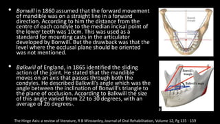
This presentation aims to shed light on the various aspects of the mandibular movement in the TMJ which is like a hinge axis. the presentation contains brief history, the various schools of thought regarding the validity and accuracy of locating the hinge axis. it talks about the various types of facebows and the various anterior and posterior points for reference employed by the arbitrary facebows.
2. • Introduction
• Definitions
• Mandibular
movements
• History
• Theories of hinge
axis
• Method of
locating hinge axis
• Conclusion
• Reference
3. Definition
• Hinge axis is defined as an imaginary line around
which the mandible may rotate within the sagittal
plane. (GPT 9)
Synonyms for hinge axis: Terminal hinge axis, Transverse axis,
Transverse horizontal axis, Transverse hinge axis
4. Mandibular movements
Rotation (hinge movement) : the action or process of rotating on
a mechanical centre or on an axis. GPT 9
Axis: a line around which a body may rotate or about which a
structure would turn if it could revolve. GPT 9
5. • Rotational movements are similar to a door hinge.
• The condyle rotates without any bodily movement.
• This happens in the lower compartment of the articular disc and the
superior surface of the condyle.
• Rotation occurs around an axis - horizontal, frontal, sagittal and can be
viewed from three reference planes - sagittal, frontal and vertical.
• Rotation in the horizontal axis occurs during mouth opening and
protrusion.
• Rotation in the frontal and sagittal axis occur as lateral movements.
6.
7. • Translation: that motion of a rigid body in which a straight line passing through any two
points always remains parallel to its initial position; the motion may be described as a
sliding or gliding motion. GPT 9
• Translatory movement: the motion of a body at any instant when all points within the
body are moving at the same velocity and in the same direction. GPT 9
• Translation occurs in the upper compartment of the articular disc and the inferior
surface of the glenoid fossa.
• In this bodily movement of the condyle occurs.
• It occurs during all lateral movements.
8. History
• The first actual kinematic location was done under the
leadership of Dr. B.B. Mc Cullum through the California
Gnathological Society and its credit for the idea was given
to Dr. Robert Harlan. The first location employed a
modified Snow facebow and it was performed for 8hrs.
New devices and concepts have evolved from this above
mentioned mechanism and its attendant theories.
• The written history of mandibular hinge axis goes back into
the first edition of Gray’s Anatomy. According to Gray and
followers the mandible moves on a hinge and forward and
lateral movements of the condyle takes place in the
glenoid fossa during movement.
The Hinge Axis: a review of literature, R B Winstanley, Journal of Oral Rehabilitation, Volume 12, Pg 135 - 159
9. • Bonwill in 1860 assumed that the forward movement
of mandible was on a straight line in a forward
direction. According to him the distance from the
centre of each condyle to the median incisal point of
the lower teeth was 10cm. This was used as a
standard for mounting casts in the articulator
developed by Bonwill. But the drawback was that the
level where the occlusal plane should be oriented
was not mentioned.
• Balkwill of England, in 1865 identified the sliding
action of the joint. He stated that the mandible
moves on an axis that passes through both the
condyles. He described Balkwill’s angle which was the
angle between the inclination of Bonwill’s triangle to
the plane of occlusion. According to Balkwill the size
of this angle varied from 22 to 30 degrees, with an
average of 26 degrees7.
The Hinge Axis: a review of literature, R B Winstanley, Journal of Oral Rehabilitation, Volume 12, Pg 135 - 159
10. • In 1899, Snow recognized the importance of hinge axis and he
contributed a facebow to transfer the axis to the articulator.
• Campione in 1905 also recognized the importance of locating
hinge axis and emphasized that, in mounting casts on an
articulator, the axis of the articulator shaft should coincide with
the mandibular transverse hinge axis of the patient.
• In 1921 Mc Collum and Stuart became convinced that
transverse axis in the condylar region about which sagittal plane
rotational movements could occur with constancy was
recordable. The scientific basis and the practical method for
locating hinge axis came through the work of Mc Collum and
Stuart in 1921 and the Gnathological Society of California and
was based on the earlier studies of Balkwill, Bennet and
Campion.
The Hinge Axis: a review of literature, R B Winstanley, Journal of Oral Rehabilitation, Volume 12, Pg 135 - 159
11. • Trapazanno and Lazari showed that more than one terminal hinge axis
existed. They used two ipsilateral styli from the same anterior clutch
supported rod. After many investigations and studies they concluded that:
• The presence of multiple hinge axis has been established
• The technique or recording terminal hinge axis requires two operators
• Relaxation of the patient during making of terminal hinge axes is essential
• Because of the presence of multiple hinge axes points, increasing or
decreasing the vertical dimension on the articulator is contraindicated
unless a new interocclusal record is made on the patient at the desired
vertical dimension
• The concept that only one terminal hinge axes exist is fallacious.
The Hinge Axis: a review of literature, R B Winstanley, Journal of Oral Rehabilitation, Volume 12, Pg 135 - 159
12. • IN 1956, Posselt stated the the hinge axis is always located within the
confines of the condylar head. Only in very few cases it approaches
the edges of the condylar head.
Terminal Hinge movement of the Mandible, Ulf Posselt; Journal of Prosthetic Dentistry, 1957
13. BIOLOGICAL SIGNIFICANCE
• It is said that there are certain proprioceptive receptors present in the
capsule of temporo-mandibular joint.
• These signals are activated only when the condyle is in the centric
relation position or the hinge position. These signals when activated
can guide the mandible during opening and closing movement. It is
for this reason that the hinge axis determination is essential.
• The capsular proprioceptors which were of only protective functional
value takes over as a guiding principle and the patient will be able to
make repeated opening and closing movements and follow the same
occlusal pattern.
Hinge Axis: Concepts, theories and clinical significance– a review, Dr. Dipankar Pal, Asian Journal of Science and Technology, 2019
14. THEORIES OF HINGE AXIS
• From the earlier experiments on hinge axis there evolved four schools
of thought:
• Absolute location of hinge axis
• Arbitrary location of hinge axis
• Non-believers in hinge axis
• Split axis rotation
A study of the Transverse axis; Arthur E. Aull, Pg. no. 469
15. ABSOLUTE LOCATION OF HINGE AXIS
This group believes in the existence and transverse hinge axis and also believes that
it can be recorded accurately. They came up with the following conclusions:
• With the use of face-bow the articulator axis can be related to the maxillary cast
in the same way as the anatomic axis is related to human maxilla.
• The mandibular hinge axis and maxillary hinge axis are coincided using a centric
relation record.
• In doing so the path of closure of mandible and the articulator will be the same.
• This helps in placement of cusps in a way that it does not collide during border
movements.
• The hinge axis is a component of every masticatory movement. This entity cannot
be disregarded
• The hinge axis relationships between the articulator and the jaw must be the
same. Otherwise the mechanical reproduction of jaw motions on the articulator
is impossible.
A study of the Transverse axis; Arthur E. Aull, Pg. no. 469
16. ARBITRARY LOCATION OF HINGE AXIS
• This group believes that accurate location of hinge axis would be of
very little value and proposes that the effortless and arbitrary location
is sufficient.
• Craddock states “But the search for the axis, in addition to being
troublesome, is of no more than academic interest, for it will never be
found to lie more than a few millimeters distant from the assumed
center in the condyle itself”.
• But according to group 1, this group fails to recognize that if the hinge
axis of the patient and the articulator are not coinciding the path of
closure will not be the same.
A study of the Transverse axis; Arthur E. Aull, Pg. no. 469
17. NON BELIEVERS
• The third group believes that hinge axis is theoretical and not
practical. It is not possible to locate hinge axis with accuracy.
• According to Lauritzen and Wolford if the terminal hinge position
could be located consistently within a radius of 1mm, this would
be more acceptable than a location with a variance of a 2 mm
radius.
• Actually, neither one is accurate, but the first is certainly more
accurate than the second. It is because the sense of perception
and delicacy of touch different for different individuals.
• Boucher commented, "The test seems to be one of the accuracy
of the machine work on the instrument rather than one of the
validity of a hinge axis registration."
A study of the Transverse axis; Arthur E. Aull, Pg. no. 470
18. SPLIT AXIS
• These were called the Transograph men.
• They believed that each condyle rotated
independently of each other.
• Slavens states, “by definition, an axis is
always a line, never a point. Again, by
definition, an axis is invariably
perpendicular to the path or plane of
rotation it controls.
• This means that the transverse axis of each
joint is a line and both of these are
perpendicular to the same plane of opening
and closing rotation”
A study of the Transverse axis; Arthur E. Aull, Pg. no. 471
19. Single transverse axis
• About 1950, Dr. William Branstad, Dr. Raymond Garvey, and the late
Dr. Robert Okey conducted an experiment to determine whether
there was one transverse axis through both condyles or an axis for
each condyle. They found that there was one transverse axis.
• Dr. Arne Lauritzen, working with a study group, repeated the same
experiment about 1957 and arrived at the same finding.
• In the fall of 1959, the Hinge Axis Committee of the Greater New York
Academy of Prosthodontics repeated this experiment and concluded
that there was only one transverse axis through both condyles.
Modern Gnathological Concepts, Victor O. Lucia; Ch 3: Hinge Axis; Pg. no 54
20. The experiment
• Clutches were cemented to the patient's teeth. A crossbar 36 inches
long was attached to the upper clutch, and another of the same
length was attached to the lower clutch.
• Four graph-lined Hags were attached to the upper bar for the purpose
of accurately locating the center of rotation.
Modern Gnathological Concepts, Victor O. Lucia; Ch 3: Hinge Axis; Pg. no 54
22. METHODS TO LOCATE HINGE AXIS
• The two popular methods of locating hinge axis are:
1. The arbitrary method
2. The kinematic method
Hinge Axis – Location, Clinical Use and Controversoes; Sunint Singh et al, Journal of Research in Dentistry, 2017
23. ARBITRARY METHOD
• Also called the Anatomic method of locating hinge axis.
• Most commonly used method because of the ease of technique.
The proponents of group 2 school of thought proposed the
arbitrary methods to locate hinge axis.
• Various anatomic landmarks were used to locate hinge axis
arbitrarily. According to them this method provided enough
accuracy for restoration of mouth, if occlusal vertical dimension
is not going to be altered to a significant extent.
24. Arbitrary Facebow
• It is the most commonly used face bow in complete denture
construction. The hinge axis is approximately located.
• The condylar rods are positioned approximately 13 mm anterior the
auditory meatus on a line running from the outer canthus of the eye
the top of the tragus also called the cantho tragal line.
• This method generally locates the rods within 5 mm of the true hinge
axis of the jaws.
• As this is an arbitrary hinge axis, errors in jaw relation may produce
occlusal discrepancies which should be corrected by minor occlusal
adjustments during insertion.
25. Facia type:
Here the hinge axis or the posterior
reference point is 13 mm anterior the
external auditory meatus and the anterior
reference point is the orbitale (midpoint of
the lower border of the orbit). The facebow
has a pointer that can be positioned at the
posterior reference point.
Earpiece type:
The posterior reference point is the
external auditory meatus and the anterior
reference point is the orbitale. The earpieces
engage into the posterior reference points
(the external auditory meatus).
26. Weinberg set up a geometric comparison using average
distances to evaluate the degree of error of axis location to
the relationship to the occlusion of the teeth. He found that
selecting the terminal hinge axis 5mm too far posteriorly with
an interocclusal record 3mm thick would produce an error of
0.1934mm at the second molar location on closure into
occlusion. The error in the anterior region would be
0.1044mm. He concluded that the transverse hinge axis
location and the subsequent face bow transfer within a 5mm
error is a practical and dependable method for orienting the
maxilla.
The transverse Hinge Axis: real or Imaginary; Lawrence A. Weinberg, 1958
27. ADVANTAGES
• Less time consuming procedure
• The technique is very simple to practice
• Uncomplicated procedure leads to reduction in of errors in location
• Records almost 5mm around the absolute location by kinematic
method which is quite considerable
• Transferring the facebow record to articulator becomes simple
• Can be used with a semi-adjustable articulator
DISADVANTAGES
• as it is not an absolute location, even an error of 5mm around true
hinge axis might lead to occlusal discrepancies, which increases the
chair side time.
28. Posterior points of referance
• Beyron point: 13mm anterior to posterior border of
centre of tragus on a line joining outer canthus of eye.
• Bergstrom point : 10mm anterior to center of
spherical insert for external auditory meatus and
7mm below FH plane
• Gysi point: This was on a line from the upper margin
of the external auditory meatus to the outer canthus
of the eye, 13 mm in front of the anterior margin of
the meatus
• Teteruck AND Lundeen’s Point: 13mm from base of
tragus to canthus.
• Snows point: 11- 13 mm anterior to the tragus.
• Denars point: 12 mm anterior to the posterior border
of tragus and 5mm inferior to the canthotragal line.
Posterior Reference Points: A Simplified Classification, Ramnath S Safiullah et al, Journal of Scientific Dentistry, 2019
29. Anterior points of reference
Orbitale
• In the skull, orbitale is the lowest point of the
infraorbital rim.
• On a patient it can be palpated through the
overlying tissue and the skin.
• One orbitale and the two posterior points that
determine the horizontal axis of rotation will define
the axis-orbital plane.
• Relating the maxillae to this plane will slightly lower
the maxillary cast anteriorly from the position that
would be established if the Frankfort horizontal
plane were used.
Wilkie, N. D. (1979). The anterior point of reference. The Journal of Prosthetic Dentistry
30. Orbitale minus 7mm
• The Frankfort horizontal plane passes through both poria and one orbital
point.
• Porion is a skull landmark, thus Sicher recommends using the midpoint of
the upper border of the external auditory meatus as the posterior cranial
landmark on a patient. Most articulators do not have a reference point
for this landmark.
• Gonzalez’ pointed out that this posterior tissue and mark on the average
lies 7 mm superior to the horizontal axis. The recommended
compensation for this discrepancy is to mark the anterior point of
reference 7 mm below orbitale on the patient or to position the orbital
pointer 7 mm above the orbital indicator of the articulator.
• Bergstrom’s’ arcon articulator automatically compensates for this error
by placing the orbital index 7 mm higher than the condylar horizontal
axis.
Anterior points of reference
Wilkie, N. D. (1979). The anterior point of reference. The Journal of Prosthetic Dentistry
31. Nasion minus 23mm
• According to Sicher,” another skull landmark, the nasion, can be
approximately located in the head as the deepest part of the
midline depression just below the level of the eyebrows.
• The nasion guide, or positioner, of the Quick Mount face-bow,
which is designed to be used with the Whip-Mix Articulator, fits
into this depression.
• This guide can be moved in and out, but not up and down, from
its attachment to the face-bow crossbar. The crossbar is located
23 mm below the midpoint of the nasion positioner.
• When the face-bow is positioned anteriorly by the nasion guide,
the crossbar will be in the approximate region of orbitale. The
face-bow crossbar and not the nasion guide is the actual
anterior reference point locator.
Anterior points of reference
Wilkie, N. D. (1979). The anterior point of reference. The Journal of Prosthetic Dentistry
32. Incisal edge plus articulator midpoint to articulator axis-
horizontal plane distance.
• Guichet” has emphasized that a logical position for the casts in the articulator
would be one which would position the plane of occlusion near the mid-
horizontal plane of the articulator.
• A deviation from this objective may position casts high or low relative to the
instrument’s upper and lower arms.
• The effect of these high or low positions may be inaccurate occlusal
relationships due to dimensional changes in the artificial stone or plaster used
for cast-mounting purposes.
Anterior points of reference
Wilkie, N. D. (1979). The anterior point of reference. The Journal of Prosthetic Dentistry
33. Alae of the Nose
• A ‘part of many complete denture techniques is to make the
tentative or the actual occlusal plane parallel with the
horizontal plane. ‘This can be achieved in two ways:
• A line from the ala of the nose to the center of the auditory
meatus describes Camper’s line.
• Knowing this, the dentist can transfer Camper’s line from the
patient to the articulator by marking the right or left ala on the
patient, setting the anterior reference pointer of the face-bow
to it, and with the face-bow, transferring the ala anteriorly and
the hinge points posteriorly, from the patient to the
articulator’s hinge or orbital indicator plane.
• A second method of establishing this relationship is to make a
wax occlusion rim parallel to Camper’s line on the face.
Anterior points of reference
Wilkie, N. D. (1979). The anterior point of reference. The Journal of Prosthetic Dentistry
34. KINEMATIC METHOD
• Two geometrical concepts are utilized:
• A line drawn through the center of a circle perpendicular
to chord meets it at its midpoint
• The line joining the center of a circle to the mid point of a
chord is perpendicular to the chord.
Wilkie, N. D. (1979). The anterior point of reference. The Journal of Prosthetic Dentistry
35. KINAMATIC FACEBOW
• This face-bow is generally used for the fabrication of fixed partial denture
and full-mouth rehabilitation.
• It is generally not used for complete denture fabrication because it requires
a long and complex procedure to record the orientation jaw relation.
• The true hinge axis should be located and marked before using the
facebow. The location of the hinge axis is then transferred to the articulator
with the face-bow.
• This face-bow requires a fully adjustable articulator to accept the true
hinge axis (THA). Since the patient's hinge axis is transferred to the
articulator, the movement of the articulator will simulate the movements
of the joint made at the terminal hinge position.
38. Clinical significance of Hinge axis
• Allows for correct recording of centric relation and its transfer to the
articulator.
• Starting point of lateral movements
• Permits a change in vertical dimension
• If transferred to articulator — teeth contact each other in the
articulator exactly as they do in the mouth.
• Helps in diagnosis and treatment planning.
39. Factors that may lead to errors in Locating
THA
• Skin Mobility
• Change in Axis
• Movement
Modern Gnathological Concepts, Victor O. Lucia; Ch 3: Hinge Axis; Pg. no 54
41. References
• The Hinge Axis: a review of literature, R B Winstanley, Journal of Oral Rehabilitation, Volume 12,
Pg 135 - 159
• Hinge Axis: Concepts, theories and clinical significance– a review, Dr. Dipankar Pal, Asian Journal
of Science and Technology, 2019
• A study of the Transverse axis; Arthur E. Aull, Pg. no. 469
• Modern Gnathological Concepts, Victor O. Lucia; Ch 3: Hinge Axis; Pg. no 54
• Hinge Axis – Location, Clinical Use and Controversoes; Sunint Singh et al, Journal of Research in
Dentistry, 2017
• The transverse Hinge Axis: real or Imaginary; Lawrence A. Weinberg, 1958
• Posterior Reference Points: A Simplified Classification, Ramnath S Safiullah et al, Journal of
Scientific Dentistry, 2019
• Wilkie, N. D. (1979). The anterior point of reference. The Journal of Prosthetic Dentistry
• Terminal Hinge movement of the Mandible, Ulf Posselt; Journal of Prosthetic Dentistry, 1957