Extra Embryonic Membranes Of Chick
•
2 likes•5,907 views

1. DEFINITION These are the membranes which do not form any part of the embryo proper but performs various functions which assist in the development of the embryo . These are discarded at the time of hatching. These membranes formed outside the embryo. 2. Types of Extra Embryonic Membranes Yolk Sac Amnion Chorion Allantois 3.Discussed Their At Time of ORIGIN It's FUNCTION After HATCHING 4. AMNIOTIC CAVITY ............................END......................................................

Recommended
More Related Content
What's hot
What's hot (20)
Similar to Extra Embryonic Membranes Of Chick
Similar to Extra Embryonic Membranes Of Chick (20)
More from DeepanshuYadav2
More from DeepanshuYadav2 (11)
Recently uploaded
Recently uploaded (20)
Extra Embryonic Membranes Of Chick
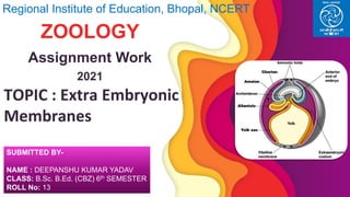
- 1. TOPIC : Extra Embryonic Membranes Regional Institute of Education, Bhopal, NCERT ZOOLOGY Assignment Work 2021 SUBMITTED BY- NAME : DEEPANSHU KUMAR YADAV CLASS: B.Sc. B.Ed. (CBZ) 6th SEMESTER ROLL No: 13
- 2. DEFINITION 2 These are the membranes which do not form any part of the embryo proper but performs various functions which assist in the development of the embryo. These are discarded at the time of hatching. These membranes formed outside the embryo.
- 3. Types of Extra Embryonic Membranes 1 2 3 4
- 4. 1
- 5. Yolk Sac ▪ From the extra embryonic splanchnopleure (endoderm on the inner and splanchnic mesoderm on the outer side) ▪ It has a wide opening into a midgut. ▪ As the development proceeds the passages of midgut is reduced to a narrow umbilical stock. ▪ Passage left on ventral side to absorb albumen. ▪ On first day mesoderm joins it and on ninth day it is fully formed. 5 ORIGIN
- 6. Yolk Sac ▪ Digest the yolk ▪ Transfer the products of digestion to the embryo. ▪ Digestive surface increased by force off the walls of the yolk sacs called yolk sac septa. ▪ In mammals yolk sac is less nutritive organ then Reptiles & Aves. ▪ In mammals it functions as embryonic haemopoietic organ (site for blood cells formation). FUNCTIONS
- 7. Yolk Sac ▪ Yolk is digested ▪ Yolk sac becomes small ▪ It is withdrawn into the intestine and the umbilicus closes AFTER HATCHING
- 8. 2
- 9. Amnion ▪ From somatopleure ( ectoderm + somatic mesoderm) ▪ Fold over the head of the embryo is called head of the embryo. ▪ The folds converge above the embryo, meet and fuse. ▪ The point of fusion is called sero-amniotic connection. ▪ Fusion results 2 membrane over the embryo: inner membrane = amnion and outer membrane is called Chorion. ORIGIN
- 10. Amnion FORMATION
- 11. Amnion ▪ Amnion protects the embryo from shock and injury. ▪ Amniotic fluid prevents its desiccation. FUNCTION
- 12. 3
- 13. Chorion ▪ From somatopleure (ectoderm + somatic mesoderm) ▪ Fusion of head fold of amnion and tail fold of amnion produces 2 membranes over the embryo. Inner layer is called amnion and outer is called Chorion. ▪ Chorion is also called as false amnion. ORIGIN
- 14. Chorion ▪ It protects the foetus. ▪ Provides place for the growth of allantois. ▪ Helps in the formation of the placenta. FUNCTION
- 15. 4
- 16. Allantois ▪ From Splanchnopleure (endoderm on inner side & Splanchnic mesoderm on the outer side). ▪ It develops from the floor of the hindgut of the foetus. ▪ It goes into choriotic cavity. ▪ Allantochorion develops allantoic arteries and veins. ▪ Allantois stock (connection between allantois and hindgut). ▪ Umbilical cord(somatopleur surrounds the allantois stock & umbilical stock). ▪ At hatching time umbilical cord breaks. ▪ Place of attachment of the umbilical cord to the body heals up. ▪ Permanent scarp, the umbilicus is formed. ORIGIN
- 17. Allantois ▪ Store insoluble nitrogenous waste matter, uric acid. ▪ Functions as extra embryonic lung. ▪ Gaseous exchange taking place between blood and external air through it. ▪ Carries on excretion, respiration and nutrition. ▪ Allantois functions as soft, elastic cushion for protecting the embryo from shock. ▪ Allantois helps in the formation of umbilical cord ORIGIN
- 18. Amniotic Cavity FUNCTONS OF AMNIOTIC CAVITY ❑ Serves as water cushion to protect the embryo. ❑ Prevents the desiccation of the embryo. ❑ Check the stagnation of blood in the embryonic blood vessels. Definition: The cavity enclosed between the embryo and amnion is called as amniotic cavity. ❑ The fluid filled in this cavity is called amniotic fluid. ❑ The embryo floats in this fluid. 18
- 19. Thank You -By DEEPANSHU KUMAR YADAV B.Sc. B.Ed. (CBZ) 6th SEMESTER ROLL No: 13 19