SlideShare a Scribd company logo
1 of 11
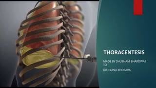
THORACENTESIS
MADE BY SHUBHAM BHARDWAJ
TO
DR. NUNU KHORAVA
Overview
 Thoracentesis is a procedure to remove fluid or air from around the lungs. A
needle is put through the chest wall into the pleural space. The pleural space is
the thin gap between the pleura of the lung and of the inner chest wall. The
pleura is a double layer of membranes that surrounds the lungs
Indications
 Congestive heart failure (CHF), the most common cause
of pleural effusion
 Viral, fungal, or bacterial infections
 Cancer
 Systemic lupus erythematosus (SLE) and other
autoimmune disease
 Inflammation of the pancreas (pancreatitis)
 A blood clot in the lung (pulmonary embolism)
 An area of pus in the pleural space (empyema)
 Liver failure
 Tuberculosis (TB)
 Pneumonia
Doctors are
also cautious
to perform
thoracentesis
on people
who:
 can’t be safely repositioned
 have bleeding disorders
 are taking blood thinners
 may have scarring from recent lung surgery
 have other conditions where potential complications
outweigh benefits
Preparing for a
thoracentesis
 tell your doctor if you:
 are currently taking medications, including blood
thinners like aspirin, clopidogrel (Plavix), or warfarin
(Coumadin)
 are allergic to any medications
 have any bleeding problems
 may be pregnant
 have lung scarring from previous procedures
 currently have any lung diseases like asthma
or emphysema
Before the
procedure
 You may have imaging tests before the procedure. These
are done to find the location of the fluid to be removed.
You may have any of the below:
• Chest X-ray
• Chest fluoroscopy
• Ultrasound
• CT scan
Procedure
1. You may be asked to remove your clothes. If so, you will
be given a hospital gown to wear. You may be asked to
remove jewelry or other objects.
2. You may be given oxygen through a nasal tube or face
mask. Your heart rate, blood pressure, and breathing will
be watched during the procedure.
3. You will be in a sitting position in a hospital bed. Your
arms will be resting on an over-bed table. This position
helps to spread out the spaces between the ribs, where
the needle is inserted. If you are not able to sit, you may
lie on your side on the edge of the bed.
4. The skin where the needle will be put in will be cleaned
with an antiseptic solution.
5. A numbing medicine (local anesthetic) will be injected in
the area.
6. When the area is numb, the healthcare provider will
put a needle between the ribs in your back. You may
feel some pressure where the needle goes in. Fluid
will slowly be withdrawn into the needle.
7. You will be asked to hold still, breathe out deeply, or
hold your breath at certain times during the procedure.
8. If there is a large amount of fluid, tubing may be
attached to the needle. This will let the fluid drain more.
The fluid will drain into a bottle or bag. In some cases, a
flexible tube (catheter) will be put in place of the needle
and the tubing will be attached for a day or two. You will
stay in the hospital until the catheter is removed.
9. When enough fluid has been removed, the needle will
be taken out. A bandage or dressing will be put on the
area.
10. Fluid samples may be sent to a lab.
11. You may have a chest X-ray taken right after the
procedure. This is to make sure your lungs are OK.
Risks
 Air in the space between the lung covering (pleural
space) that causes the lung to collapse (pneumothorax)
 Bleeding
 Infection
 Liver or spleen injury (rare)
 Your risks may vary depending on your general health
and other factors.
After the
procedure
 Your doctor will explain how to take care of the puncture
site. Make sure to contact your doctor if you begin to
have any signs of infection. Symptoms of infection
include:
 trouble breathing
 coughing up blood
 fever or chills
 pain when you take deep breaths
 redness, pain, or bleeding around the needle site
THORACENTESIS.pptx

More Related Content

Similar to THORACENTESIS.pptx

Biopsy project
Biopsy projectBiopsy project
Biopsy project
Illaiyan
 
Lung cancer presentation
Lung cancer presentationLung cancer presentation
Lung cancer presentation
Maruf Minar
 

Similar to THORACENTESIS.pptx (20)

CT GUIDED LUNG BIOPSY.pptx,lung mass is malignant (cancerous) or benign
CT GUIDED LUNG BIOPSY.pptx,lung mass is malignant (cancerous) or benignCT GUIDED LUNG BIOPSY.pptx,lung mass is malignant (cancerous) or benign
CT GUIDED LUNG BIOPSY.pptx,lung mass is malignant (cancerous) or benign
 
Thoracocentesis
ThoracocentesisThoracocentesis
Thoracocentesis
 
Swati pal ppt
Swati pal pptSwati pal ppt
Swati pal ppt
 
Biopsy project
Biopsy projectBiopsy project
Biopsy project
 
Safe suctioning
Safe suctioningSafe suctioning
Safe suctioning
 
Thoracotomy
ThoracotomyThoracotomy
Thoracotomy
 
thoracocentesis-141
thoracocentesis-141thoracocentesis-141
thoracocentesis-141
 
Pneumothorax
PneumothoraxPneumothorax
Pneumothorax
 
Pleural disease
Pleural disease Pleural disease
Pleural disease
 
Intercostal drain
Intercostal drainIntercostal drain
Intercostal drain
 
Lung cancer presentation
Lung cancer presentationLung cancer presentation
Lung cancer presentation
 
Thoracentesis 12 june 2020
Thoracentesis 12 june 2020Thoracentesis 12 june 2020
Thoracentesis 12 june 2020
 
Dilated Veins on the chest walls
Dilated Veins on the chest wallsDilated Veins on the chest walls
Dilated Veins on the chest walls
 
Tube thoracostomy
Tube thoracostomyTube thoracostomy
Tube thoracostomy
 
EMBOLISM.pptx
EMBOLISM.pptxEMBOLISM.pptx
EMBOLISM.pptx
 
Pneumothorax by Dr. Sookun Rajeev Kumar
Pneumothorax by Dr. Sookun Rajeev KumarPneumothorax by Dr. Sookun Rajeev Kumar
Pneumothorax by Dr. Sookun Rajeev Kumar
 
Tracheostomy
TracheostomyTracheostomy
Tracheostomy
 
Intravenous infusion
Intravenous infusionIntravenous infusion
Intravenous infusion
 
Pleural Fluid and Analysis of blood.pptx
Pleural Fluid and Analysis of blood.pptxPleural Fluid and Analysis of blood.pptx
Pleural Fluid and Analysis of blood.pptx
 
Chapter 11 Thoracic Injuries
Chapter 11 Thoracic InjuriesChapter 11 Thoracic Injuries
Chapter 11 Thoracic Injuries
 

More from Shubhambhardwaj437651 (6)

ASTHMA.pptx
ASTHMA.pptxASTHMA.pptx
ASTHMA.pptx
 
Diabetic ketoacidosis and hyperosmolar hyperglycemic state.pptx
Diabetic ketoacidosis and hyperosmolar hyperglycemic state.pptxDiabetic ketoacidosis and hyperosmolar hyperglycemic state.pptx
Diabetic ketoacidosis and hyperosmolar hyperglycemic state.pptx
 
occupational asthma.pptx
occupational asthma.pptxoccupational asthma.pptx
occupational asthma.pptx
 
Central hypothyroidism.pptx
Central hypothyroidism.pptxCentral hypothyroidism.pptx
Central hypothyroidism.pptx
 
STRESS DURING PANDEMIC.pptx
STRESS DURING PANDEMIC.pptxSTRESS DURING PANDEMIC.pptx
STRESS DURING PANDEMIC.pptx
 
Diabetes insipidus.pptx
Diabetes insipidus.pptxDiabetes insipidus.pptx
Diabetes insipidus.pptx
 

Recently uploaded

Quantifying Artificial Intelligence and What Comes Next!
Quantifying Artificial Intelligence and What Comes Next!Quantifying Artificial Intelligence and What Comes Next!
Quantifying Artificial Intelligence and What Comes Next!
University of Hertfordshire
 
Electricity and Circuits for Grade 9 students
Electricity and Circuits for Grade 9 studentsElectricity and Circuits for Grade 9 students
Electricity and Circuits for Grade 9 students
levieagacer
 
Warming the earth and the atmosphere.pptx
Warming the earth and the atmosphere.pptxWarming the earth and the atmosphere.pptx
Warming the earth and the atmosphere.pptx
GlendelCaroz
 
Continuum emission from within the plunging region of black hole discs
Continuum emission from within the plunging region of black hole discsContinuum emission from within the plunging region of black hole discs
Continuum emission from within the plunging region of black hole discs
Sérgio Sacani
 
HIV AND INFULENZA VIRUS PPT HIV PPT INFULENZA VIRUS PPT
HIV AND INFULENZA VIRUS PPT HIV PPT  INFULENZA VIRUS PPTHIV AND INFULENZA VIRUS PPT HIV PPT  INFULENZA VIRUS PPT

Recently uploaded (20)

Lubrication System in forced feed system
Lubrication System in forced feed systemLubrication System in forced feed system
Lubrication System in forced feed system
 
GBSN - Microbiology (Unit 7) Microbiology in Everyday Life
GBSN - Microbiology (Unit 7) Microbiology in Everyday LifeGBSN - Microbiology (Unit 7) Microbiology in Everyday Life
GBSN - Microbiology (Unit 7) Microbiology in Everyday Life
 
FORENSIC CHEMISTRY ARSON INVESTIGATION.pdf
FORENSIC CHEMISTRY ARSON INVESTIGATION.pdfFORENSIC CHEMISTRY ARSON INVESTIGATION.pdf
FORENSIC CHEMISTRY ARSON INVESTIGATION.pdf
 
Abortion uae unmarried price +27791653574 Contact Us Dubai Abu Dhabi Sharjah ...
Abortion uae unmarried price +27791653574 Contact Us Dubai Abu Dhabi Sharjah ...Abortion uae unmarried price +27791653574 Contact Us Dubai Abu Dhabi Sharjah ...
Abortion uae unmarried price +27791653574 Contact Us Dubai Abu Dhabi Sharjah ...
 
Mining Activity and Investment Opportunity in Myanmar.pptx
Mining Activity and Investment Opportunity in Myanmar.pptxMining Activity and Investment Opportunity in Myanmar.pptx
Mining Activity and Investment Opportunity in Myanmar.pptx
 
Quantifying Artificial Intelligence and What Comes Next!
Quantifying Artificial Intelligence and What Comes Next!Quantifying Artificial Intelligence and What Comes Next!
Quantifying Artificial Intelligence and What Comes Next!
 
Information science research with large language models: between science and ...
Information science research with large language models: between science and ...Information science research with large language models: between science and ...
Information science research with large language models: between science and ...
 
Costs to heap leach gold ore tailings in Karamoja region of Uganda
Costs to heap leach gold ore tailings in Karamoja region of UgandaCosts to heap leach gold ore tailings in Karamoja region of Uganda
Costs to heap leach gold ore tailings in Karamoja region of Uganda
 
Fun for mover student's book- English book for teaching.pdf
Fun for mover student's book- English book for teaching.pdfFun for mover student's book- English book for teaching.pdf
Fun for mover student's book- English book for teaching.pdf
 
Electricity and Circuits for Grade 9 students
Electricity and Circuits for Grade 9 studentsElectricity and Circuits for Grade 9 students
Electricity and Circuits for Grade 9 students
 
PARENTAL CARE IN FISHES.pptx for 5th sem
PARENTAL CARE IN FISHES.pptx for 5th semPARENTAL CARE IN FISHES.pptx for 5th sem
PARENTAL CARE IN FISHES.pptx for 5th sem
 
TEST BANK for Organic Chemistry 6th Edition.pdf
TEST BANK for Organic Chemistry 6th Edition.pdfTEST BANK for Organic Chemistry 6th Edition.pdf
TEST BANK for Organic Chemistry 6th Edition.pdf
 
Warming the earth and the atmosphere.pptx
Warming the earth and the atmosphere.pptxWarming the earth and the atmosphere.pptx
Warming the earth and the atmosphere.pptx
 
SaffronCrocusGenomicsThessalonikiOnlineMay2024TalkOnline.pptx
SaffronCrocusGenomicsThessalonikiOnlineMay2024TalkOnline.pptxSaffronCrocusGenomicsThessalonikiOnlineMay2024TalkOnline.pptx
SaffronCrocusGenomicsThessalonikiOnlineMay2024TalkOnline.pptx
 
Continuum emission from within the plunging region of black hole discs
Continuum emission from within the plunging region of black hole discsContinuum emission from within the plunging region of black hole discs
Continuum emission from within the plunging region of black hole discs
 
EU START PROJECT. START-Newsletter_Issue_4.pdf
EU START PROJECT. START-Newsletter_Issue_4.pdfEU START PROJECT. START-Newsletter_Issue_4.pdf
EU START PROJECT. START-Newsletter_Issue_4.pdf
 
X-rays from a Central “Exhaust Vent” of the Galactic Center Chimney
X-rays from a Central “Exhaust Vent” of the Galactic Center ChimneyX-rays from a Central “Exhaust Vent” of the Galactic Center Chimney
X-rays from a Central “Exhaust Vent” of the Galactic Center Chimney
 
HIV AND INFULENZA VIRUS PPT HIV PPT INFULENZA VIRUS PPT
HIV AND INFULENZA VIRUS PPT HIV PPT  INFULENZA VIRUS PPTHIV AND INFULENZA VIRUS PPT HIV PPT  INFULENZA VIRUS PPT
HIV AND INFULENZA VIRUS PPT HIV PPT INFULENZA VIRUS PPT
 
GBSN - Microbiology (Unit 6) Human and Microbial interaction
GBSN - Microbiology (Unit 6) Human and Microbial interactionGBSN - Microbiology (Unit 6) Human and Microbial interaction
GBSN - Microbiology (Unit 6) Human and Microbial interaction
 
PHOTOSYNTHETIC BACTERIA (OXYGENIC AND ANOXYGENIC)
PHOTOSYNTHETIC BACTERIA  (OXYGENIC AND ANOXYGENIC)PHOTOSYNTHETIC BACTERIA  (OXYGENIC AND ANOXYGENIC)
PHOTOSYNTHETIC BACTERIA (OXYGENIC AND ANOXYGENIC)
 

THORACENTESIS.pptx

  • 1. THORACENTESIS MADE BY SHUBHAM BHARDWAJ TO DR. NUNU KHORAVA
  • 2. Overview  Thoracentesis is a procedure to remove fluid or air from around the lungs. A needle is put through the chest wall into the pleural space. The pleural space is the thin gap between the pleura of the lung and of the inner chest wall. The pleura is a double layer of membranes that surrounds the lungs
  • 3. Indications  Congestive heart failure (CHF), the most common cause of pleural effusion  Viral, fungal, or bacterial infections  Cancer  Systemic lupus erythematosus (SLE) and other autoimmune disease  Inflammation of the pancreas (pancreatitis)  A blood clot in the lung (pulmonary embolism)  An area of pus in the pleural space (empyema)  Liver failure  Tuberculosis (TB)  Pneumonia
  • 4. Doctors are also cautious to perform thoracentesis on people who:  can’t be safely repositioned  have bleeding disorders  are taking blood thinners  may have scarring from recent lung surgery  have other conditions where potential complications outweigh benefits
  • 5. Preparing for a thoracentesis  tell your doctor if you:  are currently taking medications, including blood thinners like aspirin, clopidogrel (Plavix), or warfarin (Coumadin)  are allergic to any medications  have any bleeding problems  may be pregnant  have lung scarring from previous procedures  currently have any lung diseases like asthma or emphysema
  • 6. Before the procedure  You may have imaging tests before the procedure. These are done to find the location of the fluid to be removed. You may have any of the below: • Chest X-ray • Chest fluoroscopy • Ultrasound • CT scan
  • 7. Procedure 1. You may be asked to remove your clothes. If so, you will be given a hospital gown to wear. You may be asked to remove jewelry or other objects. 2. You may be given oxygen through a nasal tube or face mask. Your heart rate, blood pressure, and breathing will be watched during the procedure. 3. You will be in a sitting position in a hospital bed. Your arms will be resting on an over-bed table. This position helps to spread out the spaces between the ribs, where the needle is inserted. If you are not able to sit, you may lie on your side on the edge of the bed. 4. The skin where the needle will be put in will be cleaned with an antiseptic solution. 5. A numbing medicine (local anesthetic) will be injected in the area.
  • 8. 6. When the area is numb, the healthcare provider will put a needle between the ribs in your back. You may feel some pressure where the needle goes in. Fluid will slowly be withdrawn into the needle. 7. You will be asked to hold still, breathe out deeply, or hold your breath at certain times during the procedure. 8. If there is a large amount of fluid, tubing may be attached to the needle. This will let the fluid drain more. The fluid will drain into a bottle or bag. In some cases, a flexible tube (catheter) will be put in place of the needle and the tubing will be attached for a day or two. You will stay in the hospital until the catheter is removed. 9. When enough fluid has been removed, the needle will be taken out. A bandage or dressing will be put on the area. 10. Fluid samples may be sent to a lab. 11. You may have a chest X-ray taken right after the procedure. This is to make sure your lungs are OK.
  • 9. Risks  Air in the space between the lung covering (pleural space) that causes the lung to collapse (pneumothorax)  Bleeding  Infection  Liver or spleen injury (rare)  Your risks may vary depending on your general health and other factors.
  • 10. After the procedure  Your doctor will explain how to take care of the puncture site. Make sure to contact your doctor if you begin to have any signs of infection. Symptoms of infection include:  trouble breathing  coughing up blood  fever or chills  pain when you take deep breaths  redness, pain, or bleeding around the needle site