
Recommended
More Related Content
What's hot
What's hot (20)
Similar to BRONCHIECTASIS.pptx
Similar to BRONCHIECTASIS.pptx (20)
More from Radhika Kulvi
Recently uploaded
Recently uploaded (20)
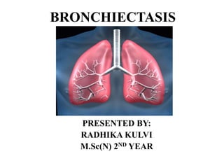
BRONCHIECTASIS.pptx
- 2. INTRODUCTION Bronchiectasis is a condition where damage tubes causes the lungs (airways) to widen or develop pouches. It makes hard to clear mucus out of lungs and can cause frequent infections. Coughing a lot with pus and mucus is the main symptom of bronchiectasis. Bronchiectasis can’t be cured but can be managed with treatment.
- 3. MEANING Bronchiectasis is the abnormal, irreversible dilatation of the bronchi.
- 4. DEFINITION Bronchiectasis (Obstructive Lung disease) is an irreversible widening (dilation) of portions of the breathing tubes or airways (bronchi) resulting from damage to the airway wall. The most common cause is severe or repeated respiratory infections, often in people who have an underlying problem with their lungs or immune system.
- 5. TYPES OF BRONCHIECTASIS CYLINDRICAL BRONCHIECTASIS VARICOSE BRONCHIECTASIS SACULAR BRONCHIECTASIS The most commonly identified morphologic type, is a smooth uniform enlargement of bronchi without focal out pouchings or tortuosity. It has irregular contours with alternating dilating and contracting lumen. Also called Cystic bronchiectasis is one of the less common morphological forms . It may be present on its own or may occur in combination with other forms of bronchiectasis .
- 7. CAUSES • Airway obstruction • Diffuse airway injury • Pulmonary infections (complications of long term Pulmonary infection) • Genetic disorder (cystic fibrosis) • Abnormal host defense • Idiopathic cause
- 8. PATHOPHYSIOLOGY Due to causative agents Damages the bronchial wall, ( loss of its supporting structure) Resulting in thick sputum that ultimately obstructs the bronchi. The walls of bronchi become permanently distended and distorted, impairing muco-ciliary clearance The retention of secretions and obstruction ultimately cause the alveoli distal (ventilation– perfusion imbalance) and hypoxemia. Bronchiectasis
- 9. CLINICAL MANIFESTATION • Chronic cough • Production of purulent sputum in copious amounts • Hemoptysis. • Clubbing of the fingers • Repeated episodes of pulmonary infection • Weight loss • Shortness of breath. • Wheezing.
- 10. DIAGNOSTIC EVALUATION • History collection • Physical examination • Chest x-ray • Sputum studies. • Pulmonary function test • Arterial blood gas studies. • Computed tomography.(CT scan) • Bronchoscopy, Bronchogram
- 11. IMMEDIATE MANAGEMENT • Oxygen Therapy • Postural drainage : To promote bronchial drainage & clear excessive secretions • Bronchoscopy: To remove mucopurulent sputum. • Chest physiotherapy, including percussion • Antimicrobial therapy Based on type of infection. • Influenza and pneumococcal vaccines . • Bronchodilators: Theophylline • Nebulizers. & Steam inhalation.
- 12. MEDICAL MANAGEMENT • Antibiotics:Amoxicillin, Levofloxacin • Bronchodialators:Albuterol, Salmeterol • Antihistamines:Citrizine, Loratadine • Corticosteroids:Fluticosone,Budesonide
- 13. SURGICAL MANAGEMENT • Segmental resection to remove a segment of a lobe. • Lobectomy : Removal of disesased part of a lobe • Pneumonectomy: Removal of an entire lung
- 14. NURSING MANAGEMENT • Assess the general condition . • Collect complete history. • Perform physical examination • Provide comfortable bed & position. • Provide nutritious diet • Provide Oxygen According To Physician Order. • Provide Psychological Support To Patient. • Provide Knowledge About Bronchiectasis. • Provide Suctioning and Maintain Hygiene of Patient • Careful monitoring of the post-operative cases • Provide Breathing & coughing exercises. • Nebulization & steam inhalation
- 15. NURSING DIAGNOSIS • Impaired gas exchange related to ventilation- perfusion imbalance. • Ineffective airway clearance related to increased mucus production. • Ineffective breathing pattern related to mucus and airway irritants. • Activity intolerance related to hypoxemia and ineffective breathing patterns.
- 16. HEALTH EDUCATION • Smoking cessation • Lifestyle modification • Nutritional balanced diet • Appropriate treatment of respiratory infection. • Personal hygiene.