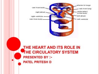
1. THE HEART AND ITS ROLE IN
THE CIRCULATORY SYSTEM
PRESENTED BY :-
PATEL PRITESH D
2.
3. The cardiovascular system consists of the heart and
blood vessels. The system is responsible for the
circulation of blood through the tissues of the body.
The heart acts as a pump and provides the force for this
circulation. Blood vessels taking blood from the heart to
the tissues are called arteries.
The largest artery in the body is called the aorta. Arising
from the heart it divides, like the branches of a tree, into
smaller and smaller branches. The smallest arteries are
called arterioles.
These thin-walled vessels are called capillaries.
Oxygen, nutrition, waste products, etc. can pass
through the walls of capillaries from blood to tissue
cells .
4. Blood from capillaries is collected
by another set of vessels that carry
it back to the heart These are
called veins .
The veins adjoining the capillaries
are very small and are called
venules.
Smaller veins join together to
form larger and larger veins .
Ultimately, the blood reaches two
large veins, the superior vena cava
and the inferior vena cava, which
pour it back into the heart.
This blood reaching the heart
through the veins has lost its
oxygen.
5. STRUCTURE
Heart is a conical hollow muscular organ situated in the
middle mediastinum ( space between the lungs ) and near
the midline of thoracic cavity .
it is enclosed with in the pericardium , it pump blood to
various part of the body .to meet their nutritive requirement
.
6. MEASUREMENTS OF HEART
Length :- 12 cm
Width :- 9 cm
Thickness :- 6 cm
Weight :- in adult female 250 gm & adult male 300 gm .
8. LAYERS OF HEART :-
Epicardium ( outer pericardium ) :-
( outer protective layers of the heart )
pericardium is the outer covering of the heart .it is
made up two layer. Which are separated by space all
pericardial cavity .
it is composed of connective tissue and fat .
the connective tissue secretes a small amount of
lubricating fluid into the pericardial cavity .
9. Middle Myocardium :-
(muscular middle layer wall of the heart )
• myocardium is responsible for pumping action of heart
• the myocardium is responsible for contractions of the heart
Inner Endocardium :-
( inner layer of the heart wall )
• it lines the cavities and valves of the heart .
• endocardium is formed by single layers of
endothelial cells
• smooth lining to reduce friction of bloodflow
10.
11.
12.
13. EXTERNAL STRUCTURE OF THE HEART :-
An apex directed downwards ,forwards and to the left.
A base ( posterior surface ) directed backwards.
Anterior , inferior ,and left lateral surfaces.
Upper, inferior ,right and left borders.
a. Apex of the heart :-
Apex of the heart is formed entirely by the left ventricle
.it is directed downwards, forwards and to the left and is
overlapped by anterior border of the left lung.it is
situated in the left fifth intercostal space 9 cm to the
leteral line .
14.
15. b. Base of the heart :- the base of the heart is also called its
posterior surface .it is formed mainly by the left atrium and by a
small part of the right artium.in relation to the base the openings
of four pulmonary veins are seen which open into the left atrium
and of the superior and inferior vena cava which open into the
right atrium .
c. Surfaces of the heart :-
the sternocostal (anterior ) surface :- of the heart is mainly
formed by the right ventricle and partly by right atrium, left
ventricle and left auricle . The left atrium is not seen as it is
covered by aorta and pulmonary trunk .
the diaphragmatic (inferior) surface :- this surface is flat and
rest on the central tendon of the diaphragm.
it is created by the right and left ventricles that are divided from
every other by the posterior interventicular groove .
16. The left ventricles create left 2/3rd of this surface and right ventricle
creates only right 1/3rd of this surface.
d. Right & left pulmonary surfaces :-
the left pulmonary surface faces the left lung .
the right pulmonary surface faces the right lung.
e. Borders :-
Separating the surfaces of the heart and its borders.
There are four main borders of the heart :-
Right border:- right atrium.
inferior border :- left ventricle and right ventricle .
left border :- left ventricle .
superior border :-right and left atrium and the great vessels.
17. Groove or sulci :-
the atrai are separated from the ventricles by a circular
atrioventicular or coronary sulcus.
18.
19. Human heart has four chembers .right and left atrium and right and
left ventricle .
Right atrium and right ventricle are seprated by tricuspid valve .
Left atrium and left ventricle are sepreted mitral valve.
20. Chambers of the heart :-
Right atrium :- it is the right upper chambers and forms
base of heart .and recieves venous blood
from all the body .
it receive de – oxygenated blood from
superior vena cava & inferior vena cava.
Right ventricle :- the right ventricle is separated from
the left ventricle by the septum.
right atrium communicates with right
ventricle through tricuspid valve .
pulmonary valve is present at the
junction of right ventricle.
the right ventricle inject blood through
pulmonary valve .
21. Left atrium :- it receive oxygenated blood from lungs through 4
(four ) pulmonary veins .
the left atrium receive oxygen rich blood which
blood in to the left ventricle through mitral valve .
Left ventricle :- the left ventricle is the largest chamber of the
heart .heart pump blood to aorta .
its wall are three time thicker then right
ventricle.
blood from left atrium enter into left ventricle to
mitral valve .
left ventricle pump oxygenated blood to all part
of the body .
22. the mitral and tricuspid valve which control blood flow from the
atrium to ventricle .
The aortic valve and pulmonary valve which control blood flow
out of the ventricles.
23. Blood Supply of Heart:
Arterial Supply: They begin with the aorta, the large artery
leaving the heart. Arteries carry oxygen-rich blood away from
the heart to all of the body's tissues. They branch several times,
becoming smaller and smaller as they carry blood farther from
the heart.
Capillaries: These are small, thin blood vessels that connect the
arteries and the veins. Their thin walls allow oxygen, nutrients,
carbon dioxide, and other waste products to pass to and from our
organ's cells.
Veins: These are blood vessels that take blood back to the heart;
this blood lacks oxygen (oxygen-poor) and is rich in waste
products that are to be excreted or removed from the body. Veins
become larger and larger as they get closer to the heart. The
superior vena cava is the large vein that brings blood from the
head and arms to the heart, and the inferior vena cava brings
blood from the abdomen and legs into the heart.
24. Right-Hand Side of the Heart:
The right-hand side of the heart receives de-oxygenated
blood from the body tissues (from the upper and lower
body via the superior vena cava and the inferior vena
cava, respectively) into the right atrium. This de-
oxygenated blood passes through the tricuspid valve
into the right ventricle. This blood is then pumped
under higher pressure from the right ventricle to the
lungs via the pulmonary artery.
25. The left-hand side of the heart:
Receives oxygenated blood from the lungs (via the
pulmonary veins) into the left atrium. This
oxygenated blood then passes through the bicuspid
valve into the left ventricle. It is then pumped to the
aorta under greater pressure. This higher pressure
ensures that the oxygenated blood leaving the heart
via the aorta is effectively delivered to other parts of
the body via the vascular system of blood vessels
(includes. arteries, arterioles and capillaries).
26. The right atrium receives deoxygenated blood from
the body and pumps it to the right ventricle.
The right ventricle gets blood from the
right atrium and pumps it to the lungs to load it with
oxygen.
The left atrium receives oxygenated blood from the
lungs and pumps it to the left ventricle.
The left ventricle is the strongest chamber of the
heart. It pumps oxygen-rich blood to the rest of the
body.
27. The four main functions of the heart are:
Pumping oxygenated blood to the other body parts.
Pumping hormones and other vital substances to
different parts of the body.
Receiving deoxygenated blood and carrying metabolic
waste products from the body and pumping it to the
lungs for oxygenation.
Maintaining blood pressure.