
Recommended
More Related Content
What's hot
What's hot (20)
Similar to Cardiovascular System
Similar to Cardiovascular System (20)
Recently uploaded
Recently uploaded (20)
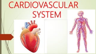
Cardiovascular System
- 2. CONTENTS Meaning Introduction Heart Blood vessels Function of cardiovascular system Points to be noted MCQS
- 3. MEANING Cardiovascular system is made up of two words ; cardiovascular and system where , CARDIOVASCULAR : relating to the circulatory system , which comprises the heart and blood vessels and carries nutrients and oxygen to the tissues and removes carbondioxide and other wastes from body SYSTEM : group of interlacting or interrelated entities that form a unified or group of organs that work together to carry out a particular task
- 4. INTRODUCTION Also known as circulatory system Is transport system carrying oxygen , nutrients , hormones and other substances to the tissue and conveying carbondioxide to the lungs and other waste products to the kidneys Consists of cardiac ( heart ) and vascular ( blood vessels )
- 5. HEART A hollow muscular organ which acts as a compression pump Is pyramidal in shape and the size of your fist Situated in middle mediastinum in between the two lungs The 2/3rd part of heart lie towards the left side of the body and remaining 1/3rd part of heart lies towards right side of body Weighs about 300gm in male and 250gm in female
- 6. COVERINGS OF THE HEART Heart is composed by a fibro serous sac called pericardium . It has two layers ; Fibrous pericardium : - an outer membrane made up of strong fibrous sheath - is conical in shape - connected to sternum anteriorly , diaphragm inferiorly and roots of great blood vessels superiorly - helps to maintain shape of the heart
- 7. Contd… Serous pericardium : - an inner layer , made up of serous membrane - has two layers ; parietal and visceral layer - parietal layer lies just inside the fibrous pericardium - visceral layer is closely related to the surface of the heart - pericardial cavity is a cavity between the parietal and visceral layers of the serous pericardium - pericardial cavity is filled with a fluid called pericardial fluid
- 8. LAYERS OF HEART The heart wall consists of three layers ; 1. Epicardium : - outermost layer formed by visceral layer of serous pericardium 2. Myocardium : - middle layer composed of specialized muscular layer known as cardiac muscle - involuntary muscle controlled by autonomic nervous system - gets the blood supply from the right and left coronary arteries
- 9. Contd… 3. Endocardium : - innermost layer consisting of a single layer of epithelial cells known as epithelium - provides a smooth lining for the blood to flow over - is continuous with the valves of the heart and blood vessels
- 10. FEATURES OF THE HEART Heart consists of following parts Chambers : - Right atrium - Left atrium - Right ventricle - Left ventricle Valves : - Right atrioventricular valve (Tricuspid valve) - Left atrioventricular valve (Bicuspid or Mitral valve) - Semilunar valve (Aortic and Pulmonic valve)
- 11. Contd… Septum : - Interatrial septum - Interventricular septum Grooves and sulci : - Atriventricular groove - Interventricular groove a. anterior b. posterior
- 12. CHAMBERS OF THE HEART Right atrium : Situated on the right part of the heart Receives deoxygenated blood from whole of the body via the superior venacava at its upper end and inferior venacava at its lower end Pumps blood to right ventricle through tricuspid valve
- 13. Contd… Right ventricle : A crescent shaped chamber situated on right side of the heart below right atrium Pumps received blood into lungs for oxygenation through pulmonary artery
- 14. Contd… Left atrium : Situated behind the right atrium Forms base of the heart Receives oxygenated blood from lungs through pulmonary veins Pumps blood into left ventricle through mitral valve
- 15. Contd… Left ventricle : Nearly a circular chamber Blunt tips forms apex of the heart Pumps oxygenated blood through out the body via aorta The wall is three times thicker as compared to right ventricle in order to pump blood to whole body
- 16. VALVES OF THE HEART Tricuspid valve : Is atrioventricular valve Present between right atrium and right ventricle Has three leaflets : anterior , septal and posterior
- 17. Contd… Mitral valve : Also known as bicuspid valve Is atrioventricular valve Present between left atrium and left ventricle Has two leaflets : anterior and posterior
- 18. Contd… Pulmonary valve : Is semilunar valve Present between right ventricle and pulmonary artery Has three flaps
- 19. Contd… Aortic valve : Is semilunar valve Present between left ventricle and aorta Has three flaps
- 20. SEPTUM OF THE HEART Interatrial septum : - divides right atrium and left atrium Interventricular septum : - divides right and left ventricles
- 21. GROOVES AND SULCI OF THE HEART Atriventricular groove : - a small circular depression on the surface of the heart which separates atrium and ventricles Interventricular groove : - separates the two ventricles on the surface of heart - has anterior and posterior parts
- 22. CONDUCTING SYSTEM OF THE HEART Is the specialized myocardium that initiate and conduct impulses for the contraction of the heart Consists of following parts ; 1. Sinoatrial node or SA node 2. Artioventricular node or AV node 3. Artioventricular bundle or AV bundle or bundle of HIS 4. The right bundle branch 5. The left bundle branch 6. The purkinje fibers
- 23. Contd… Sinoatrial node or SA node : - mass of specialized type of cells - situated in wall of right atrium near the opening of superior venacava - also known as “pacemaker” - generates an impulse and initiates the heart beat
- 24. Contd… Atrioventricular node or AV node : - situated in the lower and dorsal part of atrial septum - receives impulses generated by SA node through atrial wall
- 25. Contd… Artioventricular bundle or AV bundle or bundle of HIS : - the only muscular connection between atrial and ventricular musculatures - begins as atrioventricular node , crosses AV ring and descends to ventricular septum - divides into right and left branches at the upper border of muscular part of septum
- 26. Contd… The right bundle branch : - passes down the right side of the interventricular septum - reaches wall of right ventricles after dividing into purkinje fibers The left bundle branch : - goes to the left side of interventricular septum - distributes to left ventricle after dividing into purkinje fibers
- 27. Contd… Purkinje fibers : - thin fibers of myocardium - made from right and left bundle branches which are distributed to the wall of ventricle
- 28. ORGAN ASSOCIATED WITH THE HEART Superiorly : the great blood vessels i.e the aorta , superior venacava , pulmonary artery and pulmonary veins Inferiorly : the apex rests on the central tendon of the diaphragm Anteriorly : the sternum , ribs and intercostal muscle Posteriorly : the oesophagus , trachea , left and right bronchi , descending aorta , inferior venacava and thoracic vertebrae Laterally : the lungs – the left lung overlaps the side of the heart
- 29. BLOOD SUPPLY OF THE HEART Heart receives its blood supply from : a. Right coronary artery : - aries from the ascending aorta - larger than left coronary artery - supplies blood to the right part of heart including right atrium , right ventricle , SA node , AV node and some portion of the posterior part of left ventricle
- 30. Contd… b. Left coronary artery : - arises from the ascending aorta - supplies blood to left part of heart such as left atrium , left ventricle and some part of anterior portion of right ventricle The deoxygenated blood from heart goes to right atrium through coronary sinus .
- 31. NERVE SUPPLY OF THE HEART Is supplied by sympathetic and parasympathetic component of autonomic nervous system Sympathetic nerves increases heart rate and parasympathetic nerve decreases heart rate
- 32. BLOOD VESSELS Component of circulatory system which carries blood There are three types of blood vessels in human body according to anatomy ; 1. Artery 2. Veins 3. Capillary
- 33. Artery Blood vessels that transport blood away from heart Carries pure blood except pulmonary artery The wall of arteries consists of 3 layers from outside inwards . They are : i. Tunica adventitia (made up of connective tissue) ii. Tunica media (made up of smooth muscle) iii. Tunica intima ( made up of endothelium)
- 34. Contd… Arterial branches becomes narrower and their walls become thinner while reaching periphery called arterioles Arterioles are continued as capillaries Capillaries are very small vessels in which exchange of gases and materials takes place
- 35. Veins Blood vessels that transport blood to heart Carries deoxygenated blood except pulmonary vein Venules unite and forms veins Different veins of the body unite and forms venacava
- 36. Contd… There are two venacava in human body : 1. Superior venacava : - carries blood from upper part of body including head , neck and upper limbs 2. Inferior venacava : - carries blood from lower part of body including thorax , abdomen, pelvis and lower limbs
- 37. Capillaries Are the minute vessels from smallest arteriole Consists of a single layer of endothelial cells , through which water and other small molecular substance can pass Form a vast network of tiny vessels thus bring the nutritional materials and oxygen to the cells
- 38. FUNCTIONS OF CARDIOVASCULAR SYSTEM Circulates oxygen and removes carbondioxide Provides cell with the nutrients Removes waste products of metabolism to the excretory organs for disposal Helps regulate body temperature Protects the body from infection and blood loss
- 39. POINTS TO BE NOTED Cardiovascular system is also known as circulatory system . It is a transport system . It consists of cardio (heart) and vascular (blood vessels) . Heart is a size of your own fist . Heart weighs 300 gm in male and 250 gm in female . Heart is covered by a fibro serous sac called pericardium .
- 40. Contd… Pericardial cavity is a cavity between parietal and visceral layer which consists of pericardial fluid . Heart has four chambers ; right atrium , right ventricle , left atrium and left ventricle . Atrioventricular valves are tricuspid valve and mitral valve . Mitral valve is also known as bicuspid valve . Semilunar valves are pulmonary valve and aortic valve .
- 41. Contd… Heart is supplied with blood by right and left coronary artery . Sinoatrial node is also known as pacemaker . Arteries consists of 3 layers from outside inwards ; tunica adventitia , tunica media and tunica intima . Capillaries are very small vessels in which exchange of gases and materials takes place .
- 42. MCQS 1. Circulatory system is a a. ventilatory system b. transport system c. excretory system d. none of the above 2. The cavity between parietal layer and visceral layer of heart is called a. pericardial cavity b. pleural cavity c. parietal cavity d. none of the above 3. The innermost layer of blood vessel is a. tunica adventitia b. tunica media c. tunica intima d. none of the above
- 43. 4. The heart is supplied by a. coelic artery b. intercostal artery c. right and left coronary arteries d. mesenteric arteries 5. The wall of left ventricle is ……….. right ventricle . a. equal in thickness to b. thinner than c. thicker than d. none of the above 6. ………. is also known as pacemaker of the heart . a. sinoatrial node b. atrioventricular node c. bundle of HIS d. purkinje fibers 7. Largest vein of the body a. superior venacava b. inferior venacava c. pulmonary veins d. aorta
- 44. 8. Largest artery of the body a. pulmonary artery b. renal artery c. aorta d. carotid artey 9. Backflow of blood is prevented by a. valves b. ventricles c. atrium d. none of the above 10. Valve that is present between right ventricle and lungs is a. tricuspid valve b. aortic valve c. bicuspid valve d. pulmonic valve ANSWERS 1. B 2 . A 3 . C 4 . C 5 . C 6. A 7 . B 8 . C 9 . A 10 . D