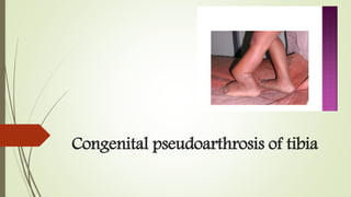
Congenital pseudoarthrosis of tibia
•Download as PPTX, PDF•
2 likes•206 views

Congenital pseudoarthrosis of tibia

Recommended
More Related Content
What's hot
What's hot (20)
Similar to Congenital pseudoarthrosis of tibia
Similar to Congenital pseudoarthrosis of tibia (20)
More from Ponnilavan Ponz
More from Ponnilavan Ponz (20)
Recently uploaded
Recently uploaded (20)
Congenital pseudoarthrosis of tibia
- 1. Congenital pseudoarthrosis of tibia
- 2. INTRODUCTION Failure of normal bone formation in the distal half of the tibia segmental defect of bone Anterolateral angulation Pathological fracture
- 3. Etiology NEUROFIBROMATOSIS: most common cause in about 40-80% cases hallmarks like café-au-lait spots and skin nodules Absent at birth and appears later Fibrous dysplasia Idiopathic
- 4. PATHOLOGY Site is usually surrounded by thickened periosteum and fibrous tissue Thickened periosteum results in hamartomatous tissue Failure of callus formation resulting in pseudoarthrosis Appearance of strangulation of bone with atrophic changes followed by avascular changes Failure of remodeling of bone results in stress fracture
- 5. Histopathology Fibrous hamartoma is the key pathology Low osteogenicity and high osteoclastogenicity Soft tissue at the pseudoarthrotic site is composed of variable of fibrous tissue, fibrocartilage and hyaline cartilage with evidence of endochondral ossification Marrow spaces are devoid of hematopoiesis This invasive fibromatosis is located in the periosteum and between broken bone ends and surrounds the tibia causing compression, osteolysis and persistence of pseudoarthrosis
- 7. ANDERSON CLASSIFICATION Dysplatic type Cystic type Late type Club foot type Angulated pseudoarthrosis
- 8. Dysplastic Diameter of tibia is narrowed Hourglass constriction is characteristic Tibia is bowed anteriorly or anterolaterally Prone to non- union and refracture Neurofibromatosis is always present in the dysplastic type It may be present at birth and develop at an average of 12 months
- 9. Cystic type Cyst like lesion in the affected segment No significant narrowing of diameter of tibia It may be present at about 8 months Neurofibromatosis not associated
- 10. Late type Affected leg shows length discrepancy After minimum trauma a stress fracture like break occurs with consequent development of pseudoarthrosis. Not associated with neurofibromatosis Anterior bowing develops between age of 4 and 12 years
- 11. Club foot type Fracture present at birth in a leg with marked anterior angulation The involved or contralateral lower limb has other associated congenital abnormalities such constriction band club foot
- 12. Angulated pseudoarthrosis Due to corrective osteotomy of anterior bowing of tibia Osteotomy results in pseudoarthrosis Therefore,simple osteotomy of anterior angulation is contraindicated
- 13. BOYDS CLASSIFICATION Type I Type II TypeIII TypeIV Type V Type VI Anterior bowing with tibia defect Pseudoarthrosis with hour glass constriction Pseudoarthrosis with bone cyst Sclerotic segments,March fracture Dysplastc fibula Interosseous neurofibroma
- 21. CLINICAL FEATURES Anterolateral,anterior rarely anteromedial bowing of the dysplastic tibia and fibula Tibia and fibula affected at the same level Bowing increases during the first two years of life and eventually develops pseudoarthrosis Patient walks with a limp or unable to walk
- 23. Criteria for diagnosis Mutiple café-au-lait spots(smooth edged,>0.5cm) Positive family history of neurofibromatosis Definite biopsy Bony lesions like pseudoarthrosis of the tibia
- 24. Radiological features Segment of tibia or tibia and fibula show hourglass thinning, sclerosis and loss of medullary cavity Angulation at two levels proximally and distally with dysplastic changes
- 27. Treatment Complete excision of the soft tissue fibromatosis at the site of pseudoarthrosis Correction of angular deformity Stimulation of bone healing Proper fixation of bone fragments
- 28. Treatment methods Ilizarov method Vascularized fibular graft Extending intramedullary nailing and bone grafting Electrical stimulation
- 31. Intramedullary rod fixation The procedure of choice for the first attempt to gain union entails resection of pseudoarthrosis, shortening and fixation with an intramedullary rod and autogenous bone grafting The procedure can be performed can be performed at an y age and rates of union of around 85% .
- 33. Complications Refracture Malalignment of tibia Limb length discrepancy Ankle valgus Ankle stiffness
- 34. Refracture Shortening Valgus deformity Splint the limb until skeletal maturity,retain intramedullary nail Union of pseudo arthrosis as early as possible, limb equalization procedure Ensure union of fibular pseudoarthrosis Retain intramedullary rod that crosses the ankle joint
- 35. Thank you