SlideShare a Scribd company logo
1 of 60
Dr/ ABD ALLAH NAZEER. MD.
Radiological imaging of Paget's disease.
Paget disease (osteitis deformans) is a chronic skeletal disorder
characterized by abnormal and excessive remodeling of bone. The
disease was named after Sir James Paget who described the condition
in 1877 in a detailed essay recognized in the medical literature as “a
lesson in accurate and lucid writing” . Paget disease is estimated to
affect approximately 3–4% of individuals older than 40 years and is
the second most common bone disease after osteoporosis that affects
the growing population of older persons in the United States.
Abnormal osseous resorption and apposition in Paget disease produce
variable clinical and imaging manifestations. Although the disease
may be asymptomatic, it can be painful or deforming and associated
with various and debilitating musculoskeletal complications.
Radiologists play a central role in the imaging diagnosis of the
process and its multifaceted manifestations. Radiography remains the
most inexpensive method to evaluate patients with Paget disease of
bone. CT, bone scintigraphy, and MRI have complementary roles in
achieving the correct diagnosis of Paget disease and its complications.
Paget disease of bone
Paget disease of the bone is a common, chronic bone
disorder characterised by excessive abnormal bone
remodelling. It frequently affects the pelvis, spine, skull
and proximal long bones and has characteristic
radiographic features.
Epidemiology
It is relatively common and can affect up to 4% of
individuals over 40 and up to 11% over the age of 80.
There may be a slight male predilection. Incidence can be
considerably higher in the United Kingdom than in other
countries. It is also common in Australia, New Zealand,
Western Europe, and the United States.
Clinical presentation
The majority (approximately three-quarters) of patients are
asymptomatic at the time of diagnosis. Presenting symptoms include:
localized pain and tenderness
increased focal temperature due to hyperaemia (due to
hypervascularity)
increased bone size: historically changing hat size was a giveaway
bowing deformities
kyphosis of the spine
decreased the range of motion
signs and symptoms relating to complications (see below)
Polyostotic disease is more prevalent than the monostotic type. The
most frequent sites of involvement are:
spine
pelvis (often asymmetric)
skull
proximal long bones
Pathology
The etiology is not entirely known, but it is a disease of
osteoclasts. Viral infection (paramyxovirus) in association
with genetic susceptibility has been postulated.
There are three stages classically described (but is part of
continuous spectrum)
lytic (incipient active): predominated by osteoclastic
activity
mixed (active): osteoblastic as well as osteoclastic activity
sclerotic/blastic (late inactive)
Markers
serum alkaline phosphatase (ALP) elevated
normal calcium and phosphorous levels
urine hydroxyproline increased
Natural history and skeletal distribution:
The primary event in Paget disease is intense focal resorption followed by
disorderly bone formation that results in overall abnormal bone
remodeling. Paget disease generally is considered a relentlessly progressive
disorder that evolves through various stages or phases of activity followed
by an inactive or quiescent stage. There is notable variability in the rate of
disease progression from one patient to another or in a single individual.
Three major phases are recognized: the lytic phase (incipient active), in
which osteoclastic resorption predominates; the mixed phase (active), in
which there is both osteoclastic and osteoblastic hyperplasia with
predominant osteoblastic activity; and finally, the blastic phase (late
inactive), in which osteoblastic activity gradually declines. These three
phases of the pagetic process—coupling abnormal osseous resorption and
apposition within the periosteal and endosteal cortical envelopes—account
for the variable radiographic appearances of the disease. As a result of this
anarchic bone behavior that produces disorganized new bone (mosaic), an
increased or decreased external bone contour and a narrowed or enlarged
marrow cavity are seen, as opposed to bone expansion that was long
regarded as a universal feature of the disease.
The anatomic distribution of Paget disease usually is
asymmetric and most commonly affects the lumbar spine (30–
75%), pelvis (30–75% of cases), sacrum (30–60%), femur (25–
35%), and cranium (25–65%). Less frequently, however,
cervical and thoracic involvement can be observed. There is a
preference for the lower extremities and a tendency for right-
sided alterations. The shoulder girdle, particularly the
proximal humerus (31%) and scapula (24%), are less
commonly affected sites. Pagetic involvement of the ribs,
fibula, and small bones in the hands and feet is infrequent.
Polyostotic disease (65–90%) is more frequent than
monostotic disease. In some patients, however, the disease is
initially or totally monostotic, a pattern that is evident in 10–
35% of cases. Monostotic Paget disease appears to
predominate in the axial skeleton, although every bone in the
skeleton can be the sole site of involvement.
Radiographic features
Plain radiograph
The early phase features osteolytic (lucent) region which is later followed by
coarsened trabeculae and bony enlargement. Sclerotic changes occur much later in
the disease process.
Skull
osteoporosis circumscripta: large, well-defined lytic lesion
cotton wool appearance: mixed lytic and sclerotic lesions of the skull
diploic widening: both inner and outer calvarial tables are involved, with the
former usually more extensively affected
Tam o'Shanter sign: frontal bone enlargement, with the appearance of the skull
falling over the facial bones, like a Tam o' Shanter hat
Spine
picture frame sign: Paget disease of the spine frequently manifests with cortical
thickening and sclerosis encasing the vertebral margins, which gives rise to the on
radiographs in mixed phase disease
squaring of vertebrae: on lateral radiographs flattening of the normal concavity of
the anterior margin of the vertebral body also adds to the rectangular appearance
vertical trabecular thickening: in Paget disease is coarser than the more delicate
pattern seen in hemangiomas with which it can be confused
Pelvis
cortical thickening and sclerosis of the ileopectineal and ischiopubic lines
acetabular protrusio
enlargement of the pubic rami and ischium
These findings are often asymmetric and for some reason, may be more commonly seen on
the right side.
Long bones
blade of grass or candle flame sign: begins as a subchondral area of lucency with
advancing tip of V-shaped osteolysis, extending towards the diaphysis
in rare cases, the disease is isolated to the diaphysis, most commonly in the tibia,
rather than subchondral bone, which can cause diagnostic confusion
lateral curvature (bowing) of the femur
anterior curvature of the tibia
Signs: Paget disease related signs include:
Tam o' Shanter sign
blade of grass sign
osteoporosis circumscripta
jigsaw pattern bone or mosaic pattern bone
picture frame vertebra
cotton wool appearance of bone
banana fracture
Looser zones
ivory vertebra sign
MRI
The overall signal characteristics are variable, likely reflecting the natural course
of the disease process in different phases.
Several major patterns of involvement have been described
dominant signal intensity in pagetic bone similar to that of fat; most common
pattern: probably corresponds to the early mixed active phase
relatively low T1 and high T2 signal alteration (also referred as a “speckled”
appearance); second most common pattern: probably corresponds to granulation
tissue, hypervascularity, and edema seen in active disease
low signal intensity on both T1 and T2 images: suggesting the presence of
compact bone or fibrous tissue; least common pattern: seen in late blastic inactive
phase
Fatty marrow signal is usually preserved in all sequences unless there is a
complication.
Bone scintigraphy
Tc99m-MDP
highly sensitive but not specific
traditionally has been said to demonstrate marked increased uptake in all
phases of the disease, although in the burnt out sclerotic quiescent phase
uptake may be normal.
Complications of Paget disease:
Common complications of Paget disease include osseous deformities,
fractures, Osteoarthrosis, basilar impression, spinal stenosis, and neurologic
abnormalities. Other less commonly encountered complications are bone
tumor, soft-tissue mass, osteomyelitis, extramedullary hematopoiesis,
rheumatoid arthritis and its variants, and crystal deposition disease.
Arthropathy Osteoarthrosis is a common complication in Paget disease. This
complication results from altered biomechanics across grossly abnormal
bones and joints, producing cartilaginous and osseous degeneration. The hip
and the knee are the articulations most frequently affected. The pattern of
narrowing of the articular space in Paget disease differs in appearance from
that in primary degenerative joint disease. With acetabular involvement,
either medial or axial joint space narrowing is seen; with involvement of both
the acetabulum and the femoral head, axial joint space loss can occur; and
with isolated femoral head involvement, superior joint space loss is noted.
Acetabular protrusion may complicate pagetic involvement of the
acetabulum. Notably, the formation of osteophytes is not a prominent
feature of the disease. Rheumatoid arthritis and its variants, as well as
crystal deposition arthropathy, also have been associated with Paget disease.
Deformity and Fracture In Paget disease, overall abnormal bone formation results in
osseous weakening, with deformity and fractures being common manifestations of the
disease. Anterior or lateral bowing of the tibias or femurs is typical. In the spine, bowing
results in kyphosis; in the hip, deformity may manifest as protrusio acetabuli.
Fracture is the most common complication of Paget disease, which can be crippling
because of the high frequency of nonunion in diseased bone. Insufficiency fractures
appear as single or multiple linear cortical radiolucent areas on the convex surface of
the long bones and are designated “banana fractures”. These pathologic fractures
usually occur in the femur, tibia, humerus, pelvis, and spine. In the femur, which is the
most common site of fracture, fractures are usually subtrochanteric in location, followed
in decreasing frequency by the upper third of the femoral shaft and the femoral neck.
Neurologic Entrapment Neurologic deficits in Paget disease may relate to
abnormalities at the base of the skull or the spine. Calvarial enlargement can cause
cranial nerve compression with neurosensory disturbances, deafness, blindness, muscle
palsies, and trigeminal neuralgia. Basilar impression, hydrocephalus, and
vertebrobasilar insufficiency are additional complications of Paget disease affecting the
craniocervical junction. Spinal stenosis, compression of the spinal cord, and cauda
equina can be caused by enlargement of the pagetic vertebral bodies or posterior
elements, spinal deformity and fracture, vertebral collapse with hemorrhage, vascular
compromise, or secondary neoplasm. Entrapment of the sciatic nerve may present
between an enlarged ischium and lesser trochanter or between the abnormal ilium and
piriformis muscle.
Neoplasms Neoplastic involvement complicating Paget disease is unusual and includes
sarcomatous transformation; giant cell tumor; and superimposed tumorous conditions,
such as metastatic disease, plasma cell myeloma, and lymphoma. The reported frequency
of sarcomatous degeneration varies according to the extent of the pagetic process. In
patients with widespread skeletal involvement, sarcomatous degeneration may occur in 5–
10% of cases, whereas in patients with less extensive skeletal disease, neoplasm may occur
in fewer than 1% of cases. Patients usually are between 55 and 80 years old, with men
affected more commonly than women (2:1 ratio). Clinical findings include focal pain,
swelling, or pathologic fracture. Although virtually any bone can be involved, the bones
most commonly affected are the femur, the pelvic bones, and the humerus and
occasionally the skull and vertebral column. Osteosarcoma predominates (50–60% of
cases) followed by malignant fibrous histiocytoma or fibrosarcoma (20–25%);
chondrosarcoma (10%); and, less commonly, lymphoma and angiosarcoma (1–3%).
The radiographic findings of malignant transformation comprise osteolysis, cortical
destruction, bony speculation, no healing fracture, and a soft-tissue mass. Osteosclerosis or
periostitis is infrequent. On bone scans, sarcomatous degeneration may appear as a “cold”
area of absent uptake, representing decreased accumulation of the tracer at the site of
neoplasm and necrosis. On CT images, neoplastic involvement in pagetic bone appears as
osseous destruction and an extra-osseous mass. MRI shows nonspecific signal intensity
characteristics of low to intermediate signal intensity on T1-weighted images and
inhomogeneous low to high signal intensity on T2-weighted images in affected bone.
Prominent inhomogeneous enhancement in pagetic bone complicated by neoplastic
disease is evident after the IV administration of contrast material.
Giant cell tumor is another type of neoplasm associated with Paget disease,
although this association is very rare and has been considered by some
authorities a giant cell reparative granuloma. The tumor almost always
occurs in the skull or the facial bones, but giant cell tumor can involve the
pagetic pelvis, the clavicle, the spine, and the tubular bones. Patients with
giant cell tumor complicating Paget disease in general are elderly persons and
have polyostotic involvement. Notably, the tumor more frequently is benign
than malignant and may show dramatic response to the use of steroids alone.
On radiographs, the tumor appears as a lytic expansile lesion with or without
an associated soft-tissue mass. MR images display an osseous segment with
the variable signal characteristics of pagetic bone and a soft-tissue
component of intermediate signal intensity on T1-weighted images and of
focal increased signal on T2-weighted images. Cystic and hemorrhagic regions
may be present in the tumor.
Although reportedly rare, metastatic disease involving either the non pagetic
bones or the pagetic bones may be associated with Paget disease. A lytic
lesion within sclerotic bone and a soft-tissue mass may be seen in patients
with metastases superimposed on Paget disease . Other neoplastic conditions
complicating Paget disease include plasma cell myeloma, lymphomas, and
leukemia's, although these associations may be random.
Frontal radiograph of right tibia of 67-year-old woman with Paget disease shows well-defined
wedge area of osteolysis (arrow) in diaphysis of tibia and blade-of-grass appearance.
Lytic phase of Paget disease (“blade-of-grass” appearance) of the appendicular skeleton in
different patients. (a) Anteroposterior radiograph of the knee in a 74-year-old man shows a
large area of lysis beginning in subchondral bone with a sharp inferior margin
(arrowheads). (b) Lateral radiograph of the upper tibia in a 41-year-old man shows a lytic
lesion in the tibial tubercle, with blade-of-grass appearance superiorly and inferiorly (arrows).
Non-complicated active Paget disease of the left distal tibia in an asymptomatic 45-year-old man with fibrovascular
marrow. (a) Anteroposterior radiograph of the ankle shows typical cortical and trabecular thickening from Paget
disease. (b) Coronal T1-weighted MR image (500/14) reveals patchy marrow replacement in the left distal tibia, although
small foci of maintained yellow marrow are seen (arrows). (c) Axial fat-suppressed T1-weighted MR image (600/15) obtained
after intravenous injection of gadolinium reveals patchy speckled enhancement in the marrow space (*) and particularly
prominent intracortical enhancement (arrowheads). (d) Axial fat-suppressed T2-weighted (5,333/84) MR image shows
heterogeneous speckled high signal intensity in the marrow (*) without cortical destruction or soft-tissue mass.
Lateral skull radiograph of 67-year-old woman with Paget disease
shows area of osteolysis (arrowheads) in frontal region that is
designated “osteoporosis circumscripta” in lytic phase of Paget disease.
Lytic phase of Paget disease of the skull in different patients with osteoporosis circumscripta. (a) Lateral radiograph of a 50-year-old man shows a well-defined area of lysis in
the frontal and occipital regions (arrowheads). (b) Lateral radiograph of a 60-year-old woman with osteoporosis reveals frontal and occipital areas of osteolysis (*) that are
more difficult to detect in this clinical setting. (c) Bone scan of the same patient as in b reveals intense uptake of radionuclide in the frontal and occipital areas, as well as in the
face. (d) Axial CT scan of a 55-year-old man with mild expansion of the head reveals a lytic lesion with sharp borders (arrows) in the frontal bone. (e) Photograph of a whole-
mounted, longitudinal section of the calvaria (H-E stain) shows calvarial expansion and extensive fibrovascular tissue in the diploic space (*).
Blastic phase of Paget disease involving the
calvaria. (a) Lateral radiograph of the skull in a
56-year-old man shows diffuse calvarial thickening
including the calvaria and maxillary sinus region,
with several areas of focal sclerosis (“cotton wool”
appearance) (arrowheads). (b) Lateral radiograph of
a skull specimen from an 85-year-old woman reveals
diffuse sclerosis and calvarial thickening with
platybasia. (c) Axial CT scan (same case as b) also
reveals the posterior thickening and mixed lysis and
sclerosis (area between arrows).
Lateral skull radiograph of 64-year-old woman with Paget disease reveals several areas of focal
sclerosis (arrowheads) that produce cotton-wool appearance. There is diffuse calvarial thickening.
Cotton wool appearance : A plain radiograph sign of late stage Paget Disease.
Its appearance is due to thickened, disorganized trabeculae which lead to
areas of sclerosis in a previously lucent area of bone – typically the skull.
Paget disease of the skull base in a 70-year-old man. Axial CT scan demonstrates
diffuse expansion and sclerosis of the bones of the skull base, characteristic of Paget
disease. Note the sparing of the maxillofacial bones, which, along with the age of the
patient, is a helpful feature in differentiating it from fibrous dysplasia.
Radiography of the skull (Panel A) showed thickening of the outer and inner tables of the cranial bones,
widening of the diploë, and a “cotton wool” appearance caused by irregular areas of sclerosis (arrows).
Computed tomography of the skull (Panel B) confirmed bony expansion, cortical bone thickening, and
irregular areas of sclerosis (arrows). These imaging findings reflect the mixed osteolytic and osteoblastic
phases of Paget's disease, resulting in accelerated bone turnover with bone deposition and expansion.
Paget's Disease with diffuse calvarial thickening.
Paget disease of skull. A and B, Axial T1-weighted (TR/TE, 400/20) (A) and T2-weighted (4300/100) (B) MR images of head
exhibit abnormal heterogeneous, decreased signal intensity in expanded calvaria. Several areas of low signal intensity
(arrowheads) representing focal osteosclerosis are seen. C, Axial CT image shows marked calvarial thickening and sclerosis.
Non complicated Paget disease of the skull in a 72-year-old woman. Axial T1-weighted (600/20) (a) and
T2-weighted (4,000/75) (b) MR images of the head show diffuse calvarial thickening that remains low
signal intensity with both pulse sequences (*). Radiograph (not shown) revealed diffuse sclerosis.
Paget disease complicated with basilar impression in 65-year-old woman. Sagittal T1-weighted MR
image (TR/TE, 500/16) displays basilar impression and compression of anterior aspect of pons. Note
marked deformity of tip of odontoid process that may be related to pagetic process. Cerebellum (C) is
also compressed. Basilar impression is diagnosed when tip of odontoid process (arrow) projects more
than 5 mm above Chamberlain line, which is drawn from hard palate (HP) to basiocciput (O). Calvaria
(arrowheads) appears expanded and displays heterogeneous, predominantly low signal intensity.
Frontal radiograph of 62-year-old man with Paget disease shows thickening
of ileopectineal (arrow) and ilio ischial (arrowhead) lines on right side of pelvis.
Coarse trabeculation and bone sclerosis are also evident as compared with
uninvolved side. Contrast medium is noted in urinary bladder.
Frontal radiograph of 57-year-old man with Paget disease reveals cortical thickening (arrowheads)
encasing entire T11 vertebral body at all four margins, which manifests as picture-frame
appearance. Vertebral body is enlarged and mildly compressed, and osteophytes are seen.
Paget disease of the spine in different patients. (a) Anteroposterior radiograph of the lumbar spine in a 45-year-old man shows subtle
vertical trabecular thickening (white arrowheads) and early picture frame appearance with condensation of trabeculae about the
superior and inferior endplates (black arrowheads). (b) Lateral radiograph of the lumbar spine in a 54-year-old woman shows cortical
thickening (picture frame appearance) about the entire vertebral body at all levels (arrowheads). (c) Sagittal CT reformatted image in a
50-year-old man shows similar trabecular thickening (arrowheads) with extension into the posterior vertebral elements (*).
Ivory vertebral body caused by Paget disease in a 50-year-old asymptomatic
man. (a)Anteroposterior radiograph shows diffuse sclerosis in the T10 vertebral
body. (b) Posterior bone scan reveals intense radionuclide uptake at the involved
vertebral level (see Fig 5d for histologic specimen of an ivory vertebral body).
Paget disease of spine in different patients. A, Axial CT scan of T11 vertebra in 57-year-old
man shows mixed lytic and sclerotic changes involving vertebral body and arch.
B, Axial CT scan of 60-year-old man shows predominant osseous sclerosis of L5 vertebral
body, ivory vertebra, that is extending into posterior osseous elements.
Non complicated Paget disease of the calcaneous in a 58-year-old asymptomatic man.
Sagittal T1-weighted (repetition time msec/echo time msec = 500/20) (a) and T2-
weighted (2,000/80)(b) MR images show cortical and trabecular thickening
(arrowheads) with maintained yellow marrow (*). In fact, there is more fat in the
affected calcaneal marrow, compared with that in the noninvolved talus.
Images obtained of 73-year-old woman with Paget disease at femur. A, Frontal radiograph of distal femur reveals
coarsened trabeculae, cross-hatched pattern (arrowheads), and cortical thickening (arrow). B, Axial T1-weighted MR
image (TR/TE, 450/16) shows thickened trabeculae (arrowheads) within maintained yellow marrow (asterisk). Cortical
thickening (arrows) is appreciated. Preservation of medullary fat in femur excludes superimposed sarcoma. C, Axial T2-
weighted MR image (3700/80) shows trabecular thickening (arrowheads) with pagetic bone marrow (asterisk) displaying
high signal intensity similar to that of fat. Fat-saturated MR image would have allowed better characterization of process.
Non complicated Paget disease of the distal femur in a 57-year-old woman. (a)Anteroposterior radiograph of the
knee shows typical cortical and trabecular thickening of the entire distal femur to subchondral bone. (b) Coronal
T1-weighted MR image (600/20) shows identical changes with maintained yellow marrow (*) between thickened
trabeculae. (c) Coronal fat-suppressed short-inversion-time inversion-recovery MR image (2,000/30/100) reveals
mild increased signal intensity in the marrow resulting from small fibrovascular elements
A, Lateral radiograph shows picture-frame appearance of Paget disease with condensation of trabeculae at
periphery of T11 vertebral body. There is mild straightening of normally concave anterior surface of bone. B and
C, Sagittal T1-weighted (TR/TE, 500/15) (B) and fat-suppressed T2-weighted (5000/90) (C) MR images display
abnormal intermediate and heterogeneous high signal intensity, respectively, in posterior aspect of T11
vertebra (arrows). Note coarsened trabeculae, in anterior aspect of vertebral body (asterisk, B). D, Sagittal fat-
suppressed T1-weighted MR image (550/19) obtained after gadolinium administration reveals marrow
enhancement (black arrowhead) and marked cortical enhancement (white arrowheads) in involved vertebra.
Mixed lytic and blastic Paget disease of the spine simulating malignant
transformation in a 60-year-old man. (a) Lateral radiograph of the thoracic
spine shows typical picture frame appearance of Paget disease with cortical
and trabecular thickening (arrowheads). (b) Axial CT scan reveals similar
changes with a focal area of lysis posteriorly (arrows) that could be caused
by lysis from Paget disease or malignant transformation. (c) Sagittal T1-
weighted (600/25) MR image shows patchy areas of maintained yellow
marrow signal intensity in the area of concern posteriorly (arrow), a finding
that excludes malignant transformation. (d) Sagittal T2-weighted
(2,500/90) MR image reveals high signal intensity in the lytic focus
posteriorly (*), but there is no epidural soft-tissue mass. This area remained
unchanged for 5 years and represents a lytic component of Paget disease.
Paget’s disease of T11 and T12 showing an increased amount of high MR signal (solid white arrow) in the posterior epidural
space at these levels on a sagittal T2-weighted, b sagittal T1-weighted and c axial T1-weighted images. This can be mistaken for
epidural lipomatosis. However, the d CT sagittal and e axial images demonstrate this to be due to the fat density (solid white
arrow) within the expanded posterior neural arch involved in PD. The axial images (c,e) were obtained at the level of the tip of
the solid arrows on sagittal images. There is also fusion of the vertebrae (dashed arrow) across the intervertebral disc. The
combination of anterior and posterior vertebral involvement in this case resulted in severe spinal canal narrowing and cord
compression. Note the high T1 signal indicating a high fatty marrow content within the pagetic T11 and T12 vertebrae.
Paget disease: Both the L1 and L2 vertebral bodies (together with their respective posterior elements)
are involved, with the vertebral bodies expanded in the antero-posterior dimension and L2 a characteristic
“picture frame” vertebra. There is involvement as well of the pelvis together with the sacral vertebral
bodies. Coarse and thickened trabeculae are particularly evident in the iliac bones bilaterally.
Paget disease complicated with degenerative joint disease in 70-year-old man. Frontal radiograph shows
pagetic involvement of right hemipelvis with trabecular thickening and sclerosis in ilium and along
ileopectineal and ilio ischial lines. Involvement of subchondral bone of ilium around acetabulum is remarkable.
Symphysis pubis appears enlarged on right side. Joint space loss in superior and axial aspects of hip and mild
protrusio acetabuli are noted. Osteophytes are not prominent. Urinary bladder stone is present.
Paget disease of the hip with osteoarthritis. Anteroposterior radiograph of the left hemipelvis shows extensive
Paget disease with osteoarthritis involving the hip. The axial pattern of narrowing of the hip (arrowheads) is
unusual and is likely caused by the underlying osseous weakening and mild protrusio acetabuli.
Anterior bone scan of 67-year-old woman with Paget disease
shows intense, increased radionuclide uptake in right hemipelvis.
Paget disease complicated with deformity and fracture. Frontal radiograph of 73-year-old woman
shows lateral bowing and bone widening of both femurs. Cortex is thickened and cortical
encroachment on medullary canal is seen. Large fracture line (arrow) extends across mid diaphysis of
left femur. Left femoral shaft displays subtle un-displaced “saw-cut” fatigue fractures (black
arrowheads). In femur, fatigue fractures typically occur along lateral cortex, where tensile forces are
greatest, leading to lateral bowing. Stress fracture (white arrowhead), which will most likely progress
to complete fracture, is barely seen in lateral cortex of right femur.
Paget disease of the spine with associated fracture
in a 50-year-old woman. (a) Lateral radiograph of
the lumbar spine shows the typical picture frame
appearance of Paget disease with severe
compression (vertebrae plana) at L4.
Paget disease with both incomplete and complete
fractures of the femur in a 70-year-old
woman. (a) Anteroposterior radiograph of the femur
shows pathognomonic changes of Paget disease: cortical
and trabecular thickening extending to subchondral bone
and subtrochanteric complete fracture. Additional
incomplete fractures (arrowheads) are seen along the
lateral convex femoral surface with callus formation.
Transport of Paget disease from iliac crest bone graft donor site to
engrafted proximal tibial area in a 64-year-old
man. (a) Anteroposterior radiograph of the knee shows a
depressed lateral tibial plateau fracture (arrow) sustained in a
motor vehicle accident. (b) Postoperative anteroposterior
radiograph shows bone graft and elevation of the depressed
fragment fixed by a single lag screw. (c, d) Follow-up
anteroposterior radiographs obtained 2 (c) and 3 (d) years later
show progressive development of characteristic Paget disease with
cortical (large arrow) and trabecular (small arrows) thickening and
a sharply defined, blade-of-grass lucent area
(arrowheads). (e) Radiograph of the pelvis reveals extensive Paget
disease throughout the pelvis and proximal femora, including the
bone graft donor site (*) in the left iliac crest.
Recurrent lytic phase of Paget disease after tibial tubercle avulsion with fixation and non-weight-bearing in a 75-
year-old man. (a) Lateral radiograph of the lower leg shows an avulsion of the patellar tendon attachment at the
tibial tubercle (black arrow). Changes of Paget disease, with trabecular and cortical thickening (white arrow), are
subtle. (b, c) Follow-up anteroposterior radiographs obtained 2 weeks (b) and 2 months (c) after screw fixation,
bracing, and non-weight-bearing reveal progressive osteolysis (white arrowheads) with apparent focal destruction of
the medial cortex (open arrow). Changes of Paget disease with trabecular (black arrowheads) and cortical (curved
arrow) thickening and sharp inferior margin of lysis (solid straight arrow) are now more apparent.
Paget disease of leg bone
Paget disease complicated with malignant transformation in 81-year-old man with long history of
disease. Analysis of biopsy specimen proved lesion to be osteosarcoma. A, Radiograph shows
extensive bone destruction with osteolysis and cortical loss in distal portion of humerus. Remainder
of bone shows changes of Paget disease. Associated soft-tissue mass is readily apparent.
B, Corresponding transverse CT image reveals pagetic involvement of humerus.
Sarcomatous transformation to osteosarcoma of Paget disease of the humerus in a 77-year-old
woman. (a) Anteroposterior radiograph of the humerus shows typical cortical and trabecular thickening from Paget
disease involving the entire humerus and lateral bowing. Area of medullary sclerosis with mineralization in the soft
tissue is also seen (arrowheads). (b) Sagittal T2-weighted (3,500/105) MR image reveals focal marrow replacement
and associated soft-tissue mass (*). (c)Anteroposterior radiograph and photograph of the coronally sectioned gross
specimen show the osteosarcoma (*) in the humeral mid shaft and adjacent soft tissues (arrow).
Sarcomatous transformation of Paget disease in the tibia to malignant fibrous histiocytoma in a 75-year-old
woman. (a) Lateral radiograph of the tibia shows typical changes of Paget disease, with cortical and trabecular thickening
involving the entire bone and anterior bowing. Focal destruction of the anterior cortex is seen superiorly, with a soft-tissue
mass (arrowhead). (b) T1-weighted (500/20) MR image shows focal marrow replacement at the proximal tibial site of
malignant transformation with cortical destruction and associated soft-tissue mass (*). Yellow marrow signal intensity is seen
throughout the remainder of the tibia. (c) T1-weighted (500/20) MR image obtained with gadolinium shows marked
enhancement. T2-weighted MR image (not shown) revealed similar high signal intensity in the sarcoma.
GCT of the face associated with Paget disease. (a) Axial CT scan
shows characteristic thickening and sclerosis of the diploic space
(arrows) caused by Paget disease. Expansile mass involving the
maxillary sinus (*) represents the associated GCT. (b, c) Coronal
T1-weighted (500/20)(b) and T2-weighted (2,500/80) (c) MR
images obtained after intravenous injection of gadolinium also
reveal Paget disease with calvarial thickening and maintained
yellow marrow signal intensity (arrowheads). The maxillary GCT
shows focal expansion, intermediate signal intensity with both
pulse sequences, and mild diffuse enhancement (*).
Whole Body Bone Scan indicating Paget's disease.
Bone scan of a patient who has Paget's disease. The images were
acquired 2.5 h after injection of 925 MBq of 99mTc-methylene diphosphate.
Polyostotic Paget disease with multiple static planar bone Scintigraphic images showing increased tracer uptake
in skull, multiple vertebrae (Mickey Mouse sign), pelvis on both sides, sacrum, and distal one-third of right femur.
Paget disease of the skull.
Mickey Mouse Sign-increased radiotracer uptake on bone
scan in pedicles and spinous process in Paget's disease.
Conclusion:
Paget disease is a common skeletal disorder of middle-aged
and elderly persons characterized by excessive and abnormal
remodeling of bone. The disease varies considerably in severity
and evolves through various phases of activity, followed by an
inactive phase. The radiographic features of Paget disease are
virtually diagnostic, including an initial osteolytic phase and a
subsequent osteosclerotic phase. Bone enlargement with
increased radiodensity, accentuated trabecular pattern, and
deformity is typical. Complications associated with Paget
disease are pathologic fractures, neurologic symptoms,
skeletal deformities, articular derangements, and secondary
neoplasms. CT and MRI help delineate pagetic bone changes
and have prove extremely useful in the diagnosis of
sarcomatous transformation, which constitutes the most
dreaded complication of the disease.
Thank You.

More Related Content

What's hot

Imaging in arthritis
Imaging in arthritisImaging in arthritis
Imaging in arthritisNavni Garg
 
Diagnostic Imaging of Mandible & Maxilla
Diagnostic Imaging of Mandible & MaxillaDiagnostic Imaging of Mandible & Maxilla
Diagnostic Imaging of Mandible & MaxillaMohamed M.A. Zaitoun
 
Presentation1.pptx, radiological imaging of skeletal dysplasia
Presentation1.pptx, radiological imaging of skeletal dysplasiaPresentation1.pptx, radiological imaging of skeletal dysplasia
Presentation1.pptx, radiological imaging of skeletal dysplasiaAbdellah Nazeer
 
Bone tumor radiological approach
Bone tumor radiological approachBone tumor radiological approach
Bone tumor radiological approachSitanshu Barik
 
Diagnostic Imaging of Bone Infection
Diagnostic Imaging of Bone InfectionDiagnostic Imaging of Bone Infection
Diagnostic Imaging of Bone InfectionMohamed M.A. Zaitoun
 
Presentation1.pptx, interpretation of x ray on bone tumour.
Presentation1.pptx, interpretation of x ray on bone tumour.Presentation1.pptx, interpretation of x ray on bone tumour.
Presentation1.pptx, interpretation of x ray on bone tumour.Abdellah Nazeer
 
Metabolic & endocrine disorders affecting bone (Radiology)
Metabolic & endocrine disorders affecting bone (Radiology)Metabolic & endocrine disorders affecting bone (Radiology)
Metabolic & endocrine disorders affecting bone (Radiology)Dr.Santosh Atreya
 
Presentation1.pptx, imaging of the lower urnary system
Presentation1.pptx, imaging of the lower urnary systemPresentation1.pptx, imaging of the lower urnary system
Presentation1.pptx, imaging of the lower urnary systemAbdellah Nazeer
 
Presentation1.pptx, radiological imaging of salivary glands diseases.
Presentation1.pptx, radiological imaging of salivary glands diseases.Presentation1.pptx, radiological imaging of salivary glands diseases.
Presentation1.pptx, radiological imaging of salivary glands diseases.Abdellah Nazeer
 
Osteosarcoma Radiology Review
Osteosarcoma Radiology ReviewOsteosarcoma Radiology Review
Osteosarcoma Radiology ReviewRajesh Venunath
 
Avascular necrosis of Hip Xray
Avascular necrosis of Hip XrayAvascular necrosis of Hip Xray
Avascular necrosis of Hip XrayGaurav Singh
 
Imaging of inflammatory arthritis ppt
Imaging of inflammatory arthritis pptImaging of inflammatory arthritis ppt
Imaging of inflammatory arthritis pptNaba Kumar Barman
 
Avascular necrosis Radiology
Avascular necrosis RadiologyAvascular necrosis Radiology
Avascular necrosis Radiologyrajss007
 
Introduction skeletal radiology(11月20.)
Introduction   skeletal radiology(11月20.)Introduction   skeletal radiology(11月20.)
Introduction skeletal radiology(11月20.)ghalan
 
Malignant bone tumours
Malignant bone tumoursMalignant bone tumours
Malignant bone tumoursArchana Koshy
 

What's hot (20)

Periosteal reaction
Periosteal reactionPeriosteal reaction
Periosteal reaction
 
Imaging in arthritis
Imaging in arthritisImaging in arthritis
Imaging in arthritis
 
Diagnostic Imaging of Mandible & Maxilla
Diagnostic Imaging of Mandible & MaxillaDiagnostic Imaging of Mandible & Maxilla
Diagnostic Imaging of Mandible & Maxilla
 
Radiology of Bone Tumours
Radiology of Bone TumoursRadiology of Bone Tumours
Radiology of Bone Tumours
 
Presentation1.pptx, radiological imaging of skeletal dysplasia
Presentation1.pptx, radiological imaging of skeletal dysplasiaPresentation1.pptx, radiological imaging of skeletal dysplasia
Presentation1.pptx, radiological imaging of skeletal dysplasia
 
Diagnostic Imaging of Bone Tumors
Diagnostic Imaging of Bone TumorsDiagnostic Imaging of Bone Tumors
Diagnostic Imaging of Bone Tumors
 
Bone tumor radiological approach
Bone tumor radiological approachBone tumor radiological approach
Bone tumor radiological approach
 
Diagnostic Imaging of Bone Infection
Diagnostic Imaging of Bone InfectionDiagnostic Imaging of Bone Infection
Diagnostic Imaging of Bone Infection
 
Presentation1.pptx, interpretation of x ray on bone tumour.
Presentation1.pptx, interpretation of x ray on bone tumour.Presentation1.pptx, interpretation of x ray on bone tumour.
Presentation1.pptx, interpretation of x ray on bone tumour.
 
Metabolic & endocrine disorders affecting bone (Radiology)
Metabolic & endocrine disorders affecting bone (Radiology)Metabolic & endocrine disorders affecting bone (Radiology)
Metabolic & endocrine disorders affecting bone (Radiology)
 
Osteopetrosis
OsteopetrosisOsteopetrosis
Osteopetrosis
 
Presentation1.pptx, imaging of the lower urnary system
Presentation1.pptx, imaging of the lower urnary systemPresentation1.pptx, imaging of the lower urnary system
Presentation1.pptx, imaging of the lower urnary system
 
Presentation1.pptx, radiological imaging of salivary glands diseases.
Presentation1.pptx, radiological imaging of salivary glands diseases.Presentation1.pptx, radiological imaging of salivary glands diseases.
Presentation1.pptx, radiological imaging of salivary glands diseases.
 
Osteosarcoma Radiology Review
Osteosarcoma Radiology ReviewOsteosarcoma Radiology Review
Osteosarcoma Radiology Review
 
Jaw lesion radiology ppt
Jaw lesion  radiology pptJaw lesion  radiology ppt
Jaw lesion radiology ppt
 
Avascular necrosis of Hip Xray
Avascular necrosis of Hip XrayAvascular necrosis of Hip Xray
Avascular necrosis of Hip Xray
 
Imaging of inflammatory arthritis ppt
Imaging of inflammatory arthritis pptImaging of inflammatory arthritis ppt
Imaging of inflammatory arthritis ppt
 
Avascular necrosis Radiology
Avascular necrosis RadiologyAvascular necrosis Radiology
Avascular necrosis Radiology
 
Introduction skeletal radiology(11月20.)
Introduction   skeletal radiology(11月20.)Introduction   skeletal radiology(11月20.)
Introduction skeletal radiology(11月20.)
 
Malignant bone tumours
Malignant bone tumoursMalignant bone tumours
Malignant bone tumours
 

Viewers also liked

Pagets disease of bone
Pagets disease of bonePagets disease of bone
Pagets disease of bonePankaj Kaira
 
Paget's disease of bone -
Paget's disease of bone - Paget's disease of bone -
Paget's disease of bone - vinod naneria
 
Pagets disease-of-bone-by dr.athar
Pagets disease-of-bone-by dr.atharPagets disease-of-bone-by dr.athar
Pagets disease-of-bone-by dr.atharMuhammad Khan
 
Paget’S Disease Of Bone
Paget’S Disease Of BonePaget’S Disease Of Bone
Paget’S Disease Of Boneguestab6748
 
Pagests disease,eosinophilic granuloma,heterotopic ossification
Pagests disease,eosinophilic granuloma,heterotopic ossificationPagests disease,eosinophilic granuloma,heterotopic ossification
Pagests disease,eosinophilic granuloma,heterotopic ossificationluay hassan
 
Congenital anomalies and Normal skeletal variants- Cervical spine
Congenital anomalies and Normal skeletal variants-  Cervical spineCongenital anomalies and Normal skeletal variants-  Cervical spine
Congenital anomalies and Normal skeletal variants- Cervical spineSanal Kumar
 
Tumor mega prosthesis
Tumor mega prosthesisTumor mega prosthesis
Tumor mega prosthesisSrinath Gupta
 
Pagets disease (Imran Khan Salarzai)
Pagets disease (Imran Khan Salarzai)Pagets disease (Imran Khan Salarzai)
Pagets disease (Imran Khan Salarzai)Mr.imran khan TNO
 
Paget's Disease of the Breast
Paget's Disease of the BreastPaget's Disease of the Breast
Paget's Disease of the BreastKETAN VAGHOLKAR
 
Congenital Anomalies Of Spine And Spinal Cord
Congenital Anomalies Of Spine And Spinal CordCongenital Anomalies Of Spine And Spinal Cord
Congenital Anomalies Of Spine And Spinal CordRoshan Valentine
 
Bone tumours and principles of limb salvage surgery
Bone tumours and principles of limb salvage surgeryBone tumours and principles of limb salvage surgery
Bone tumours and principles of limb salvage surgeryPaudel Sushil
 
Paget's disease group project
Paget's disease   group projectPaget's disease   group project
Paget's disease group projectchristenbiddle
 
Fibro osseous lesion
Fibro osseous lesionFibro osseous lesion
Fibro osseous lesionKapil Malik
 
Presentation1.pptx, radiological imaging of congenital anomalies of the spine...
Presentation1.pptx, radiological imaging of congenital anomalies of the spine...Presentation1.pptx, radiological imaging of congenital anomalies of the spine...
Presentation1.pptx, radiological imaging of congenital anomalies of the spine...Abdellah Nazeer
 
Osteoporosis, Osteomalacia, Paget's disease
Osteoporosis, Osteomalacia, Paget's diseaseOsteoporosis, Osteomalacia, Paget's disease
Osteoporosis, Osteomalacia, Paget's diseaseCarmela Domocmat
 
Fibro osseous lesions of jaw
Fibro osseous lesions of jawFibro osseous lesions of jaw
Fibro osseous lesions of jawShivani Shivu
 

Viewers also liked (20)

Pagets disease of bone
Pagets disease of bonePagets disease of bone
Pagets disease of bone
 
Paget's disease of bone -
Paget's disease of bone - Paget's disease of bone -
Paget's disease of bone -
 
Signs in Paget’s disease
Signs in Paget’s diseaseSigns in Paget’s disease
Signs in Paget’s disease
 
paget's disease
paget's diseasepaget's disease
paget's disease
 
Pagets disease-of-bone-by dr.athar
Pagets disease-of-bone-by dr.atharPagets disease-of-bone-by dr.athar
Pagets disease-of-bone-by dr.athar
 
Paget’S Disease Of Bone
Paget’S Disease Of BonePaget’S Disease Of Bone
Paget’S Disease Of Bone
 
1
11
1
 
Pagests disease,eosinophilic granuloma,heterotopic ossification
Pagests disease,eosinophilic granuloma,heterotopic ossificationPagests disease,eosinophilic granuloma,heterotopic ossification
Pagests disease,eosinophilic granuloma,heterotopic ossification
 
Congenital anomalies and Normal skeletal variants- Cervical spine
Congenital anomalies and Normal skeletal variants-  Cervical spineCongenital anomalies and Normal skeletal variants-  Cervical spine
Congenital anomalies and Normal skeletal variants- Cervical spine
 
Tumor mega prosthesis
Tumor mega prosthesisTumor mega prosthesis
Tumor mega prosthesis
 
Pagets disease (Imran Khan Salarzai)
Pagets disease (Imran Khan Salarzai)Pagets disease (Imran Khan Salarzai)
Pagets disease (Imran Khan Salarzai)
 
Thigh & Hip
Thigh & HipThigh & Hip
Thigh & Hip
 
Paget's Disease of the Breast
Paget's Disease of the BreastPaget's Disease of the Breast
Paget's Disease of the Breast
 
Congenital Anomalies Of Spine And Spinal Cord
Congenital Anomalies Of Spine And Spinal CordCongenital Anomalies Of Spine And Spinal Cord
Congenital Anomalies Of Spine And Spinal Cord
 
Bone tumours and principles of limb salvage surgery
Bone tumours and principles of limb salvage surgeryBone tumours and principles of limb salvage surgery
Bone tumours and principles of limb salvage surgery
 
Paget's disease group project
Paget's disease   group projectPaget's disease   group project
Paget's disease group project
 
Fibro osseous lesion
Fibro osseous lesionFibro osseous lesion
Fibro osseous lesion
 
Presentation1.pptx, radiological imaging of congenital anomalies of the spine...
Presentation1.pptx, radiological imaging of congenital anomalies of the spine...Presentation1.pptx, radiological imaging of congenital anomalies of the spine...
Presentation1.pptx, radiological imaging of congenital anomalies of the spine...
 
Osteoporosis, Osteomalacia, Paget's disease
Osteoporosis, Osteomalacia, Paget's diseaseOsteoporosis, Osteomalacia, Paget's disease
Osteoporosis, Osteomalacia, Paget's disease
 
Fibro osseous lesions of jaw
Fibro osseous lesions of jawFibro osseous lesions of jaw
Fibro osseous lesions of jaw
 

Similar to Presentation1.pptx, radiological imaging of paget disease.

Arthritis and arthroplasty- dr. Mahmoud Abdel Kareem
Arthritis and arthroplasty- dr. Mahmoud Abdel KareemArthritis and arthroplasty- dr. Mahmoud Abdel Kareem
Arthritis and arthroplasty- dr. Mahmoud Abdel KareemAhmed-shedeed
 
Dr.salah.radiology.bone and joints disease
Dr.salah.radiology.bone and joints diseaseDr.salah.radiology.bone and joints disease
Dr.salah.radiology.bone and joints diseasestudent
 
D. Firas lecture minimum muhadharaty require
D. Firas lecture minimum muhadharaty requireD. Firas lecture minimum muhadharaty require
D. Firas lecture minimum muhadharaty requirehussainAltaher
 
Pagetsdiseaseofthebone
Pagetsdiseaseofthebone Pagetsdiseaseofthebone
Pagetsdiseaseofthebone Shiva P
 
Paget’s disease of the bone
Paget’s disease of the bonePaget’s disease of the bone
Paget’s disease of the boneHari Prasath
 
Seronegative spondyloarthropathies
Seronegative spondyloarthropathiesSeronegative spondyloarthropathies
Seronegative spondyloarthropathiesairwave12
 
D Firas Joint Imaging-1 (Muhadharaty).pptx
D Firas Joint Imaging-1 (Muhadharaty).pptxD Firas Joint Imaging-1 (Muhadharaty).pptx
D Firas Joint Imaging-1 (Muhadharaty).pptxhussainAltaher
 
Orthopedic Aspects Of Metabolic Bone Disease By Xiu
Orthopedic Aspects Of Metabolic Bone Disease By XiuOrthopedic Aspects Of Metabolic Bone Disease By Xiu
Orthopedic Aspects Of Metabolic Bone Disease By XiuXiu Srithammasit
 
osteoarthritis
osteoarthritisosteoarthritis
osteoarthritisdeddytri26
 
SERONEGATIVE SPONDYLOARTHROPATHY.pptx
SERONEGATIVE SPONDYLOARTHROPATHY.pptxSERONEGATIVE SPONDYLOARTHROPATHY.pptx
SERONEGATIVE SPONDYLOARTHROPATHY.pptxNihal Ahmed
 
joints Radiological evaluation pdf.pdf
joints Radiological evaluation pdf.pdfjoints Radiological evaluation pdf.pdf
joints Radiological evaluation pdf.pdfIhtPostPlanAll
 

Similar to Presentation1.pptx, radiological imaging of paget disease. (20)

Arthritis and arthroplasty- dr. Mahmoud Abdel Kareem
Arthritis and arthroplasty- dr. Mahmoud Abdel KareemArthritis and arthroplasty- dr. Mahmoud Abdel Kareem
Arthritis and arthroplasty- dr. Mahmoud Abdel Kareem
 
Mm and pagets
Mm and pagetsMm and pagets
Mm and pagets
 
Dr.salah.radiology.bone and joints disease
Dr.salah.radiology.bone and joints diseaseDr.salah.radiology.bone and joints disease
Dr.salah.radiology.bone and joints disease
 
Radiology 5th year, 3rd lecture (Dr. Salah Mohammad Fatih)
Radiology 5th year, 3rd lecture (Dr. Salah Mohammad Fatih)Radiology 5th year, 3rd lecture (Dr. Salah Mohammad Fatih)
Radiology 5th year, 3rd lecture (Dr. Salah Mohammad Fatih)
 
D. Firas lecture minimum muhadharaty require
D. Firas lecture minimum muhadharaty requireD. Firas lecture minimum muhadharaty require
D. Firas lecture minimum muhadharaty require
 
Pagetsdiseaseofthebone
Pagetsdiseaseofthebone Pagetsdiseaseofthebone
Pagetsdiseaseofthebone
 
Paget’s disease of the bone
Paget’s disease of the bonePaget’s disease of the bone
Paget’s disease of the bone
 
Seronegative spondyloarthropathies
Seronegative spondyloarthropathiesSeronegative spondyloarthropathies
Seronegative spondyloarthropathies
 
D Firas Joint Imaging-1 (Muhadharaty).pptx
D Firas Joint Imaging-1 (Muhadharaty).pptxD Firas Joint Imaging-1 (Muhadharaty).pptx
D Firas Joint Imaging-1 (Muhadharaty).pptx
 
Orthopedic Aspects Of Metabolic Bone Disease By Xiu
Orthopedic Aspects Of Metabolic Bone Disease By XiuOrthopedic Aspects Of Metabolic Bone Disease By Xiu
Orthopedic Aspects Of Metabolic Bone Disease By Xiu
 
Paget disease
Paget diseasePaget disease
Paget disease
 
Bone disorders
Bone disordersBone disorders
Bone disorders
 
Osteosarcoma
Osteosarcoma Osteosarcoma
Osteosarcoma
 
osteoarthritis
osteoarthritisosteoarthritis
osteoarthritis
 
Soft tissue s
Soft tissue sSoft tissue s
Soft tissue s
 
Psoriatic arthritis
Psoriatic arthritisPsoriatic arthritis
Psoriatic arthritis
 
SERONEGATIVE SPONDYLOARTHROPATHY.pptx
SERONEGATIVE SPONDYLOARTHROPATHY.pptxSERONEGATIVE SPONDYLOARTHROPATHY.pptx
SERONEGATIVE SPONDYLOARTHROPATHY.pptx
 
Bones And Muscles
Bones And MusclesBones And Muscles
Bones And Muscles
 
Bones And Muscles
Bones And MusclesBones And Muscles
Bones And Muscles
 
joints Radiological evaluation pdf.pdf
joints Radiological evaluation pdf.pdfjoints Radiological evaluation pdf.pdf
joints Radiological evaluation pdf.pdf
 

More from Abdellah Nazeer

Muculoskeletal Pediatic Imaging..pptx
Muculoskeletal Pediatic Imaging..pptxMuculoskeletal Pediatic Imaging..pptx
Muculoskeletal Pediatic Imaging..pptxAbdellah Nazeer
 
Presentation1, Ultrasound of the bowel loops and the lymph nodes..pptx
Presentation1, Ultrasound of the bowel loops and the lymph nodes..pptxPresentation1, Ultrasound of the bowel loops and the lymph nodes..pptx
Presentation1, Ultrasound of the bowel loops and the lymph nodes..pptxAbdellah Nazeer
 
Presentation1 Short cases MD..pptx
Presentation1 Short cases MD..pptxPresentation1 Short cases MD..pptx
Presentation1 Short cases MD..pptxAbdellah Nazeer
 
Presentation1, MD MCQ Cases..pptx
Presentation1, MD MCQ Cases..pptxPresentation1, MD MCQ Cases..pptx
Presentation1, MD MCQ Cases..pptxAbdellah Nazeer
 
Presentation1, Short Cases Quiz..pptx
Presentation1, Short Cases Quiz..pptxPresentation1, Short Cases Quiz..pptx
Presentation1, Short Cases Quiz..pptxAbdellah Nazeer
 
Presentation1, radiological imaging of lateral hindfoot impingement.
Presentation1, radiological imaging of lateral hindfoot impingement.Presentation1, radiological imaging of lateral hindfoot impingement.
Presentation1, radiological imaging of lateral hindfoot impingement.Abdellah Nazeer
 
Presentation2, radiological anatomy of the liver and spleen.
Presentation2, radiological anatomy of the liver and spleen.Presentation2, radiological anatomy of the liver and spleen.
Presentation2, radiological anatomy of the liver and spleen.Abdellah Nazeer
 
Presentation1, artifacts and pitfalls of the wrist and elbow joints.
Presentation1, artifacts and pitfalls of the wrist and elbow joints.Presentation1, artifacts and pitfalls of the wrist and elbow joints.
Presentation1, artifacts and pitfalls of the wrist and elbow joints.Abdellah Nazeer
 
Presentation1, artifact and pitfalls of the knee, hip and ankle joints.
Presentation1, artifact and pitfalls of the knee, hip and ankle joints.Presentation1, artifact and pitfalls of the knee, hip and ankle joints.
Presentation1, artifact and pitfalls of the knee, hip and ankle joints.Abdellah Nazeer
 
Presentation1, radiological imaging of artifact and pitfalls in shoulder join...
Presentation1, radiological imaging of artifact and pitfalls in shoulder join...Presentation1, radiological imaging of artifact and pitfalls in shoulder join...
Presentation1, radiological imaging of artifact and pitfalls in shoulder join...Abdellah Nazeer
 
Presentation1, radiological imaging of internal abdominal hernia.
Presentation1, radiological imaging of internal abdominal hernia.Presentation1, radiological imaging of internal abdominal hernia.
Presentation1, radiological imaging of internal abdominal hernia.Abdellah Nazeer
 
Presentation11, radiological imaging of ovarian torsion.
Presentation11, radiological imaging of ovarian torsion.Presentation11, radiological imaging of ovarian torsion.
Presentation11, radiological imaging of ovarian torsion.Abdellah Nazeer
 
Presentation1, musculoskeletal anatomy.
Presentation1, musculoskeletal anatomy.Presentation1, musculoskeletal anatomy.
Presentation1, musculoskeletal anatomy.Abdellah Nazeer
 
Presentation1, new mri techniques in the diagnosis and monitoring of multiple...
Presentation1, new mri techniques in the diagnosis and monitoring of multiple...Presentation1, new mri techniques in the diagnosis and monitoring of multiple...
Presentation1, new mri techniques in the diagnosis and monitoring of multiple...Abdellah Nazeer
 
Presentation1, radiological application of diffusion weighted mri in neck mas...
Presentation1, radiological application of diffusion weighted mri in neck mas...Presentation1, radiological application of diffusion weighted mri in neck mas...
Presentation1, radiological application of diffusion weighted mri in neck mas...Abdellah Nazeer
 
Presentation1, radiological application of diffusion weighted images in breas...
Presentation1, radiological application of diffusion weighted images in breas...Presentation1, radiological application of diffusion weighted images in breas...
Presentation1, radiological application of diffusion weighted images in breas...Abdellah Nazeer
 
Presentation1, radiological application of diffusion weighted images in abdom...
Presentation1, radiological application of diffusion weighted images in abdom...Presentation1, radiological application of diffusion weighted images in abdom...
Presentation1, radiological application of diffusion weighted images in abdom...Abdellah Nazeer
 
Presentation1, radiological application of diffusion weighted imges in neuror...
Presentation1, radiological application of diffusion weighted imges in neuror...Presentation1, radiological application of diffusion weighted imges in neuror...
Presentation1, radiological application of diffusion weighted imges in neuror...Abdellah Nazeer
 
Presentation1, mr physics.
Presentation1, mr physics.Presentation1, mr physics.
Presentation1, mr physics.Abdellah Nazeer
 
Presentation1. ct physics.
Presentation1. ct physics.Presentation1. ct physics.
Presentation1. ct physics.Abdellah Nazeer
 

More from Abdellah Nazeer (20)

Muculoskeletal Pediatic Imaging..pptx
Muculoskeletal Pediatic Imaging..pptxMuculoskeletal Pediatic Imaging..pptx
Muculoskeletal Pediatic Imaging..pptx
 
Presentation1, Ultrasound of the bowel loops and the lymph nodes..pptx
Presentation1, Ultrasound of the bowel loops and the lymph nodes..pptxPresentation1, Ultrasound of the bowel loops and the lymph nodes..pptx
Presentation1, Ultrasound of the bowel loops and the lymph nodes..pptx
 
Presentation1 Short cases MD..pptx
Presentation1 Short cases MD..pptxPresentation1 Short cases MD..pptx
Presentation1 Short cases MD..pptx
 
Presentation1, MD MCQ Cases..pptx
Presentation1, MD MCQ Cases..pptxPresentation1, MD MCQ Cases..pptx
Presentation1, MD MCQ Cases..pptx
 
Presentation1, Short Cases Quiz..pptx
Presentation1, Short Cases Quiz..pptxPresentation1, Short Cases Quiz..pptx
Presentation1, Short Cases Quiz..pptx
 
Presentation1, radiological imaging of lateral hindfoot impingement.
Presentation1, radiological imaging of lateral hindfoot impingement.Presentation1, radiological imaging of lateral hindfoot impingement.
Presentation1, radiological imaging of lateral hindfoot impingement.
 
Presentation2, radiological anatomy of the liver and spleen.
Presentation2, radiological anatomy of the liver and spleen.Presentation2, radiological anatomy of the liver and spleen.
Presentation2, radiological anatomy of the liver and spleen.
 
Presentation1, artifacts and pitfalls of the wrist and elbow joints.
Presentation1, artifacts and pitfalls of the wrist and elbow joints.Presentation1, artifacts and pitfalls of the wrist and elbow joints.
Presentation1, artifacts and pitfalls of the wrist and elbow joints.
 
Presentation1, artifact and pitfalls of the knee, hip and ankle joints.
Presentation1, artifact and pitfalls of the knee, hip and ankle joints.Presentation1, artifact and pitfalls of the knee, hip and ankle joints.
Presentation1, artifact and pitfalls of the knee, hip and ankle joints.
 
Presentation1, radiological imaging of artifact and pitfalls in shoulder join...
Presentation1, radiological imaging of artifact and pitfalls in shoulder join...Presentation1, radiological imaging of artifact and pitfalls in shoulder join...
Presentation1, radiological imaging of artifact and pitfalls in shoulder join...
 
Presentation1, radiological imaging of internal abdominal hernia.
Presentation1, radiological imaging of internal abdominal hernia.Presentation1, radiological imaging of internal abdominal hernia.
Presentation1, radiological imaging of internal abdominal hernia.
 
Presentation11, radiological imaging of ovarian torsion.
Presentation11, radiological imaging of ovarian torsion.Presentation11, radiological imaging of ovarian torsion.
Presentation11, radiological imaging of ovarian torsion.
 
Presentation1, musculoskeletal anatomy.
Presentation1, musculoskeletal anatomy.Presentation1, musculoskeletal anatomy.
Presentation1, musculoskeletal anatomy.
 
Presentation1, new mri techniques in the diagnosis and monitoring of multiple...
Presentation1, new mri techniques in the diagnosis and monitoring of multiple...Presentation1, new mri techniques in the diagnosis and monitoring of multiple...
Presentation1, new mri techniques in the diagnosis and monitoring of multiple...
 
Presentation1, radiological application of diffusion weighted mri in neck mas...
Presentation1, radiological application of diffusion weighted mri in neck mas...Presentation1, radiological application of diffusion weighted mri in neck mas...
Presentation1, radiological application of diffusion weighted mri in neck mas...
 
Presentation1, radiological application of diffusion weighted images in breas...
Presentation1, radiological application of diffusion weighted images in breas...Presentation1, radiological application of diffusion weighted images in breas...
Presentation1, radiological application of diffusion weighted images in breas...
 
Presentation1, radiological application of diffusion weighted images in abdom...
Presentation1, radiological application of diffusion weighted images in abdom...Presentation1, radiological application of diffusion weighted images in abdom...
Presentation1, radiological application of diffusion weighted images in abdom...
 
Presentation1, radiological application of diffusion weighted imges in neuror...
Presentation1, radiological application of diffusion weighted imges in neuror...Presentation1, radiological application of diffusion weighted imges in neuror...
Presentation1, radiological application of diffusion weighted imges in neuror...
 
Presentation1, mr physics.
Presentation1, mr physics.Presentation1, mr physics.
Presentation1, mr physics.
 
Presentation1. ct physics.
Presentation1. ct physics.Presentation1. ct physics.
Presentation1. ct physics.
 

Recently uploaded

VIP Call Girls Pune Vrinda 9907093804 Short 1500 Night 6000 Best call girls S...
VIP Call Girls Pune Vrinda 9907093804 Short 1500 Night 6000 Best call girls S...VIP Call Girls Pune Vrinda 9907093804 Short 1500 Night 6000 Best call girls S...
VIP Call Girls Pune Vrinda 9907093804 Short 1500 Night 6000 Best call girls S...Miss joya
 
Hemostasis Physiology and Clinical correlations by Dr Faiza.pdf
Hemostasis Physiology and Clinical correlations by Dr Faiza.pdfHemostasis Physiology and Clinical correlations by Dr Faiza.pdf
Hemostasis Physiology and Clinical correlations by Dr Faiza.pdfMedicoseAcademics
 
Call Girls Jp Nagar Just Call 7001305949 Top Class Call Girl Service Available
Call Girls Jp Nagar Just Call 7001305949 Top Class Call Girl Service AvailableCall Girls Jp Nagar Just Call 7001305949 Top Class Call Girl Service Available
Call Girls Jp Nagar Just Call 7001305949 Top Class Call Girl Service Availablenarwatsonia7
 
Housewife Call Girls Bangalore - Call 7001305949 Rs-3500 with A/C Room Cash o...
Housewife Call Girls Bangalore - Call 7001305949 Rs-3500 with A/C Room Cash o...Housewife Call Girls Bangalore - Call 7001305949 Rs-3500 with A/C Room Cash o...
Housewife Call Girls Bangalore - Call 7001305949 Rs-3500 with A/C Room Cash o...narwatsonia7
 
Call Girl Bangalore Nandini 7001305949 Independent Escort Service Bangalore
Call Girl Bangalore Nandini 7001305949 Independent Escort Service BangaloreCall Girl Bangalore Nandini 7001305949 Independent Escort Service Bangalore
Call Girl Bangalore Nandini 7001305949 Independent Escort Service Bangalorenarwatsonia7
 
Call Girl Lucknow Mallika 7001305949 Independent Escort Service Lucknow
Call Girl Lucknow Mallika 7001305949 Independent Escort Service LucknowCall Girl Lucknow Mallika 7001305949 Independent Escort Service Lucknow
Call Girl Lucknow Mallika 7001305949 Independent Escort Service Lucknownarwatsonia7
 
Russian Call Girls Chickpet - 7001305949 Booking and charges genuine rate for...
Russian Call Girls Chickpet - 7001305949 Booking and charges genuine rate for...Russian Call Girls Chickpet - 7001305949 Booking and charges genuine rate for...
Russian Call Girls Chickpet - 7001305949 Booking and charges genuine rate for...narwatsonia7
 
Call Girls Service In Shyam Nagar Whatsapp 8445551418 Independent Escort Service
Call Girls Service In Shyam Nagar Whatsapp 8445551418 Independent Escort ServiceCall Girls Service In Shyam Nagar Whatsapp 8445551418 Independent Escort Service
Call Girls Service In Shyam Nagar Whatsapp 8445551418 Independent Escort Serviceparulsinha
 
Call Girls ITPL Just Call 7001305949 Top Class Call Girl Service Available
Call Girls ITPL Just Call 7001305949 Top Class Call Girl Service AvailableCall Girls ITPL Just Call 7001305949 Top Class Call Girl Service Available
Call Girls ITPL Just Call 7001305949 Top Class Call Girl Service Availablenarwatsonia7
 
Call Girl Service Bidadi - For 7001305949 Cheap & Best with original Photos
Call Girl Service Bidadi - For 7001305949 Cheap & Best with original PhotosCall Girl Service Bidadi - For 7001305949 Cheap & Best with original Photos
Call Girl Service Bidadi - For 7001305949 Cheap & Best with original Photosnarwatsonia7
 
Ahmedabad Call Girls CG Road 🔝9907093804 Short 1500 💋 Night 6000
Ahmedabad Call Girls CG Road 🔝9907093804  Short 1500  💋 Night 6000Ahmedabad Call Girls CG Road 🔝9907093804  Short 1500  💋 Night 6000
Ahmedabad Call Girls CG Road 🔝9907093804 Short 1500 💋 Night 6000aliya bhat
 
Call Girls Service Nandiambakkam | 7001305949 At Low Cost Cash Payment Booking
Call Girls Service Nandiambakkam | 7001305949 At Low Cost Cash Payment BookingCall Girls Service Nandiambakkam | 7001305949 At Low Cost Cash Payment Booking
Call Girls Service Nandiambakkam | 7001305949 At Low Cost Cash Payment BookingNehru place Escorts
 
VIP Call Girls Lucknow Nandini 7001305949 Independent Escort Service Lucknow
VIP Call Girls Lucknow Nandini 7001305949 Independent Escort Service LucknowVIP Call Girls Lucknow Nandini 7001305949 Independent Escort Service Lucknow
VIP Call Girls Lucknow Nandini 7001305949 Independent Escort Service Lucknownarwatsonia7
 
Vip Call Girls Anna Salai Chennai 👉 8250192130 ❣️💯 Top Class Girls Available
Vip Call Girls Anna Salai Chennai 👉 8250192130 ❣️💯 Top Class Girls AvailableVip Call Girls Anna Salai Chennai 👉 8250192130 ❣️💯 Top Class Girls Available
Vip Call Girls Anna Salai Chennai 👉 8250192130 ❣️💯 Top Class Girls AvailableNehru place Escorts
 
Call Girls Jayanagar Just Call 7001305949 Top Class Call Girl Service Available
Call Girls Jayanagar Just Call 7001305949 Top Class Call Girl Service AvailableCall Girls Jayanagar Just Call 7001305949 Top Class Call Girl Service Available
Call Girls Jayanagar Just Call 7001305949 Top Class Call Girl Service Availablenarwatsonia7
 
High Profile Call Girls Jaipur Vani 8445551418 Independent Escort Service Jaipur
High Profile Call Girls Jaipur Vani 8445551418 Independent Escort Service JaipurHigh Profile Call Girls Jaipur Vani 8445551418 Independent Escort Service Jaipur
High Profile Call Girls Jaipur Vani 8445551418 Independent Escort Service Jaipurparulsinha
 
Low Rate Call Girls Pune Esha 9907093804 Short 1500 Night 6000 Best call girl...
Low Rate Call Girls Pune Esha 9907093804 Short 1500 Night 6000 Best call girl...Low Rate Call Girls Pune Esha 9907093804 Short 1500 Night 6000 Best call girl...
Low Rate Call Girls Pune Esha 9907093804 Short 1500 Night 6000 Best call girl...Miss joya
 

Recently uploaded (20)

VIP Call Girls Pune Vrinda 9907093804 Short 1500 Night 6000 Best call girls S...
VIP Call Girls Pune Vrinda 9907093804 Short 1500 Night 6000 Best call girls S...VIP Call Girls Pune Vrinda 9907093804 Short 1500 Night 6000 Best call girls S...
VIP Call Girls Pune Vrinda 9907093804 Short 1500 Night 6000 Best call girls S...
 
Hemostasis Physiology and Clinical correlations by Dr Faiza.pdf
Hemostasis Physiology and Clinical correlations by Dr Faiza.pdfHemostasis Physiology and Clinical correlations by Dr Faiza.pdf
Hemostasis Physiology and Clinical correlations by Dr Faiza.pdf
 
Call Girls Jp Nagar Just Call 7001305949 Top Class Call Girl Service Available
Call Girls Jp Nagar Just Call 7001305949 Top Class Call Girl Service AvailableCall Girls Jp Nagar Just Call 7001305949 Top Class Call Girl Service Available
Call Girls Jp Nagar Just Call 7001305949 Top Class Call Girl Service Available
 
Escort Service Call Girls In Sarita Vihar,, 99530°56974 Delhi NCR
Escort Service Call Girls In Sarita Vihar,, 99530°56974 Delhi NCREscort Service Call Girls In Sarita Vihar,, 99530°56974 Delhi NCR
Escort Service Call Girls In Sarita Vihar,, 99530°56974 Delhi NCR
 
Housewife Call Girls Bangalore - Call 7001305949 Rs-3500 with A/C Room Cash o...
Housewife Call Girls Bangalore - Call 7001305949 Rs-3500 with A/C Room Cash o...Housewife Call Girls Bangalore - Call 7001305949 Rs-3500 with A/C Room Cash o...
Housewife Call Girls Bangalore - Call 7001305949 Rs-3500 with A/C Room Cash o...
 
Call Girl Bangalore Nandini 7001305949 Independent Escort Service Bangalore
Call Girl Bangalore Nandini 7001305949 Independent Escort Service BangaloreCall Girl Bangalore Nandini 7001305949 Independent Escort Service Bangalore
Call Girl Bangalore Nandini 7001305949 Independent Escort Service Bangalore
 
Call Girl Lucknow Mallika 7001305949 Independent Escort Service Lucknow
Call Girl Lucknow Mallika 7001305949 Independent Escort Service LucknowCall Girl Lucknow Mallika 7001305949 Independent Escort Service Lucknow
Call Girl Lucknow Mallika 7001305949 Independent Escort Service Lucknow
 
Russian Call Girls Chickpet - 7001305949 Booking and charges genuine rate for...
Russian Call Girls Chickpet - 7001305949 Booking and charges genuine rate for...Russian Call Girls Chickpet - 7001305949 Booking and charges genuine rate for...
Russian Call Girls Chickpet - 7001305949 Booking and charges genuine rate for...
 
Call Girls Service In Shyam Nagar Whatsapp 8445551418 Independent Escort Service
Call Girls Service In Shyam Nagar Whatsapp 8445551418 Independent Escort ServiceCall Girls Service In Shyam Nagar Whatsapp 8445551418 Independent Escort Service
Call Girls Service In Shyam Nagar Whatsapp 8445551418 Independent Escort Service
 
sauth delhi call girls in Bhajanpura 🔝 9953056974 🔝 escort Service
sauth delhi call girls in Bhajanpura 🔝 9953056974 🔝 escort Servicesauth delhi call girls in Bhajanpura 🔝 9953056974 🔝 escort Service
sauth delhi call girls in Bhajanpura 🔝 9953056974 🔝 escort Service
 
Call Girls ITPL Just Call 7001305949 Top Class Call Girl Service Available
Call Girls ITPL Just Call 7001305949 Top Class Call Girl Service AvailableCall Girls ITPL Just Call 7001305949 Top Class Call Girl Service Available
Call Girls ITPL Just Call 7001305949 Top Class Call Girl Service Available
 
Call Girl Service Bidadi - For 7001305949 Cheap & Best with original Photos
Call Girl Service Bidadi - For 7001305949 Cheap & Best with original PhotosCall Girl Service Bidadi - For 7001305949 Cheap & Best with original Photos
Call Girl Service Bidadi - For 7001305949 Cheap & Best with original Photos
 
Ahmedabad Call Girls CG Road 🔝9907093804 Short 1500 💋 Night 6000
Ahmedabad Call Girls CG Road 🔝9907093804  Short 1500  💋 Night 6000Ahmedabad Call Girls CG Road 🔝9907093804  Short 1500  💋 Night 6000
Ahmedabad Call Girls CG Road 🔝9907093804 Short 1500 💋 Night 6000
 
Call Girls Service Nandiambakkam | 7001305949 At Low Cost Cash Payment Booking
Call Girls Service Nandiambakkam | 7001305949 At Low Cost Cash Payment BookingCall Girls Service Nandiambakkam | 7001305949 At Low Cost Cash Payment Booking
Call Girls Service Nandiambakkam | 7001305949 At Low Cost Cash Payment Booking
 
VIP Call Girls Lucknow Nandini 7001305949 Independent Escort Service Lucknow
VIP Call Girls Lucknow Nandini 7001305949 Independent Escort Service LucknowVIP Call Girls Lucknow Nandini 7001305949 Independent Escort Service Lucknow
VIP Call Girls Lucknow Nandini 7001305949 Independent Escort Service Lucknow
 
Vip Call Girls Anna Salai Chennai 👉 8250192130 ❣️💯 Top Class Girls Available
Vip Call Girls Anna Salai Chennai 👉 8250192130 ❣️💯 Top Class Girls AvailableVip Call Girls Anna Salai Chennai 👉 8250192130 ❣️💯 Top Class Girls Available
Vip Call Girls Anna Salai Chennai 👉 8250192130 ❣️💯 Top Class Girls Available
 
Russian Call Girls in Delhi Tanvi ➡️ 9711199012 💋📞 Independent Escort Service...
Russian Call Girls in Delhi Tanvi ➡️ 9711199012 💋📞 Independent Escort Service...Russian Call Girls in Delhi Tanvi ➡️ 9711199012 💋📞 Independent Escort Service...
Russian Call Girls in Delhi Tanvi ➡️ 9711199012 💋📞 Independent Escort Service...
 
Call Girls Jayanagar Just Call 7001305949 Top Class Call Girl Service Available
Call Girls Jayanagar Just Call 7001305949 Top Class Call Girl Service AvailableCall Girls Jayanagar Just Call 7001305949 Top Class Call Girl Service Available
Call Girls Jayanagar Just Call 7001305949 Top Class Call Girl Service Available
 
High Profile Call Girls Jaipur Vani 8445551418 Independent Escort Service Jaipur
High Profile Call Girls Jaipur Vani 8445551418 Independent Escort Service JaipurHigh Profile Call Girls Jaipur Vani 8445551418 Independent Escort Service Jaipur
High Profile Call Girls Jaipur Vani 8445551418 Independent Escort Service Jaipur
 
Low Rate Call Girls Pune Esha 9907093804 Short 1500 Night 6000 Best call girl...
Low Rate Call Girls Pune Esha 9907093804 Short 1500 Night 6000 Best call girl...Low Rate Call Girls Pune Esha 9907093804 Short 1500 Night 6000 Best call girl...
Low Rate Call Girls Pune Esha 9907093804 Short 1500 Night 6000 Best call girl...
 

Presentation1.pptx, radiological imaging of paget disease.

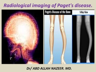
  • 1. Dr/ ABD ALLAH NAZEER. MD. Radiological imaging of Paget's disease.
  • 2. Paget disease (osteitis deformans) is a chronic skeletal disorder characterized by abnormal and excessive remodeling of bone. The disease was named after Sir James Paget who described the condition in 1877 in a detailed essay recognized in the medical literature as “a lesson in accurate and lucid writing” . Paget disease is estimated to affect approximately 3–4% of individuals older than 40 years and is the second most common bone disease after osteoporosis that affects the growing population of older persons in the United States. Abnormal osseous resorption and apposition in Paget disease produce variable clinical and imaging manifestations. Although the disease may be asymptomatic, it can be painful or deforming and associated with various and debilitating musculoskeletal complications. Radiologists play a central role in the imaging diagnosis of the process and its multifaceted manifestations. Radiography remains the most inexpensive method to evaluate patients with Paget disease of bone. CT, bone scintigraphy, and MRI have complementary roles in achieving the correct diagnosis of Paget disease and its complications.
  • 3. Paget disease of bone Paget disease of the bone is a common, chronic bone disorder characterised by excessive abnormal bone remodelling. It frequently affects the pelvis, spine, skull and proximal long bones and has characteristic radiographic features. Epidemiology It is relatively common and can affect up to 4% of individuals over 40 and up to 11% over the age of 80. There may be a slight male predilection. Incidence can be considerably higher in the United Kingdom than in other countries. It is also common in Australia, New Zealand, Western Europe, and the United States.
  • 4. Clinical presentation The majority (approximately three-quarters) of patients are asymptomatic at the time of diagnosis. Presenting symptoms include: localized pain and tenderness increased focal temperature due to hyperaemia (due to hypervascularity) increased bone size: historically changing hat size was a giveaway bowing deformities kyphosis of the spine decreased the range of motion signs and symptoms relating to complications (see below) Polyostotic disease is more prevalent than the monostotic type. The most frequent sites of involvement are: spine pelvis (often asymmetric) skull proximal long bones
  • 5. Pathology The etiology is not entirely known, but it is a disease of osteoclasts. Viral infection (paramyxovirus) in association with genetic susceptibility has been postulated. There are three stages classically described (but is part of continuous spectrum) lytic (incipient active): predominated by osteoclastic activity mixed (active): osteoblastic as well as osteoclastic activity sclerotic/blastic (late inactive) Markers serum alkaline phosphatase (ALP) elevated normal calcium and phosphorous levels urine hydroxyproline increased
  • 6. Natural history and skeletal distribution: The primary event in Paget disease is intense focal resorption followed by disorderly bone formation that results in overall abnormal bone remodeling. Paget disease generally is considered a relentlessly progressive disorder that evolves through various stages or phases of activity followed by an inactive or quiescent stage. There is notable variability in the rate of disease progression from one patient to another or in a single individual. Three major phases are recognized: the lytic phase (incipient active), in which osteoclastic resorption predominates; the mixed phase (active), in which there is both osteoclastic and osteoblastic hyperplasia with predominant osteoblastic activity; and finally, the blastic phase (late inactive), in which osteoblastic activity gradually declines. These three phases of the pagetic process—coupling abnormal osseous resorption and apposition within the periosteal and endosteal cortical envelopes—account for the variable radiographic appearances of the disease. As a result of this anarchic bone behavior that produces disorganized new bone (mosaic), an increased or decreased external bone contour and a narrowed or enlarged marrow cavity are seen, as opposed to bone expansion that was long regarded as a universal feature of the disease.
  • 7. The anatomic distribution of Paget disease usually is asymmetric and most commonly affects the lumbar spine (30– 75%), pelvis (30–75% of cases), sacrum (30–60%), femur (25– 35%), and cranium (25–65%). Less frequently, however, cervical and thoracic involvement can be observed. There is a preference for the lower extremities and a tendency for right- sided alterations. The shoulder girdle, particularly the proximal humerus (31%) and scapula (24%), are less commonly affected sites. Pagetic involvement of the ribs, fibula, and small bones in the hands and feet is infrequent. Polyostotic disease (65–90%) is more frequent than monostotic disease. In some patients, however, the disease is initially or totally monostotic, a pattern that is evident in 10– 35% of cases. Monostotic Paget disease appears to predominate in the axial skeleton, although every bone in the skeleton can be the sole site of involvement.
  • 8. Radiographic features Plain radiograph The early phase features osteolytic (lucent) region which is later followed by coarsened trabeculae and bony enlargement. Sclerotic changes occur much later in the disease process. Skull osteoporosis circumscripta: large, well-defined lytic lesion cotton wool appearance: mixed lytic and sclerotic lesions of the skull diploic widening: both inner and outer calvarial tables are involved, with the former usually more extensively affected Tam o'Shanter sign: frontal bone enlargement, with the appearance of the skull falling over the facial bones, like a Tam o' Shanter hat Spine picture frame sign: Paget disease of the spine frequently manifests with cortical thickening and sclerosis encasing the vertebral margins, which gives rise to the on radiographs in mixed phase disease squaring of vertebrae: on lateral radiographs flattening of the normal concavity of the anterior margin of the vertebral body also adds to the rectangular appearance vertical trabecular thickening: in Paget disease is coarser than the more delicate pattern seen in hemangiomas with which it can be confused
  • 9. Pelvis cortical thickening and sclerosis of the ileopectineal and ischiopubic lines acetabular protrusio enlargement of the pubic rami and ischium These findings are often asymmetric and for some reason, may be more commonly seen on the right side. Long bones blade of grass or candle flame sign: begins as a subchondral area of lucency with advancing tip of V-shaped osteolysis, extending towards the diaphysis in rare cases, the disease is isolated to the diaphysis, most commonly in the tibia, rather than subchondral bone, which can cause diagnostic confusion lateral curvature (bowing) of the femur anterior curvature of the tibia Signs: Paget disease related signs include: Tam o' Shanter sign blade of grass sign osteoporosis circumscripta jigsaw pattern bone or mosaic pattern bone picture frame vertebra cotton wool appearance of bone banana fracture Looser zones ivory vertebra sign
  • 10. MRI The overall signal characteristics are variable, likely reflecting the natural course of the disease process in different phases. Several major patterns of involvement have been described dominant signal intensity in pagetic bone similar to that of fat; most common pattern: probably corresponds to the early mixed active phase relatively low T1 and high T2 signal alteration (also referred as a “speckled” appearance); second most common pattern: probably corresponds to granulation tissue, hypervascularity, and edema seen in active disease low signal intensity on both T1 and T2 images: suggesting the presence of compact bone or fibrous tissue; least common pattern: seen in late blastic inactive phase Fatty marrow signal is usually preserved in all sequences unless there is a complication. Bone scintigraphy Tc99m-MDP highly sensitive but not specific traditionally has been said to demonstrate marked increased uptake in all phases of the disease, although in the burnt out sclerotic quiescent phase uptake may be normal.
  • 11. Complications of Paget disease: Common complications of Paget disease include osseous deformities, fractures, Osteoarthrosis, basilar impression, spinal stenosis, and neurologic abnormalities. Other less commonly encountered complications are bone tumor, soft-tissue mass, osteomyelitis, extramedullary hematopoiesis, rheumatoid arthritis and its variants, and crystal deposition disease. Arthropathy Osteoarthrosis is a common complication in Paget disease. This complication results from altered biomechanics across grossly abnormal bones and joints, producing cartilaginous and osseous degeneration. The hip and the knee are the articulations most frequently affected. The pattern of narrowing of the articular space in Paget disease differs in appearance from that in primary degenerative joint disease. With acetabular involvement, either medial or axial joint space narrowing is seen; with involvement of both the acetabulum and the femoral head, axial joint space loss can occur; and with isolated femoral head involvement, superior joint space loss is noted. Acetabular protrusion may complicate pagetic involvement of the acetabulum. Notably, the formation of osteophytes is not a prominent feature of the disease. Rheumatoid arthritis and its variants, as well as crystal deposition arthropathy, also have been associated with Paget disease.
  • 12. Deformity and Fracture In Paget disease, overall abnormal bone formation results in osseous weakening, with deformity and fractures being common manifestations of the disease. Anterior or lateral bowing of the tibias or femurs is typical. In the spine, bowing results in kyphosis; in the hip, deformity may manifest as protrusio acetabuli. Fracture is the most common complication of Paget disease, which can be crippling because of the high frequency of nonunion in diseased bone. Insufficiency fractures appear as single or multiple linear cortical radiolucent areas on the convex surface of the long bones and are designated “banana fractures”. These pathologic fractures usually occur in the femur, tibia, humerus, pelvis, and spine. In the femur, which is the most common site of fracture, fractures are usually subtrochanteric in location, followed in decreasing frequency by the upper third of the femoral shaft and the femoral neck. Neurologic Entrapment Neurologic deficits in Paget disease may relate to abnormalities at the base of the skull or the spine. Calvarial enlargement can cause cranial nerve compression with neurosensory disturbances, deafness, blindness, muscle palsies, and trigeminal neuralgia. Basilar impression, hydrocephalus, and vertebrobasilar insufficiency are additional complications of Paget disease affecting the craniocervical junction. Spinal stenosis, compression of the spinal cord, and cauda equina can be caused by enlargement of the pagetic vertebral bodies or posterior elements, spinal deformity and fracture, vertebral collapse with hemorrhage, vascular compromise, or secondary neoplasm. Entrapment of the sciatic nerve may present between an enlarged ischium and lesser trochanter or between the abnormal ilium and piriformis muscle.
  • 13. Neoplasms Neoplastic involvement complicating Paget disease is unusual and includes sarcomatous transformation; giant cell tumor; and superimposed tumorous conditions, such as metastatic disease, plasma cell myeloma, and lymphoma. The reported frequency of sarcomatous degeneration varies according to the extent of the pagetic process. In patients with widespread skeletal involvement, sarcomatous degeneration may occur in 5– 10% of cases, whereas in patients with less extensive skeletal disease, neoplasm may occur in fewer than 1% of cases. Patients usually are between 55 and 80 years old, with men affected more commonly than women (2:1 ratio). Clinical findings include focal pain, swelling, or pathologic fracture. Although virtually any bone can be involved, the bones most commonly affected are the femur, the pelvic bones, and the humerus and occasionally the skull and vertebral column. Osteosarcoma predominates (50–60% of cases) followed by malignant fibrous histiocytoma or fibrosarcoma (20–25%); chondrosarcoma (10%); and, less commonly, lymphoma and angiosarcoma (1–3%). The radiographic findings of malignant transformation comprise osteolysis, cortical destruction, bony speculation, no healing fracture, and a soft-tissue mass. Osteosclerosis or periostitis is infrequent. On bone scans, sarcomatous degeneration may appear as a “cold” area of absent uptake, representing decreased accumulation of the tracer at the site of neoplasm and necrosis. On CT images, neoplastic involvement in pagetic bone appears as osseous destruction and an extra-osseous mass. MRI shows nonspecific signal intensity characteristics of low to intermediate signal intensity on T1-weighted images and inhomogeneous low to high signal intensity on T2-weighted images in affected bone. Prominent inhomogeneous enhancement in pagetic bone complicated by neoplastic disease is evident after the IV administration of contrast material.
  • 14. Giant cell tumor is another type of neoplasm associated with Paget disease, although this association is very rare and has been considered by some authorities a giant cell reparative granuloma. The tumor almost always occurs in the skull or the facial bones, but giant cell tumor can involve the pagetic pelvis, the clavicle, the spine, and the tubular bones. Patients with giant cell tumor complicating Paget disease in general are elderly persons and have polyostotic involvement. Notably, the tumor more frequently is benign than malignant and may show dramatic response to the use of steroids alone. On radiographs, the tumor appears as a lytic expansile lesion with or without an associated soft-tissue mass. MR images display an osseous segment with the variable signal characteristics of pagetic bone and a soft-tissue component of intermediate signal intensity on T1-weighted images and of focal increased signal on T2-weighted images. Cystic and hemorrhagic regions may be present in the tumor. Although reportedly rare, metastatic disease involving either the non pagetic bones or the pagetic bones may be associated with Paget disease. A lytic lesion within sclerotic bone and a soft-tissue mass may be seen in patients with metastases superimposed on Paget disease . Other neoplastic conditions complicating Paget disease include plasma cell myeloma, lymphomas, and leukemia's, although these associations may be random.
  • 15. Frontal radiograph of right tibia of 67-year-old woman with Paget disease shows well-defined wedge area of osteolysis (arrow) in diaphysis of tibia and blade-of-grass appearance.
  • 16. Lytic phase of Paget disease (“blade-of-grass” appearance) of the appendicular skeleton in different patients. (a) Anteroposterior radiograph of the knee in a 74-year-old man shows a large area of lysis beginning in subchondral bone with a sharp inferior margin (arrowheads). (b) Lateral radiograph of the upper tibia in a 41-year-old man shows a lytic lesion in the tibial tubercle, with blade-of-grass appearance superiorly and inferiorly (arrows).
  • 17. Non-complicated active Paget disease of the left distal tibia in an asymptomatic 45-year-old man with fibrovascular marrow. (a) Anteroposterior radiograph of the ankle shows typical cortical and trabecular thickening from Paget disease. (b) Coronal T1-weighted MR image (500/14) reveals patchy marrow replacement in the left distal tibia, although small foci of maintained yellow marrow are seen (arrows). (c) Axial fat-suppressed T1-weighted MR image (600/15) obtained after intravenous injection of gadolinium reveals patchy speckled enhancement in the marrow space (*) and particularly prominent intracortical enhancement (arrowheads). (d) Axial fat-suppressed T2-weighted (5,333/84) MR image shows heterogeneous speckled high signal intensity in the marrow (*) without cortical destruction or soft-tissue mass.
  • 18. Lateral skull radiograph of 67-year-old woman with Paget disease shows area of osteolysis (arrowheads) in frontal region that is designated “osteoporosis circumscripta” in lytic phase of Paget disease.
  • 19. Lytic phase of Paget disease of the skull in different patients with osteoporosis circumscripta. (a) Lateral radiograph of a 50-year-old man shows a well-defined area of lysis in the frontal and occipital regions (arrowheads). (b) Lateral radiograph of a 60-year-old woman with osteoporosis reveals frontal and occipital areas of osteolysis (*) that are more difficult to detect in this clinical setting. (c) Bone scan of the same patient as in b reveals intense uptake of radionuclide in the frontal and occipital areas, as well as in the face. (d) Axial CT scan of a 55-year-old man with mild expansion of the head reveals a lytic lesion with sharp borders (arrows) in the frontal bone. (e) Photograph of a whole- mounted, longitudinal section of the calvaria (H-E stain) shows calvarial expansion and extensive fibrovascular tissue in the diploic space (*).
  • 20. Blastic phase of Paget disease involving the calvaria. (a) Lateral radiograph of the skull in a 56-year-old man shows diffuse calvarial thickening including the calvaria and maxillary sinus region, with several areas of focal sclerosis (“cotton wool” appearance) (arrowheads). (b) Lateral radiograph of a skull specimen from an 85-year-old woman reveals diffuse sclerosis and calvarial thickening with platybasia. (c) Axial CT scan (same case as b) also reveals the posterior thickening and mixed lysis and sclerosis (area between arrows).
  • 21. Lateral skull radiograph of 64-year-old woman with Paget disease reveals several areas of focal sclerosis (arrowheads) that produce cotton-wool appearance. There is diffuse calvarial thickening.
  • 22. Cotton wool appearance : A plain radiograph sign of late stage Paget Disease. Its appearance is due to thickened, disorganized trabeculae which lead to areas of sclerosis in a previously lucent area of bone – typically the skull.
  • 23. Paget disease of the skull base in a 70-year-old man. Axial CT scan demonstrates diffuse expansion and sclerosis of the bones of the skull base, characteristic of Paget disease. Note the sparing of the maxillofacial bones, which, along with the age of the patient, is a helpful feature in differentiating it from fibrous dysplasia.
  • 24. Radiography of the skull (Panel A) showed thickening of the outer and inner tables of the cranial bones, widening of the diploë, and a “cotton wool” appearance caused by irregular areas of sclerosis (arrows). Computed tomography of the skull (Panel B) confirmed bony expansion, cortical bone thickening, and irregular areas of sclerosis (arrows). These imaging findings reflect the mixed osteolytic and osteoblastic phases of Paget's disease, resulting in accelerated bone turnover with bone deposition and expansion.
  • 25. Paget's Disease with diffuse calvarial thickening.
  • 26. Paget disease of skull. A and B, Axial T1-weighted (TR/TE, 400/20) (A) and T2-weighted (4300/100) (B) MR images of head exhibit abnormal heterogeneous, decreased signal intensity in expanded calvaria. Several areas of low signal intensity (arrowheads) representing focal osteosclerosis are seen. C, Axial CT image shows marked calvarial thickening and sclerosis.
  • 27. Non complicated Paget disease of the skull in a 72-year-old woman. Axial T1-weighted (600/20) (a) and T2-weighted (4,000/75) (b) MR images of the head show diffuse calvarial thickening that remains low signal intensity with both pulse sequences (*). Radiograph (not shown) revealed diffuse sclerosis.
  • 28. Paget disease complicated with basilar impression in 65-year-old woman. Sagittal T1-weighted MR image (TR/TE, 500/16) displays basilar impression and compression of anterior aspect of pons. Note marked deformity of tip of odontoid process that may be related to pagetic process. Cerebellum (C) is also compressed. Basilar impression is diagnosed when tip of odontoid process (arrow) projects more than 5 mm above Chamberlain line, which is drawn from hard palate (HP) to basiocciput (O). Calvaria (arrowheads) appears expanded and displays heterogeneous, predominantly low signal intensity.
  • 29.
  • 30. Frontal radiograph of 62-year-old man with Paget disease shows thickening of ileopectineal (arrow) and ilio ischial (arrowhead) lines on right side of pelvis. Coarse trabeculation and bone sclerosis are also evident as compared with uninvolved side. Contrast medium is noted in urinary bladder.
  • 31. Frontal radiograph of 57-year-old man with Paget disease reveals cortical thickening (arrowheads) encasing entire T11 vertebral body at all four margins, which manifests as picture-frame appearance. Vertebral body is enlarged and mildly compressed, and osteophytes are seen.
  • 32. Paget disease of the spine in different patients. (a) Anteroposterior radiograph of the lumbar spine in a 45-year-old man shows subtle vertical trabecular thickening (white arrowheads) and early picture frame appearance with condensation of trabeculae about the superior and inferior endplates (black arrowheads). (b) Lateral radiograph of the lumbar spine in a 54-year-old woman shows cortical thickening (picture frame appearance) about the entire vertebral body at all levels (arrowheads). (c) Sagittal CT reformatted image in a 50-year-old man shows similar trabecular thickening (arrowheads) with extension into the posterior vertebral elements (*).
  • 33. Ivory vertebral body caused by Paget disease in a 50-year-old asymptomatic man. (a)Anteroposterior radiograph shows diffuse sclerosis in the T10 vertebral body. (b) Posterior bone scan reveals intense radionuclide uptake at the involved vertebral level (see Fig 5d for histologic specimen of an ivory vertebral body).
  • 34. Paget disease of spine in different patients. A, Axial CT scan of T11 vertebra in 57-year-old man shows mixed lytic and sclerotic changes involving vertebral body and arch. B, Axial CT scan of 60-year-old man shows predominant osseous sclerosis of L5 vertebral body, ivory vertebra, that is extending into posterior osseous elements.
  • 35. Non complicated Paget disease of the calcaneous in a 58-year-old asymptomatic man. Sagittal T1-weighted (repetition time msec/echo time msec = 500/20) (a) and T2- weighted (2,000/80)(b) MR images show cortical and trabecular thickening (arrowheads) with maintained yellow marrow (*). In fact, there is more fat in the affected calcaneal marrow, compared with that in the noninvolved talus.
  • 36. Images obtained of 73-year-old woman with Paget disease at femur. A, Frontal radiograph of distal femur reveals coarsened trabeculae, cross-hatched pattern (arrowheads), and cortical thickening (arrow). B, Axial T1-weighted MR image (TR/TE, 450/16) shows thickened trabeculae (arrowheads) within maintained yellow marrow (asterisk). Cortical thickening (arrows) is appreciated. Preservation of medullary fat in femur excludes superimposed sarcoma. C, Axial T2- weighted MR image (3700/80) shows trabecular thickening (arrowheads) with pagetic bone marrow (asterisk) displaying high signal intensity similar to that of fat. Fat-saturated MR image would have allowed better characterization of process.
  • 37. Non complicated Paget disease of the distal femur in a 57-year-old woman. (a)Anteroposterior radiograph of the knee shows typical cortical and trabecular thickening of the entire distal femur to subchondral bone. (b) Coronal T1-weighted MR image (600/20) shows identical changes with maintained yellow marrow (*) between thickened trabeculae. (c) Coronal fat-suppressed short-inversion-time inversion-recovery MR image (2,000/30/100) reveals mild increased signal intensity in the marrow resulting from small fibrovascular elements
  • 38. A, Lateral radiograph shows picture-frame appearance of Paget disease with condensation of trabeculae at periphery of T11 vertebral body. There is mild straightening of normally concave anterior surface of bone. B and C, Sagittal T1-weighted (TR/TE, 500/15) (B) and fat-suppressed T2-weighted (5000/90) (C) MR images display abnormal intermediate and heterogeneous high signal intensity, respectively, in posterior aspect of T11 vertebra (arrows). Note coarsened trabeculae, in anterior aspect of vertebral body (asterisk, B). D, Sagittal fat- suppressed T1-weighted MR image (550/19) obtained after gadolinium administration reveals marrow enhancement (black arrowhead) and marked cortical enhancement (white arrowheads) in involved vertebra.
  • 39. Mixed lytic and blastic Paget disease of the spine simulating malignant transformation in a 60-year-old man. (a) Lateral radiograph of the thoracic spine shows typical picture frame appearance of Paget disease with cortical and trabecular thickening (arrowheads). (b) Axial CT scan reveals similar changes with a focal area of lysis posteriorly (arrows) that could be caused by lysis from Paget disease or malignant transformation. (c) Sagittal T1- weighted (600/25) MR image shows patchy areas of maintained yellow marrow signal intensity in the area of concern posteriorly (arrow), a finding that excludes malignant transformation. (d) Sagittal T2-weighted (2,500/90) MR image reveals high signal intensity in the lytic focus posteriorly (*), but there is no epidural soft-tissue mass. This area remained unchanged for 5 years and represents a lytic component of Paget disease.
  • 40. Paget’s disease of T11 and T12 showing an increased amount of high MR signal (solid white arrow) in the posterior epidural space at these levels on a sagittal T2-weighted, b sagittal T1-weighted and c axial T1-weighted images. This can be mistaken for epidural lipomatosis. However, the d CT sagittal and e axial images demonstrate this to be due to the fat density (solid white arrow) within the expanded posterior neural arch involved in PD. The axial images (c,e) were obtained at the level of the tip of the solid arrows on sagittal images. There is also fusion of the vertebrae (dashed arrow) across the intervertebral disc. The combination of anterior and posterior vertebral involvement in this case resulted in severe spinal canal narrowing and cord compression. Note the high T1 signal indicating a high fatty marrow content within the pagetic T11 and T12 vertebrae.
  • 41. Paget disease: Both the L1 and L2 vertebral bodies (together with their respective posterior elements) are involved, with the vertebral bodies expanded in the antero-posterior dimension and L2 a characteristic “picture frame” vertebra. There is involvement as well of the pelvis together with the sacral vertebral bodies. Coarse and thickened trabeculae are particularly evident in the iliac bones bilaterally.
  • 42. Paget disease complicated with degenerative joint disease in 70-year-old man. Frontal radiograph shows pagetic involvement of right hemipelvis with trabecular thickening and sclerosis in ilium and along ileopectineal and ilio ischial lines. Involvement of subchondral bone of ilium around acetabulum is remarkable. Symphysis pubis appears enlarged on right side. Joint space loss in superior and axial aspects of hip and mild protrusio acetabuli are noted. Osteophytes are not prominent. Urinary bladder stone is present.
  • 43. Paget disease of the hip with osteoarthritis. Anteroposterior radiograph of the left hemipelvis shows extensive Paget disease with osteoarthritis involving the hip. The axial pattern of narrowing of the hip (arrowheads) is unusual and is likely caused by the underlying osseous weakening and mild protrusio acetabuli.
  • 44. Anterior bone scan of 67-year-old woman with Paget disease shows intense, increased radionuclide uptake in right hemipelvis.
  • 45. Paget disease complicated with deformity and fracture. Frontal radiograph of 73-year-old woman shows lateral bowing and bone widening of both femurs. Cortex is thickened and cortical encroachment on medullary canal is seen. Large fracture line (arrow) extends across mid diaphysis of left femur. Left femoral shaft displays subtle un-displaced “saw-cut” fatigue fractures (black arrowheads). In femur, fatigue fractures typically occur along lateral cortex, where tensile forces are greatest, leading to lateral bowing. Stress fracture (white arrowhead), which will most likely progress to complete fracture, is barely seen in lateral cortex of right femur.
  • 46. Paget disease of the spine with associated fracture in a 50-year-old woman. (a) Lateral radiograph of the lumbar spine shows the typical picture frame appearance of Paget disease with severe compression (vertebrae plana) at L4. Paget disease with both incomplete and complete fractures of the femur in a 70-year-old woman. (a) Anteroposterior radiograph of the femur shows pathognomonic changes of Paget disease: cortical and trabecular thickening extending to subchondral bone and subtrochanteric complete fracture. Additional incomplete fractures (arrowheads) are seen along the lateral convex femoral surface with callus formation.
  • 47. Transport of Paget disease from iliac crest bone graft donor site to engrafted proximal tibial area in a 64-year-old man. (a) Anteroposterior radiograph of the knee shows a depressed lateral tibial plateau fracture (arrow) sustained in a motor vehicle accident. (b) Postoperative anteroposterior radiograph shows bone graft and elevation of the depressed fragment fixed by a single lag screw. (c, d) Follow-up anteroposterior radiographs obtained 2 (c) and 3 (d) years later show progressive development of characteristic Paget disease with cortical (large arrow) and trabecular (small arrows) thickening and a sharply defined, blade-of-grass lucent area (arrowheads). (e) Radiograph of the pelvis reveals extensive Paget disease throughout the pelvis and proximal femora, including the bone graft donor site (*) in the left iliac crest.
  • 48. Recurrent lytic phase of Paget disease after tibial tubercle avulsion with fixation and non-weight-bearing in a 75- year-old man. (a) Lateral radiograph of the lower leg shows an avulsion of the patellar tendon attachment at the tibial tubercle (black arrow). Changes of Paget disease, with trabecular and cortical thickening (white arrow), are subtle. (b, c) Follow-up anteroposterior radiographs obtained 2 weeks (b) and 2 months (c) after screw fixation, bracing, and non-weight-bearing reveal progressive osteolysis (white arrowheads) with apparent focal destruction of the medial cortex (open arrow). Changes of Paget disease with trabecular (black arrowheads) and cortical (curved arrow) thickening and sharp inferior margin of lysis (solid straight arrow) are now more apparent.
  • 49. Paget disease of leg bone
  • 50. Paget disease complicated with malignant transformation in 81-year-old man with long history of disease. Analysis of biopsy specimen proved lesion to be osteosarcoma. A, Radiograph shows extensive bone destruction with osteolysis and cortical loss in distal portion of humerus. Remainder of bone shows changes of Paget disease. Associated soft-tissue mass is readily apparent. B, Corresponding transverse CT image reveals pagetic involvement of humerus.
  • 51. Sarcomatous transformation to osteosarcoma of Paget disease of the humerus in a 77-year-old woman. (a) Anteroposterior radiograph of the humerus shows typical cortical and trabecular thickening from Paget disease involving the entire humerus and lateral bowing. Area of medullary sclerosis with mineralization in the soft tissue is also seen (arrowheads). (b) Sagittal T2-weighted (3,500/105) MR image reveals focal marrow replacement and associated soft-tissue mass (*). (c)Anteroposterior radiograph and photograph of the coronally sectioned gross specimen show the osteosarcoma (*) in the humeral mid shaft and adjacent soft tissues (arrow).
  • 52. Sarcomatous transformation of Paget disease in the tibia to malignant fibrous histiocytoma in a 75-year-old woman. (a) Lateral radiograph of the tibia shows typical changes of Paget disease, with cortical and trabecular thickening involving the entire bone and anterior bowing. Focal destruction of the anterior cortex is seen superiorly, with a soft-tissue mass (arrowhead). (b) T1-weighted (500/20) MR image shows focal marrow replacement at the proximal tibial site of malignant transformation with cortical destruction and associated soft-tissue mass (*). Yellow marrow signal intensity is seen throughout the remainder of the tibia. (c) T1-weighted (500/20) MR image obtained with gadolinium shows marked enhancement. T2-weighted MR image (not shown) revealed similar high signal intensity in the sarcoma.
  • 53. GCT of the face associated with Paget disease. (a) Axial CT scan shows characteristic thickening and sclerosis of the diploic space (arrows) caused by Paget disease. Expansile mass involving the maxillary sinus (*) represents the associated GCT. (b, c) Coronal T1-weighted (500/20)(b) and T2-weighted (2,500/80) (c) MR images obtained after intravenous injection of gadolinium also reveal Paget disease with calvarial thickening and maintained yellow marrow signal intensity (arrowheads). The maxillary GCT shows focal expansion, intermediate signal intensity with both pulse sequences, and mild diffuse enhancement (*).
  • 54. Whole Body Bone Scan indicating Paget's disease.
  • 55. Bone scan of a patient who has Paget's disease. The images were acquired 2.5 h after injection of 925 MBq of 99mTc-methylene diphosphate.
  • 56. Polyostotic Paget disease with multiple static planar bone Scintigraphic images showing increased tracer uptake in skull, multiple vertebrae (Mickey Mouse sign), pelvis on both sides, sacrum, and distal one-third of right femur.
  • 57. Paget disease of the skull.
  • 58. Mickey Mouse Sign-increased radiotracer uptake on bone scan in pedicles and spinous process in Paget's disease.
  • 59. Conclusion: Paget disease is a common skeletal disorder of middle-aged and elderly persons characterized by excessive and abnormal remodeling of bone. The disease varies considerably in severity and evolves through various phases of activity, followed by an inactive phase. The radiographic features of Paget disease are virtually diagnostic, including an initial osteolytic phase and a subsequent osteosclerotic phase. Bone enlargement with increased radiodensity, accentuated trabecular pattern, and deformity is typical. Complications associated with Paget disease are pathologic fractures, neurologic symptoms, skeletal deformities, articular derangements, and secondary neoplasms. CT and MRI help delineate pagetic bone changes and have prove extremely useful in the diagnosis of sarcomatous transformation, which constitutes the most dreaded complication of the disease.