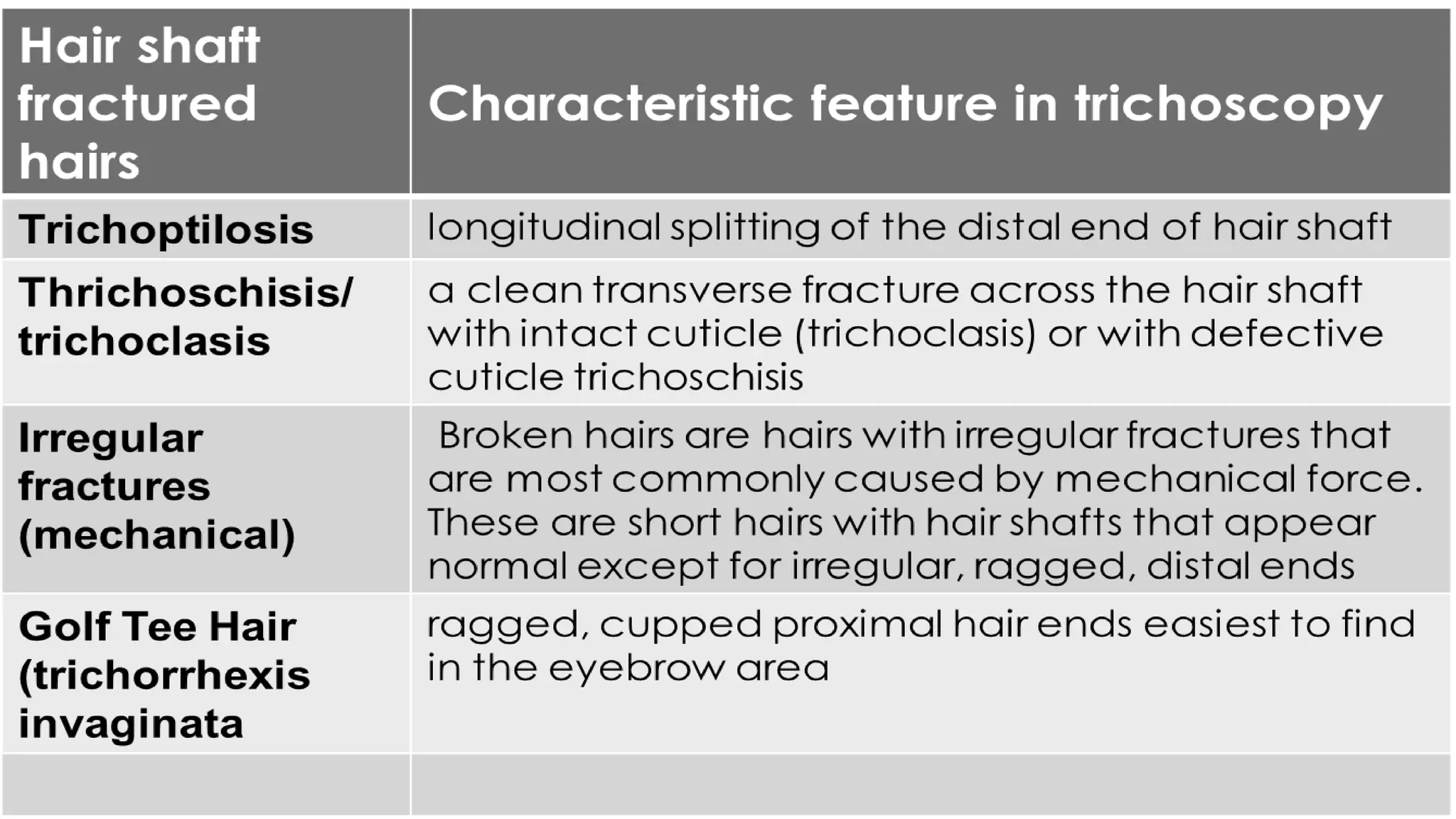
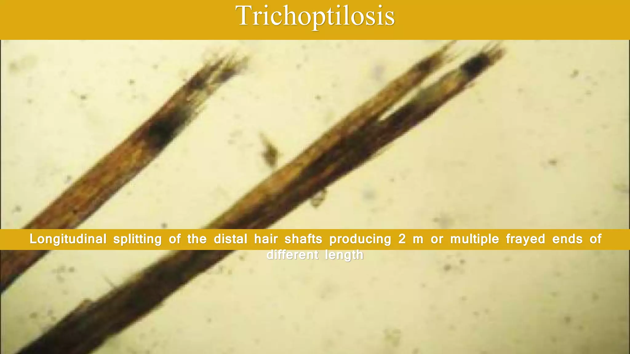
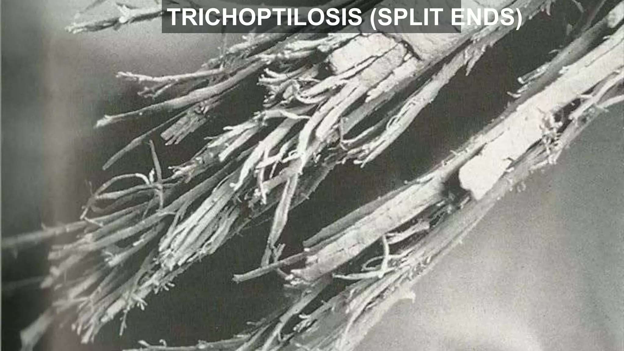
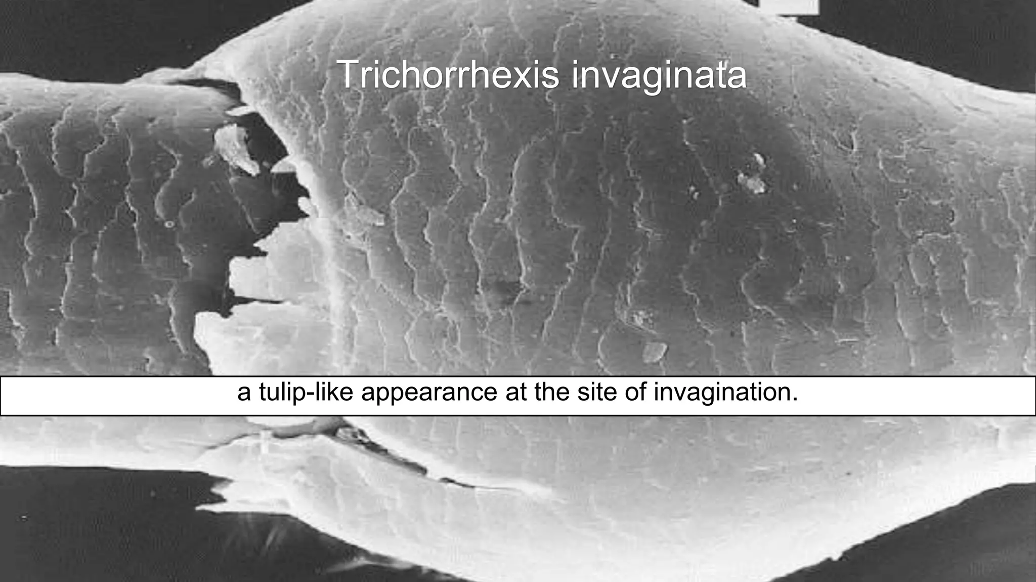
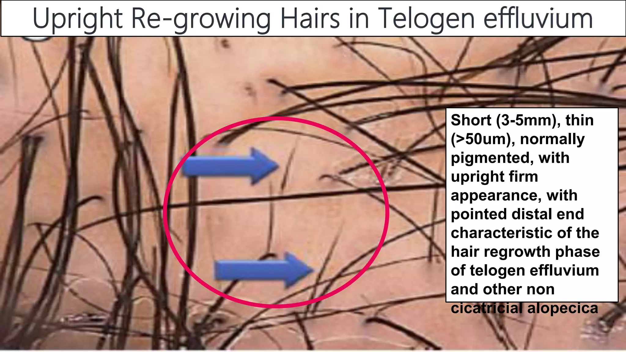
Trichoptilosis by Hair Styling Procedure
It is a symptom of hair shaft damage (“hair weathering”) that may be caused
by hair-damaging environmental factors and cosmetic procedures
A long longitudinal break
Trichoptilosis
Longitudinal splitting of the distal hair shafts producing 2 m or multiple frayed ends of
different length
TRICHOPTILOSIS (SPLIT ENDS)
Trichoclasis
A clean transverse fracture across the hair shaft bounded only by intact
cuticle develop secondary to conditions that weaken the hair
Trichoschisis
A complete clean transverse fractures across the hair shaft with Localized
Absence of the Cuticle commonly associated with trichothiodystrophy
Note cuticular damage, which leads to transverse fractures in the hair shaft. Polarized
light would help to differentiate trichoschisis from trichothiodystrophy
Trichoschisis
Irregular, ragged distal end in trichotillomania
Broken Hairs
Hairs are broken
at different levels
above the scalp
Golf tee hairs
Golf tee hairs
Golf tee hairs
the distal end of the
broken hair will have a
concave, cupped
appearance
Netherton’s
syndrome
Netherton syndrome
the expanded proximal end of an invaginate node after abreak has occurred
Golf tee hair’ appearance
NORMAL HAIR ENDS
SPLINT ENDS AND PAINT BRUSH APPEARANCEN
Tightly curled, with knots and breakage and in cross section, it is flattened or
Hair Shaft in African American
Trichorrhexis nodosa and trichoptilosis in a broken shaft
Trichonodosis (Hair Knotting)
observed in patients with short curly hair either spontaneously or in response to
scratching with little clinical signifcance
An acquired, transient condition as an effect of scratching or hair styling
procedures
TRICHONODOSIS(HAIR KNOTTING)
Trichonodosis
an incidental finding of little clinical significance in short curly hair
Hair cast (Peripilar kerating cast)
firm, white, freely movable tubular masses that encircle the hair shafts
Cylindrical and hyaline mass involving the hair shaft
Hair cast (peripilar cast)
Hair casts may be a
secondary phenomenon in
the course of psoriasis,
seborrheic dermatitis, or
lichen planopilaris.
Idiopathic hair
casts may be
observed in healthy
individuals
White nodules along
affected hair shafts results
from hair weathering at
free ends of long hair
Acquired Trichorrhexis nodosa
Causes of TN include mechanical,
chemical, and thermal damage,
mainly as a result of hairstyling and
processing.
Trichorrhexis Nodosa
At the site of these nodular thickenings, hairs bend with a rounded edge
Trichorrhexis Nodosa
Polarizing microscope
Broken hairs leave brush-like ends with numerous small fibers at the distal end of
the hair shaft
Acquired Trichorrhexis Nodosa
Brush like tip
Light microscopy of trichorrhexis nodosa
A broken, tapered end
The characteristic findings of
light microscopy are
sometimes obscured at the
distal end of the hair shaft.
Trichorrhexis nodosa
The disruption of the cuticle and the splaying out of the cortical cells.
Trichorrhexis Invaginata)
Hair shaft invaginates at several points
along the shaft irregularly and appears as
multiple small nodules spaced along the
shaft at irregular intervals.
Bamboo Hairs
Netherton syndrome.
bamboo, matchstick, and golf-tee hairs
‘‘regularly bended ribbon sign
bamboo-like appearance at
site of invagination
Trichorrhexis invaginata
Trichorrhexis Invaginata
Pathognomonic of Netherton syndrome
High-magnification
trichoscopy shows an
invagination of the
distal portion of the
hair shaft into its
proximal portion
forming a ball-in-cup
appearance
Trichorrhexis invaginata
a tulip-like appearance at the site of invagination.
Upright Re-growing Hairs in Telogen effluvium
Short (3-5mm), thin
(>50um), normally
pigmented, with
upright firm
appearance, with
pointed distal end
characteristic of the
hair regrowth phase
of telogen effluvium
and other non
cicatricial alopecica
Very short < 3mm, Very thin
< (30um), hypopigmented,
non medullated, weak
appearance with blurred
distal ends
Vellus hairs
Hair blocks are very short hair
with horizontal distal end
i-Hairs are hair
blocks with dark
distal end
TINEA CAPITIES
Tulip hairs in trichotillomania at low Magnifications
light-colored hair shafts with dark distal ends
Broken hairs with a
diagonal fracture surface
and a cuticle detached at
the distal end
Slightly thinner
at the base than
at the distal end
Tulip hairs at higher Magnifications
both cicatricial and non cicatricial alopecia reflecting
different pathologic mechanisms and histopathology
Broom fibers
Broom hairs in non cicatricial alopecia
Multiple short full-thickness hairs emerging from one follicular unit in a patient who
Flame hairs in in active trichotillomania
residues from recently pulled hairs
Typical monilethrix hairs with
regularly distributed nodes
and internodes
the defect is in the
internodes (constricted
sections)
Nodosities
correspond to the
normal hair caliber
Monilethrix
Regular nodes correspond to
normal hair shaft thickness,
contain medulla whereas the
internodes are narrowing
with no medulla
Monilethrix
Hairs are bent regularly at
multiple locations and have a
tendency to fracture at
constriction sites
Monilethrix of Eyebrows
Regular spaced elliptical nodes
Pseudo-monilethrix
Irregularly spaced nodes
wider than the
normal shaft,
occurring near
the site of
overlap of the
hairs
Some authors consider it an optical illusions
Denting of the hair at the site of pressure from an overlapping hair
Pseudo-monilethrix
Pseudomonilethrix .
Rectangular indentations on one side of the hair shaft must be differentiated from
irregular fusiform narrowins of Pohl Pinkus constrictions
These constrictions occur most commonly when the metabolic and mitotic activity of the follicle are
rapidly and repeatedly suppressed by an external or internal factor
Pohl-Pinkus Constrictions
Monilethrix like hairs
Pohl-Pinkus constrictions
• Alopecia areata
• Chemotherapy
• Blood loss
• Severe nutritional
deficiencies
• Cicatricial alopecia
• Locoalized hereditary
hypotrichosis
Pohl-Pinkus constrictions or monilethrix like, the nodules appear wider
and the internodes has the same width as the hair shaft
Regularly distributed constrictions in monilethrix where the
nodes are same as hair shaft and internodes are contrictions
Differentiate monilethrix from monilethrix like
Monilethrix – like congenital hypotrichosis
spaces between narrowing are significantly
shorter and more superficial compared with
true monilethrix
almost invisible in
thicker hairs
Exclamation and Micro-exclamation Mark Hairs
Short hairs that are thin and hypopigmented at the proximal end and thicker and
darker at the distal end sometimes shorter than 1-2mm
Exclamation and Micro-exclamation Mark Hairs
• Alopecia areata
• chemotherapy
• anagen effluvium
• Trichotillomania
Tapered hairs (Hair Coudability)
hairs that are thin at the proximal end and become normal distally are called tapered
hairs when the hair is longer than one dermoscopic field of view (×70)
Not pathognomonic for alopecia areata
Tapered hairs (Hair coudability)
Very long exclamation mark or Pohl-Pinkus constrictions
Re-growing Pigtail hair
Short, regularly twisted hairs (circular or oval) with tapered ends
result from rapid hair
regrowth, before full
recovery of the hair
follicle. .
Re-growing pigtail hairs
Although most characteristic for alopecia areata, regrowing pigtail hairs also may be
present in cicatricial alopecia
Comma Hair and Corkscrew
Comma hairs with multiple
twists form corkscrew- like
structures.
characterized by
homogeneous thickness and
pigmentation of the hair shaft
and a sharp diagonal end
short comma-like hairs
are a hallmark of tinea capitis in patients of all skin phototypes
corkscrew hairs
Comma hairs often coexist
with corkscrew hairs.
Comma Hairs
the presence of multiple comma hairs in focal alopecia is
pathognomonic for tinea capitis
Coiled hairs in trichotillomania
After fracturing, the remaining part of the hair coils
irregularly at the fracture site giving different
appearance
Coiled or Hook like hair
differ from re-growing pigtail hairs in their irregular, oval appearance and blunt end.
Occasionally, they may have a hook-like
appearance
Zigzag or Z hairs
Any disease that cause focal weakening of the hair shaft
Pili torti
Twists of hair shafts along the long axis, usually through an angle of180
Pili torti has numerous causes, both inherited and acquired
Pili Torti in low magnification
Hair shafts slightly bent at
different angles at irregular
intervals
PILIT TORTI HAIR
A common, nonspecific symptom associated with many types of hair loss, both
inherited and acquired.
Monilethrix / Pili Torti
repeated dilations and constrictions of
the hair shaft
relatively flat shape of the
hair fiber with regular twists
Monilethrix
Pili Torti
a cyclical short, wavy hair that recalls a characteristic “moving snake” image;
Woolly hair
Woolly hair
Tightly coiled curls often thin and
hypopigmented hair shaft
resembling a crawling snake with
short wave cycles with increased
fragility
Hairs are flat, thinner, curly twisted, elliptical or oval in cross section some
hairs may have trichorrhexis nodos
Woolly hair
Pili Annulati
Bands are nearly the width of a hair and their borders are not clear-cut,
Pili Annulati
Air spaces in the cortexof the hair appear darker in light microscopy.
Pili Annulati
Pili Annulati with reflected light
Banded appearance of hair
Tinea capitis caused by Microsporum canis
Hairs with thin transverse bands (Morse code hairs)
Acquired hair shaft dystrophy in cicatricial alopecia
The most common reason for this condition is perifollicular fibrosis. Before a
follicle is destroyed, it produces various types of dystrophic hair shafts usually in
whitish or milky red areas with decrease of hair follicles number
Acquired hair shaft dystrophy in cicatricial alopecia .
it produces various types of dystrophic hair shafts.
emerge in whitish or milky red areas with a
decreased number of hair follicles
perifollicular fibrosis
Acquired hair shaft dystrophy in cicatricial alopecia .
The hair is short and very thick compared with other hairs in this patient with dissecting
cellulitis
Braiding damages hair shaft cuticle.
Broken Hair Shaft
The gas-filled bubbles are caused by excess physical stress on the hair shaft, appear
as white oval spaces with swiss chees structure
Light microscopy of Bubble Hair
HAIR TRICHOTHIODYSTROPHY
Cross-sectional microscopy demonstrating reniform and other abnormal shapes.
Un-combable Hair
Many cross-sections of hair fibers show typical triangular shapes
Uncombable hair syndrome
Shrinking tube technique
Pili trianguli et canaliculi on light microscopy
canal-like longitudinal depressions along the hair shaft
Triangular cross-section and canalicular depression or gutter along one side
Uncombable hair syndrome
Uncombable Hair Pili trianguli et canaliculi
Canal-like longitudinal depressions along the hair shaft
Bubble hair
Bubbles form within the hair cortex and medulla as a result of high temperatures from
Trichothiodystrophy
Flattened hair shafts, one twisting like a ribbon.
Tricho-thiodystrophy
Several areas of trichoschisis.
Flattened shaft twisted through 360°
Trichothiodystrophy
Trichoschisis
Trichothiodystrophy Light Microscopy
Trichothiodystrophy (TTD)
under polarizing microscopy, it displays a diagnostic alternating light and dark
banding pattern, called ‘‘tiger tail banding
Trichothiodystrophy TTD
Trichoschisis
Trichorrhexis nodosa-like fraying
showing absence of root sheaths and ruffled cuticle.
Loose Anagen Syndrome
Loose anagen hair with misshapen darkly pigmented bulb, short twisted segment
immediately distal to the bulb, absent inner root sheath, and“ ruffled” cuticle
Loose Anagen Syndrome
Mitotic arrest narrows the shafts and hair breaks when they reach
the scalp surface
Anagen arrest with chemotherapy
Examination of the hair shaft under polarized light microscopy
Normal hair
Minimal weathering of the cuticle with chipped cuticle cells.
Weathering of the cuticle with lifting of the cuticle cells.
severe weathering and loss of the cuticle.
Irregular ridging and fluting and disordered cuticle scale pattern
Trichothiodystrophy.
Infestations
(Pediculus Humanus Capitis
Nits attached to hair shaft
The head louse egg or nit is 0.8 mm in length
Head Louse Egg or Nit
able to differentiate lice nits containing nymphs from empty nits of hatched parasites
and from amorphous pseudonits and hair casts.
Empty nits
Incomplete Nit Attached to a Hair
Male human scalp louse
Dermatoscopy of pediculosis pubis
White Piedra
Nodules measure 1 to 1.5 mm in diameter; they are fusiform and of soft
consistency, mainly attached to the distal portions of the hair
Hair shafts appear coated by yellow or beige sheaths.
Occasionally red, green or light brown, soft, loose
White Piedra (Trichosporon beigelii)
Hard brown to black nodules, firmly attached
Black piedra (Piedra Hortae)
Trichorrhexis Nodosa
Trichomycosis Axillaris
concretions on the shafts of axillary and (less often) pubic hairs. The hair shafts develop
adherent yellow, red or black nodules or cylindrical sheaths that can be seen with the
naked eye
Superficial Corynebacterial Infection
Dirty scalp : prepuberal children Use
Use of scalp camouflage products Scalp staining
Scalp staining from hair dyes
Perifollicular Evaluation of hair follicle opening
• Normal
• Empty
• Fibrotic
• Biologic Material
Normal hair and scalp findings on trichoscopy
Number of hairs emerging from one follicular unit
One hair per follicular unit in androgenetic alopecia usually less than 30/% but
decreased in telogen effluvium and androgenic alopecia
Aniso-trichosis
Two hairs per follicular unit in
a healthy individual .
The number of hairs in a follicular unit
varies from one to three in healthy
persons
Some authors may consider this number borderline normal
Small hair tufts in
lichen planopilaris
tufts of five to seven hairs
The number of hairs in one follicular unit is increased in
tufted folliculitis
Large Tufted hairs is charachterisitic folliculitis decalvans
a tuft containing more than 20 hairs walled by a widened, hyperkeratotic, scaly
hair follicle opening
Broom hairs in non cicatricial alopecia
Multiple short full-thickness hairs emerging from one follicular unit
in a patient who otherwise has long hair
Multiple hair shafts emerging from the
same follicular ostium in a brush-like
pattern can differentiate pili multigemini
from compound follicles
Black dots
Black dots are residues of pigmented hairs that have
been broken or destroyed at the level of the scalp.
• Alopecia areata
• Tinea capitis
• Dissecting Cellulitis
• Tinea capitis
• Trichotillomania
Black dots and short vellus hairs
Alopecia areata in dark skinned individuals
Black dots are not
present in healthy
individuals or in
patients with PHL or
telogen effluvium
Yellow dots are hair follicle openings that contain keratosebaceous material. They may
be present in alopecia areata, DLE, and female PHL
Yellow dots, appearing as large 3-D soap bubbles imposed over dark dystrophic hairs,
are specific
Dissecting cellulitis
Large yellow dots (follicular keratotic plugs) are characteristic.
Discoid lupus erythematosus.
Dermoscopy guided biopsy, the area shows numerous yellow keratotic plugs
Chronic Cutaneous Lupus Erythematosus (CCLE)
White Dots
Pinpoint white dots correspond to hair follicle openings and eccrine gland openings,
regardless of hair loss seen on the normal scalp of dark skin phenotype
White Dots
White dots may appear as fibrotic white dots or pinpoint white dots
Chronic Cutaneous Lupus Erythematosus (CCLE)
Branching capillaries
White patches
Keratin plugs White dots
Blue grey dots
White Dots
Classic white dots represent areas of perifollicular fibrosis and are observed most
commonly in lichen planopilaris
Lichen Planopilaris
White Dots
Lichen Planopilaris
perifollicular scaleswhite dots
blue-grey dots
"target" pattern
Central, Centrifugal, cicatricial (CCCA)
single hairs or group of two hairs surrounded by a peripilar gray-white halo
shows reduced hair density
small pinpoint white dots
Red dots have been described in DLE and in individuals with vitiligo. Pink-grey and
grey dots have been observed in the eyebrow area of patients with frontal fibrosing
alopecia
Red. Pink, Grey
Dyschromia due to pigment incontinence secondary to interface dermatitis
and prominent follicular red dots; a good prognostic for possible hair regrowth
Discoid lupus erythematosus.
EPIDERMAL SCALING
Normal
psoriasis
Seborrheic
dermatitis
Diffuse
Lichen
plano-pilaris
Frontal
fibrosing
Alopecia
FOLLICULITI
S
DECALVANS
Peri
follicular
Epidermal scaling is a common finding in healthy individuals or associated with use of
hair cosmetic
Psoriasis
Discoid Lupus Erythematosus
multiple thick arborizing vessels
large yellow dots
fine scaling
Seborrheic dermatitis
Common Dandruff
Perifollicular scaling with some scales forming tubular structures around the emerging
hair shaft.
Lichen Planopilari (LPP) or
Frontal fibrosing alopecia (FFA)
TRACTION ALOPECIA
Dermatoscopy is very helpful to establish if hair style causing traction. It shows
small casts, mobile, cylindrical structures enveloping the proximal hair shaft and
may be mistaken for nits.
Tufted hairs in Folliculitis Decalvans .
Perifollicular scaling with the formation of tubular scaly structures around hair shafts is
observed in LPP (white) and in folliculitis decalvans (yellow)
NORMAL SCALP
Scalp color in dark-skinned individuals can vary from light brown to dark black
Another typical feature is
a perifollicular pigmented
network or honeycomb
pattern
A characteristic feature of the
pigmented scalp is the
presence of pin point white
dots corresponds to acro-
syringeal and follicular
openings
Sun exposure
Androgenic Alopecia
Peripilar sign
A brown halo, roughly 1 mm in diameter, around the emergence of the hair shaft
Ludwig type I hair loss patient
Brown peripilar signs
The presence of a pigmented network in a honeycomb pattern is an important feature
to distinguish from DLE because inflammatory process in the former is restricted to the
hair follicles
Lichen Planopilaris
Dyschromia due to pigment incontinence secondary to interface dermatitis
and prominent follicular red dots.
Discoid lupus erythematosus.
FRD was proposed as a normal trichoscopic feature, appearing to be related to the
rich vasculature that naturally envelops the normal hair follicle.
Follicular Red Dots (FRD)
Discoid lupus erythematosus.
The red dots are erythematous polycyclic, concentric structures, regularly
distributed around the follicular ostia and denotes good prognostic for hair
regrowth
markedly reduced follicular ostia, scale, erythema, and perifollicular blue-gray dots.
Frontal fibrosing alopecia
Discoid lupus erythematosus
The absence of pigmented network indicates destruction of the interfollicular
Speckled blue-gray dots
milky red areas and follicular plugging
The milky-red (strawberry ice cream) color is
indicative of fibrosis of recent onset
Absence of follicular openings manifesting as
empty yellow or black dots
Cicatricial alopecia
Evaluation of Blood Vessels
Appearance of cutaneous microvessels may vary in type, arrangement, and
number,is of special importance in differential diagnosis of inflammatory scalp
disease such as psoriasis, seborrheic dermatitis, lichen planopilaris and DLE
Several inflammatory scalp disorders are characterized by a
specific pattern of blood vessel arrangement on trichoscopy
Blood vessels
are C-shaped or slightly curved vessels with no branching. They are common in in
flammatory scalp diseases, such as seborrheic dermatitis and psoriasis, but also may be
observed in healthy individuals
Comma vessels or Linear Vessels
Hairpin elongated vessels are characteristic of cicatricial alopecia and are
observed most frequently in classic lichen planopilaris and folliculitis decalvans. This
vessel type typically is found in the perifollicular area, especially adjacent to tufted hairs
Hairpin ( linear looped ) Elongated vessels
Thin arborizing vessels usually are thinner than the average hair on the trichoscopic image. They are a
normal finding in the scalp, especially in the occipital and temporal areas . They are distributed regularly
between follicular units. Some authors suggest that thin arborizing vessels are characteristic of seborrheic
dermatitis; however, thus far, studies have not compared patients with seborrheic dermatitis with healthy
controls
Thin arborizing vessels
SEBORRHEIC DERMATITIS
the most common patterns are arborizing vessels and atypical red
vessels in the absence of red Dots and globules.
Lace-like vessels ( arrow ) are a combination of serpentine and looped vessels. These capillaries have a
serpentine shape and open ends that usually are spaced widely. These vessels are most characteristic of
psoriasis and correspond to the tortuous and dilated blood vessels within the elongated dermal papillae
seen on histopathology
Lace - like vessels
Glomerular (i.e., coiled or twisted) vessels ( arrows ), are most characteristic of psoriasis.
They usually are regularly arranged in lines or circles ( rings ). At lower magnification,
these circles appear as “red globular rings”
Glomerular ( coiled ) vessels
. Helical (corkscrew) vessels ( arrows ) are linear vessels twisted along a central axis. They are observed
rarely in scalp skin and never in healthy individuals. In noncancerous lesions, linear helical vessels are a
marker of severe inflammation and are observed in severe scalp psoriasis, pemphigus, and cutaneous T-
cell lymphoma.
Helical vessels or Corkscrew Vessels
A psoriatic plaque: dilated, elongated, “bushy” capillaries, homogeneously distributed
(twisted loop in 100% help to differentiate it from seborrheic dermatitis
PSORIASIS
Discoid Lupus Erythematosus
multiple thick arborizing vessels
large yellow dots
fine scaling
Yellow dots with radial, thin arborizing
vessels emerging from the dot are
considered characteristic
Characteristic large yellow dots and arborizing vessels
Discoid Lupus Erythematosus
large erythematous nodules on the occipital area with few remaining hairs.
DISCHARGE
BACTERIAL
INFECTIONS
TINEA
CAPITIS
FOLLICULITIES
DECALVAN
DISSECTINH
CELLULITIS
Dissecting folliculitis
Cultures from the pustules usually reveal Staphylococcus aureus
Folliculitisdecalvans
Perifollicular pustules are seen in active lesions.
DISCHARGE
HAIR SHAFT DISORDERS WITHOUT
INCREASED FRAGILITY
Scanning electron micrograph of hair surface
Eyebrows, eyelashes, and other body hair also may be evaluated
with trichoscopy with 0.9% NaCl as an immersion fluid

Hair Loss and Trichoscopy

  • 5.
    Trichoptilosis by HairStyling Procedure It is a symptom of hair shaft damage (“hair weathering”) that may be caused by hair-damaging environmental factors and cosmetic procedures A long longitudinal break
  • 6.
    Trichoptilosis Longitudinal splitting ofthe distal hair shafts producing 2 m or multiple frayed ends of different length
  • 7.
  • 8.
    Trichoclasis A clean transversefracture across the hair shaft bounded only by intact cuticle develop secondary to conditions that weaken the hair
  • 9.
    Trichoschisis A complete cleantransverse fractures across the hair shaft with Localized Absence of the Cuticle commonly associated with trichothiodystrophy
  • 10.
    Note cuticular damage,which leads to transverse fractures in the hair shaft. Polarized light would help to differentiate trichoschisis from trichothiodystrophy Trichoschisis
  • 11.
    Irregular, ragged distalend in trichotillomania Broken Hairs Hairs are broken at different levels above the scalp
  • 12.
    Golf tee hairs Golftee hairs Golf tee hairs the distal end of the broken hair will have a concave, cupped appearance Netherton’s syndrome
  • 13.
    Netherton syndrome the expandedproximal end of an invaginate node after abreak has occurred Golf tee hair’ appearance
  • 14.
  • 15.
    SPLINT ENDS ANDPAINT BRUSH APPEARANCEN
  • 18.
    Tightly curled, withknots and breakage and in cross section, it is flattened or Hair Shaft in African American
  • 19.
    Trichorrhexis nodosa andtrichoptilosis in a broken shaft
  • 20.
    Trichonodosis (Hair Knotting) observedin patients with short curly hair either spontaneously or in response to scratching with little clinical signifcance
  • 21.
    An acquired, transientcondition as an effect of scratching or hair styling procedures TRICHONODOSIS(HAIR KNOTTING)
  • 22.
    Trichonodosis an incidental findingof little clinical significance in short curly hair
  • 23.
    Hair cast (Peripilarkerating cast) firm, white, freely movable tubular masses that encircle the hair shafts
  • 24.
    Cylindrical and hyalinemass involving the hair shaft Hair cast (peripilar cast) Hair casts may be a secondary phenomenon in the course of psoriasis, seborrheic dermatitis, or lichen planopilaris. Idiopathic hair casts may be observed in healthy individuals
  • 25.
    White nodules along affectedhair shafts results from hair weathering at free ends of long hair Acquired Trichorrhexis nodosa Causes of TN include mechanical, chemical, and thermal damage, mainly as a result of hairstyling and processing.
  • 26.
    Trichorrhexis Nodosa At thesite of these nodular thickenings, hairs bend with a rounded edge
  • 27.
  • 28.
    Broken hairs leavebrush-like ends with numerous small fibers at the distal end of the hair shaft
  • 29.
  • 30.
    Light microscopy oftrichorrhexis nodosa A broken, tapered end The characteristic findings of light microscopy are sometimes obscured at the distal end of the hair shaft.
  • 31.
    Trichorrhexis nodosa The disruptionof the cuticle and the splaying out of the cortical cells.
  • 32.
    Trichorrhexis Invaginata) Hair shaftinvaginates at several points along the shaft irregularly and appears as multiple small nodules spaced along the shaft at irregular intervals. Bamboo Hairs
  • 33.
    Netherton syndrome. bamboo, matchstick,and golf-tee hairs ‘‘regularly bended ribbon sign
  • 34.
    bamboo-like appearance at siteof invagination Trichorrhexis invaginata
  • 35.
  • 36.
    Trichorrhexis Invaginata Pathognomonic ofNetherton syndrome High-magnification trichoscopy shows an invagination of the distal portion of the hair shaft into its proximal portion forming a ball-in-cup appearance
  • 37.
    Trichorrhexis invaginata a tulip-likeappearance at the site of invagination.
  • 40.
    Upright Re-growing Hairsin Telogen effluvium Short (3-5mm), thin (>50um), normally pigmented, with upright firm appearance, with pointed distal end characteristic of the hair regrowth phase of telogen effluvium and other non cicatricial alopecica
  • 41.
    Very short <3mm, Very thin < (30um), hypopigmented, non medullated, weak appearance with blurred distal ends Vellus hairs
  • 43.
    Hair blocks arevery short hair with horizontal distal end i-Hairs are hair blocks with dark distal end TINEA CAPITIES
  • 44.
    Tulip hairs intrichotillomania at low Magnifications light-colored hair shafts with dark distal ends
  • 45.
    Broken hairs witha diagonal fracture surface and a cuticle detached at the distal end Slightly thinner at the base than at the distal end Tulip hairs at higher Magnifications
  • 46.
    both cicatricial andnon cicatricial alopecia reflecting different pathologic mechanisms and histopathology Broom fibers
  • 47.
    Broom hairs innon cicatricial alopecia Multiple short full-thickness hairs emerging from one follicular unit in a patient who
  • 48.
    Flame hairs inin active trichotillomania residues from recently pulled hairs
  • 51.
    Typical monilethrix hairswith regularly distributed nodes and internodes the defect is in the internodes (constricted sections) Nodosities correspond to the normal hair caliber
  • 52.
    Monilethrix Regular nodes correspondto normal hair shaft thickness, contain medulla whereas the internodes are narrowing with no medulla
  • 53.
    Monilethrix Hairs are bentregularly at multiple locations and have a tendency to fracture at constriction sites
  • 54.
    Monilethrix of Eyebrows Regularspaced elliptical nodes
  • 55.
    Pseudo-monilethrix Irregularly spaced nodes widerthan the normal shaft, occurring near the site of overlap of the hairs Some authors consider it an optical illusions
  • 56.
    Denting of thehair at the site of pressure from an overlapping hair Pseudo-monilethrix
  • 57.
    Pseudomonilethrix . Rectangular indentationson one side of the hair shaft must be differentiated from irregular fusiform narrowins of Pohl Pinkus constrictions
  • 58.
    These constrictions occurmost commonly when the metabolic and mitotic activity of the follicle are rapidly and repeatedly suppressed by an external or internal factor Pohl-Pinkus Constrictions Monilethrix like hairs
  • 59.
    Monilethrix like hairs Pohl-Pinkusconstrictions • Alopecia areata • Chemotherapy • Blood loss • Severe nutritional deficiencies • Cicatricial alopecia • Locoalized hereditary hypotrichosis
  • 60.
    Pohl-Pinkus constrictions ormonilethrix like, the nodules appear wider and the internodes has the same width as the hair shaft Regularly distributed constrictions in monilethrix where the nodes are same as hair shaft and internodes are contrictions Differentiate monilethrix from monilethrix like
  • 61.
    Monilethrix – likecongenital hypotrichosis spaces between narrowing are significantly shorter and more superficial compared with true monilethrix almost invisible in thicker hairs
  • 62.
    Exclamation and Micro-exclamationMark Hairs Short hairs that are thin and hypopigmented at the proximal end and thicker and darker at the distal end sometimes shorter than 1-2mm
  • 63.
    Exclamation and Micro-exclamationMark Hairs • Alopecia areata • chemotherapy • anagen effluvium • Trichotillomania
  • 64.
    Tapered hairs (HairCoudability) hairs that are thin at the proximal end and become normal distally are called tapered hairs when the hair is longer than one dermoscopic field of view (×70) Not pathognomonic for alopecia areata
  • 65.
    Tapered hairs (Haircoudability) Very long exclamation mark or Pohl-Pinkus constrictions
  • 68.
    Re-growing Pigtail hair Short,regularly twisted hairs (circular or oval) with tapered ends
  • 69.
    result from rapidhair regrowth, before full recovery of the hair follicle. . Re-growing pigtail hairs Although most characteristic for alopecia areata, regrowing pigtail hairs also may be present in cicatricial alopecia
  • 70.
    Comma Hair andCorkscrew Comma hairs with multiple twists form corkscrew- like structures. characterized by homogeneous thickness and pigmentation of the hair shaft and a sharp diagonal end short comma-like hairs
  • 71.
    are a hallmarkof tinea capitis in patients of all skin phototypes corkscrew hairs Comma hairs often coexist with corkscrew hairs.
  • 72.
    Comma Hairs the presenceof multiple comma hairs in focal alopecia is pathognomonic for tinea capitis
  • 73.
    Coiled hairs intrichotillomania After fracturing, the remaining part of the hair coils irregularly at the fracture site giving different appearance
  • 74.
    Coiled or Hooklike hair differ from re-growing pigtail hairs in their irregular, oval appearance and blunt end. Occasionally, they may have a hook-like appearance
  • 75.
    Zigzag or Zhairs Any disease that cause focal weakening of the hair shaft
  • 76.
    Pili torti Twists ofhair shafts along the long axis, usually through an angle of180
  • 77.
    Pili torti hasnumerous causes, both inherited and acquired Pili Torti in low magnification Hair shafts slightly bent at different angles at irregular intervals
  • 78.
    PILIT TORTI HAIR Acommon, nonspecific symptom associated with many types of hair loss, both inherited and acquired.
  • 79.
    Monilethrix / PiliTorti repeated dilations and constrictions of the hair shaft relatively flat shape of the hair fiber with regular twists Monilethrix Pili Torti
  • 80.
    a cyclical short,wavy hair that recalls a characteristic “moving snake” image; Woolly hair
  • 81.
    Woolly hair Tightly coiledcurls often thin and hypopigmented hair shaft resembling a crawling snake with short wave cycles with increased fragility
  • 82.
    Hairs are flat,thinner, curly twisted, elliptical or oval in cross section some hairs may have trichorrhexis nodos Woolly hair
  • 84.
    Pili Annulati Bands arenearly the width of a hair and their borders are not clear-cut,
  • 85.
  • 86.
    Air spaces inthe cortexof the hair appear darker in light microscopy. Pili Annulati
  • 87.
    Pili Annulati withreflected light Banded appearance of hair
  • 88.
    Tinea capitis causedby Microsporum canis Hairs with thin transverse bands (Morse code hairs)
  • 89.
    Acquired hair shaftdystrophy in cicatricial alopecia The most common reason for this condition is perifollicular fibrosis. Before a follicle is destroyed, it produces various types of dystrophic hair shafts usually in whitish or milky red areas with decrease of hair follicles number
  • 90.
    Acquired hair shaftdystrophy in cicatricial alopecia . it produces various types of dystrophic hair shafts. emerge in whitish or milky red areas with a decreased number of hair follicles perifollicular fibrosis
  • 91.
    Acquired hair shaftdystrophy in cicatricial alopecia . The hair is short and very thick compared with other hairs in this patient with dissecting cellulitis
  • 92.
    Braiding damages hairshaft cuticle.
  • 93.
  • 94.
    The gas-filled bubblesare caused by excess physical stress on the hair shaft, appear as white oval spaces with swiss chees structure Light microscopy of Bubble Hair
  • 95.
  • 96.
    Cross-sectional microscopy demonstratingreniform and other abnormal shapes. Un-combable Hair
  • 97.
    Many cross-sections ofhair fibers show typical triangular shapes Uncombable hair syndrome Shrinking tube technique
  • 98.
    Pili trianguli etcanaliculi on light microscopy canal-like longitudinal depressions along the hair shaft
  • 99.
    Triangular cross-section andcanalicular depression or gutter along one side Uncombable hair syndrome
  • 100.
    Uncombable Hair Pilitrianguli et canaliculi Canal-like longitudinal depressions along the hair shaft
  • 101.
    Bubble hair Bubbles formwithin the hair cortex and medulla as a result of high temperatures from
  • 102.
  • 103.
    Flattened hair shafts,one twisting like a ribbon. Tricho-thiodystrophy Several areas of trichoschisis.
  • 104.
    Flattened shaft twistedthrough 360° Trichothiodystrophy
  • 105.
  • 106.
    Trichothiodystrophy (TTD) under polarizingmicroscopy, it displays a diagnostic alternating light and dark banding pattern, called ‘‘tiger tail banding
  • 107.
  • 108.
    showing absence ofroot sheaths and ruffled cuticle. Loose Anagen Syndrome
  • 109.
    Loose anagen hairwith misshapen darkly pigmented bulb, short twisted segment immediately distal to the bulb, absent inner root sheath, and“ ruffled” cuticle Loose Anagen Syndrome
  • 110.
    Mitotic arrest narrowsthe shafts and hair breaks when they reach the scalp surface Anagen arrest with chemotherapy
  • 111.
    Examination of thehair shaft under polarized light microscopy Normal hair
  • 112.
    Minimal weathering ofthe cuticle with chipped cuticle cells.
  • 113.
    Weathering of thecuticle with lifting of the cuticle cells.
  • 114.
    severe weathering andloss of the cuticle.
  • 115.
    Irregular ridging andfluting and disordered cuticle scale pattern Trichothiodystrophy.
  • 117.
  • 118.
    Nits attached tohair shaft
  • 119.
    The head louseegg or nit is 0.8 mm in length Head Louse Egg or Nit
  • 120.
    able to differentiatelice nits containing nymphs from empty nits of hatched parasites and from amorphous pseudonits and hair casts. Empty nits
  • 121.
  • 122.
  • 123.
  • 126.
    White Piedra Nodules measure1 to 1.5 mm in diameter; they are fusiform and of soft consistency, mainly attached to the distal portions of the hair Hair shafts appear coated by yellow or beige sheaths.
  • 127.
    Occasionally red, greenor light brown, soft, loose White Piedra (Trichosporon beigelii)
  • 128.
    Hard brown toblack nodules, firmly attached Black piedra (Piedra Hortae)
  • 129.
  • 130.
    Trichomycosis Axillaris concretions onthe shafts of axillary and (less often) pubic hairs. The hair shafts develop adherent yellow, red or black nodules or cylindrical sheaths that can be seen with the naked eye Superficial Corynebacterial Infection
  • 131.
    Dirty scalp :prepuberal children Use
  • 132.
    Use of scalpcamouflage products Scalp staining
  • 133.
  • 136.
    Perifollicular Evaluation ofhair follicle opening • Normal • Empty • Fibrotic • Biologic Material
  • 137.
    Normal hair andscalp findings on trichoscopy
  • 138.
    Number of hairsemerging from one follicular unit
  • 139.
    One hair perfollicular unit in androgenetic alopecia usually less than 30/% but decreased in telogen effluvium and androgenic alopecia
  • 140.
  • 141.
    Two hairs perfollicular unit in a healthy individual . The number of hairs in a follicular unit varies from one to three in healthy persons
  • 142.
    Some authors mayconsider this number borderline normal Small hair tufts in lichen planopilaris tufts of five to seven hairs The number of hairs in one follicular unit is increased in tufted folliculitis
  • 143.
    Large Tufted hairsis charachterisitic folliculitis decalvans a tuft containing more than 20 hairs walled by a widened, hyperkeratotic, scaly hair follicle opening
  • 144.
    Broom hairs innon cicatricial alopecia Multiple short full-thickness hairs emerging from one follicular unit in a patient who otherwise has long hair Multiple hair shafts emerging from the same follicular ostium in a brush-like pattern can differentiate pili multigemini from compound follicles
  • 145.
    Black dots Black dotsare residues of pigmented hairs that have been broken or destroyed at the level of the scalp. • Alopecia areata • Tinea capitis • Dissecting Cellulitis • Tinea capitis • Trichotillomania
  • 146.
    Black dots andshort vellus hairs Alopecia areata in dark skinned individuals Black dots are not present in healthy individuals or in patients with PHL or telogen effluvium
  • 147.
    Yellow dots arehair follicle openings that contain keratosebaceous material. They may be present in alopecia areata, DLE, and female PHL
  • 148.
    Yellow dots, appearingas large 3-D soap bubbles imposed over dark dystrophic hairs, are specific Dissecting cellulitis
  • 149.
    Large yellow dots(follicular keratotic plugs) are characteristic. Discoid lupus erythematosus.
  • 150.
    Dermoscopy guided biopsy,the area shows numerous yellow keratotic plugs Chronic Cutaneous Lupus Erythematosus (CCLE)
  • 151.
    White Dots Pinpoint whitedots correspond to hair follicle openings and eccrine gland openings, regardless of hair loss seen on the normal scalp of dark skin phenotype
  • 152.
    White Dots White dotsmay appear as fibrotic white dots or pinpoint white dots
  • 153.
    Chronic Cutaneous LupusErythematosus (CCLE) Branching capillaries White patches Keratin plugs White dots Blue grey dots
  • 154.
    White Dots Classic whitedots represent areas of perifollicular fibrosis and are observed most commonly in lichen planopilaris Lichen Planopilaris White Dots
  • 155.
    Lichen Planopilaris perifollicular scaleswhitedots blue-grey dots "target" pattern
  • 156.
    Central, Centrifugal, cicatricial(CCCA) single hairs or group of two hairs surrounded by a peripilar gray-white halo shows reduced hair density small pinpoint white dots
  • 157.
    Red dots havebeen described in DLE and in individuals with vitiligo. Pink-grey and grey dots have been observed in the eyebrow area of patients with frontal fibrosing alopecia Red. Pink, Grey
  • 158.
    Dyschromia due topigment incontinence secondary to interface dermatitis and prominent follicular red dots; a good prognostic for possible hair regrowth Discoid lupus erythematosus.
  • 161.
  • 162.
    Epidermal scaling isa common finding in healthy individuals or associated with use of hair cosmetic Psoriasis
  • 163.
    Discoid Lupus Erythematosus multiplethick arborizing vessels large yellow dots fine scaling
  • 164.
  • 165.
  • 166.
    Perifollicular scaling withsome scales forming tubular structures around the emerging hair shaft. Lichen Planopilari (LPP) or Frontal fibrosing alopecia (FFA)
  • 167.
    TRACTION ALOPECIA Dermatoscopy isvery helpful to establish if hair style causing traction. It shows small casts, mobile, cylindrical structures enveloping the proximal hair shaft and may be mistaken for nits.
  • 168.
    Tufted hairs inFolliculitis Decalvans . Perifollicular scaling with the formation of tubular scaly structures around hair shafts is observed in LPP (white) and in folliculitis decalvans (yellow)
  • 171.
    NORMAL SCALP Scalp colorin dark-skinned individuals can vary from light brown to dark black Another typical feature is a perifollicular pigmented network or honeycomb pattern A characteristic feature of the pigmented scalp is the presence of pin point white dots corresponds to acro- syringeal and follicular openings
  • 172.
  • 173.
    Androgenic Alopecia Peripilar sign Abrown halo, roughly 1 mm in diameter, around the emergence of the hair shaft
  • 174.
    Ludwig type Ihair loss patient Brown peripilar signs
  • 175.
    The presence ofa pigmented network in a honeycomb pattern is an important feature to distinguish from DLE because inflammatory process in the former is restricted to the hair follicles Lichen Planopilaris
  • 176.
    Dyschromia due topigment incontinence secondary to interface dermatitis and prominent follicular red dots. Discoid lupus erythematosus.
  • 177.
    FRD was proposedas a normal trichoscopic feature, appearing to be related to the rich vasculature that naturally envelops the normal hair follicle. Follicular Red Dots (FRD)
  • 178.
    Discoid lupus erythematosus. Thered dots are erythematous polycyclic, concentric structures, regularly distributed around the follicular ostia and denotes good prognostic for hair regrowth
  • 179.
    markedly reduced follicularostia, scale, erythema, and perifollicular blue-gray dots. Frontal fibrosing alopecia
  • 180.
    Discoid lupus erythematosus Theabsence of pigmented network indicates destruction of the interfollicular Speckled blue-gray dots milky red areas and follicular plugging
  • 181.
    The milky-red (strawberryice cream) color is indicative of fibrosis of recent onset Absence of follicular openings manifesting as empty yellow or black dots Cicatricial alopecia
  • 182.
    Evaluation of BloodVessels Appearance of cutaneous microvessels may vary in type, arrangement, and number,is of special importance in differential diagnosis of inflammatory scalp disease such as psoriasis, seborrheic dermatitis, lichen planopilaris and DLE
  • 183.
    Several inflammatory scalpdisorders are characterized by a specific pattern of blood vessel arrangement on trichoscopy Blood vessels
  • 186.
    are C-shaped orslightly curved vessels with no branching. They are common in in flammatory scalp diseases, such as seborrheic dermatitis and psoriasis, but also may be observed in healthy individuals Comma vessels or Linear Vessels
  • 187.
    Hairpin elongated vesselsare characteristic of cicatricial alopecia and are observed most frequently in classic lichen planopilaris and folliculitis decalvans. This vessel type typically is found in the perifollicular area, especially adjacent to tufted hairs Hairpin ( linear looped ) Elongated vessels
  • 188.
    Thin arborizing vesselsusually are thinner than the average hair on the trichoscopic image. They are a normal finding in the scalp, especially in the occipital and temporal areas . They are distributed regularly between follicular units. Some authors suggest that thin arborizing vessels are characteristic of seborrheic dermatitis; however, thus far, studies have not compared patients with seborrheic dermatitis with healthy controls Thin arborizing vessels
  • 189.
    SEBORRHEIC DERMATITIS the mostcommon patterns are arborizing vessels and atypical red vessels in the absence of red Dots and globules.
  • 190.
    Lace-like vessels (arrow ) are a combination of serpentine and looped vessels. These capillaries have a serpentine shape and open ends that usually are spaced widely. These vessels are most characteristic of psoriasis and correspond to the tortuous and dilated blood vessels within the elongated dermal papillae seen on histopathology Lace - like vessels
  • 191.
    Glomerular (i.e., coiledor twisted) vessels ( arrows ), are most characteristic of psoriasis. They usually are regularly arranged in lines or circles ( rings ). At lower magnification, these circles appear as “red globular rings” Glomerular ( coiled ) vessels
  • 192.
    . Helical (corkscrew)vessels ( arrows ) are linear vessels twisted along a central axis. They are observed rarely in scalp skin and never in healthy individuals. In noncancerous lesions, linear helical vessels are a marker of severe inflammation and are observed in severe scalp psoriasis, pemphigus, and cutaneous T- cell lymphoma. Helical vessels or Corkscrew Vessels
  • 193.
    A psoriatic plaque:dilated, elongated, “bushy” capillaries, homogeneously distributed (twisted loop in 100% help to differentiate it from seborrheic dermatitis PSORIASIS
  • 194.
    Discoid Lupus Erythematosus multiplethick arborizing vessels large yellow dots fine scaling Yellow dots with radial, thin arborizing vessels emerging from the dot are considered characteristic
  • 195.
    Characteristic large yellowdots and arborizing vessels Discoid Lupus Erythematosus
  • 196.
    large erythematous noduleson the occipital area with few remaining hairs. DISCHARGE BACTERIAL INFECTIONS TINEA CAPITIS FOLLICULITIES DECALVAN DISSECTINH CELLULITIS Dissecting folliculitis
  • 197.
    Cultures from thepustules usually reveal Staphylococcus aureus Folliculitisdecalvans Perifollicular pustules are seen in active lesions. DISCHARGE
  • 198.
    HAIR SHAFT DISORDERSWITHOUT INCREASED FRAGILITY Scanning electron micrograph of hair surface
  • 199.
    Eyebrows, eyelashes, andother body hair also may be evaluated with trichoscopy with 0.9% NaCl as an immersion fluid