Mri components
•Download as PPTX, PDF•
12 likes•7,604 views

basic and brief but informative knowledge about how MRI works and what are its components ... easy to understand as well as presenting during lectures and in classes . share it

Recommended
More Related Content
What's hot
What's hot (20)
Viewers also liked
Viewers also liked (16)
Similar to Mri components
Similar to Mri components (20)
More from Syed Hammad .
Recently uploaded
Recently uploaded (20)
Mri components
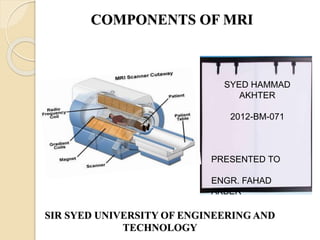
- 1. COMPONENTS OF MRI SIR SYED UNIVERSITY OF ENGINEERING AND TECHNOLOGY SYED HAMMAD AKHTER 2012-BM-071 PRESENTED TO ENGR. FAHAD AKBER
- 2. Component sMAGNET S Super conducting magnet •Very high voltages are used •Wire is winded on coil & electrically applied to the ends •More voltage greater the field Permanent magnet •Heavy weight •Full of strength •Low cost •Internal core is made of it that generate mag field all the time Resistive magnet •Internal x-axis is controlled by resistive magnet •Portion of imaging is controlled
- 3. GRADIENT COIL Have a magnetic field that changes temporally and able to variate its field . Contained with in magnetic assembly Magnetic field produced must be distorted or altered with gradient coil Imaging magnet contains three separate set of gradient coils produced in 3 directions (x,y,z directions) SHIELDING Magnetic field shielding (shield by conducting material like iron) (Improves homogeneity by protecting from interference) RF shielding (enclosing in material like copper to block external RF interference) COMPUTER Data acquisition control (acquisition of RF signal from patient body . Sequence of RF pulse is transmitted to the body) Image reconstruction (computer use collected data during acquisition process to create or construct image by fourier transform) Image storage (image are stored in the computer for future viewing)
- 4. RADIO FREQUENCY SYSTEMSignals are used to transmit the image from the patients body in MRI process. RF energy is used is a form of non ionizing radiation RF COILS These are located with in Magnet Function as antennae for both transmitting and receiving signals from tissues. Three basic types of RF coils used according different anatomical regions. √ Head = for head region √ Body = for thorax and abdominal region √ Surface = for small regions (limbs) : Generates RF energy in the form of RF pulses Applied to coil and transmitted to patient’s body Absorbed by the tissues Short time after RF pulse transmission resonating tissue will respond by returning Signal Provide data from which image is reconstructed Resulting image is display of RF signal RF TRANSMITTER RF RECIEVER
- 5. MRI IMAGES