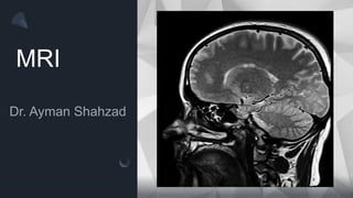
3. Introduction to MRI
• A procedure in which radio waves and a powerful magnet linked to a computer
are used to create detailed pictures of areas inside the body. These pictures can
show the difference between normal and diseased tissue.
• MRI is especially useful for imaging the brain, the spine, the soft tissue of joints,
and the inside of bones. Also called NMRI, nuclear magnetic resonance
imaging.
• MRI scanning is a non-invasive and painless but much expensive procedure.
4. Introduction to MRI
• An MRI scan uses a large magnet, radio waves, and a computer to create a
detailed, cross-sectional image of internal organs and structures.
• The scanner itself typically resembles a large tube with a table in the middle,
allowing the patient to slide in.
• An MRI scan differs from CT scans and X-rays, as it does not use potentially
harmful ionizing radiation.
5.
6.
7. Principle of MRI
• Magnetic resonance imaging (MRI) systems provide highly detailed images of
tissue in the body. The systems detect and process the signals generated when
hydrogen atoms, which are abundant in tissue, are placed in a strong magnetic
field and excited by a resonant magnetic excitation pulse.
• Purpose is to align H protons in H2O (little magnets)
9. Common nuclei with NMR properties
Since hydrogen protons are the most abundant in human
body, we use its this property to get MRI images.
10. A Single Proton
+
+
+
There is electric charge
on the surface of the proton,
thus creating a small current
loop and generating
magnetic moment m.
The proton also has
mass which
generates an
angular momentum
J when it is spinning.
J
m
Thus proton “magnet” differs from the magnetic bar in that it
also possesses angular momentum caused by spinning.
38. T1-weighted image
• T1 images can be thought of as a map
of proton energy within fatty tissues of
the body
• Fatty tissues include subcutaneous fat
(SC fat) and bone marrow of the
vertebral bodies
• Cerebrospinal fluid (CSF) contains no
fat – so it appears black on T1-
weighted images
39. T2-weighted image
• T2 images are a map of proton energy
within fatty AND water-based tissues of
the body
• Fatty tissue is distinguished from water-
based tissue by comparing with the T1
images – anything that is bright on the
T2 images but dark on the T1 images is
fluid-based tissue
40.
41. T1 and T2 weighted image – Pathology
(spine)
• Loss of the normal high signal in
the bone marrow indicates loss of
normal fatty tissue and increased
water content
• Abnormal low signal on T1 images
frequently indicates a pathological
process such as trauma, infection,
or cancer
• The same areas are whiter than
usual on this T2 image indicating
increased water content
• Abnormal brightness on a T2
image indicates a disease
process such as trauma,
infection, or cancer
42.
43.
44.
45. Studying an MRI
• T1 and T2 weighted images represent the core types of MR images.
• The different sequences tell you what is in the lesion and how it is behaving. Using
these features, the location of the lesion, and the clinical history, we can make a
diagnosis.
• Anatomy, as with all scans, is key. MRIs produce a very clear view of structures;
therefore strong anatomical knowledge is particularly helpful.
• Spend time looking at normal scans. The more you become familiar with what is
normal, the easier it is to see when things are abnormal.
• Always compare both sides of the scan – pathology is rarely bilateral.
• Be methodical!
46. Indications of an MRI
• Brain – indications include stroke, temporal lobe epilepsy, infection,
inflammation, tumour, multiple sclerosis (MS), dementia, post-trauma, metabolic
disorders, congenital malformations, internal auditory canal pathology, vascular
pathology, pituitary fossa pathology, nerve palsies and metabolic disorders.
• Spinal cord – indications include radiculopathy, myelopathy, MS, inflammation,
infection, tumour, congenital malformation, postoperative investigation and post-
trauma.
• Musculoskeletal (MSK) – indications include all MSK system: joints for
derangement, infection, inflammation, post-trauma, tumour and vascular
pathologies. Plain films are still very useful.
47. Indications of an MRI
• Abdomen and pelvis – investigates pathology of the various organs including
tumours, vascular pathologies, infection, inflammation, congenital abnormalities
and metabolic disorders. Used for detection of local invasion of rectal, prostatic
and cervical carcinomas, and assessing the anatomy in peri-anal fistulae.
• Cardiac – indications include ischemia, tumor, infiltrative diseases, congenital
malformation and cardiomyopathy
• Vascular studies – increasingly being carried out without contrast medium (so
with no risk of contrast allergy or nephrogenic systemic fibrosis (NSF)
(see Gadolinium contrast medium (MRI contrast agents)), as new techniques
are developed.
• Pregnancy – indications for the placental position and invasion, as well as
reviewing foetal anomalies, particularly cerebral.
48. MRI with contrast
• Gadolinium contrast media is used for similar cases to iodinated contrast in
computed tomography (CT), but only totals approximately 30% of studies.
• MRI is mainly done as MSK, spine and cerebral MRI scans, for indications
which are most frequently ca not usually requiring a contrast injection.
49. Contraindications of MRI
• Pacemaker, defibrillator or wires other than sternal wires – the exact mechanism of
malfunction is not certain, but death has been recorded. More recent studies report
minimal complications or malfunction of modern pacemakers, but caution is required.
There is now at least one model of pacemaker that is marketed as MRI safe; however,
scanning is to be completed under strict guidelines. These include switching the device
off before the commencement of the procedure, and the presence of specialized
medical personnel and company representatives. Scanning is carried out only on
specific body areas under strict field regulations, and so should be considered only in
extreme cases.
• Metallic foreign body in the eye – these might move or heat during scanning resulting in
serious eye injury. They require exclusion using orbital X-rays on the day of scanning
(or before the day of scanning, providing no further eye injuries with metal fragments
occur in the interim between the X-ray and the MRI).
50. Contraindications of MRI
• Deep brain stimulator – possible thermal injury along the wires, malfunction of device.
• Swan-Ganz catheter – wire causes melting of adjacent catheter and malfunction.
• Bullets or gunshot pellets – near great vessels or vital organs, such as the lungs, heart
or brain, which might move because of insufficient adjacent scar/tissue and cause
damage.
• Cerebral aneurysm clips – if magnetic, can move. Also not scanned if type unknown.
• Cochlear implant – malfunction.
• Magnetic dental implants – loss of magnetic hold to keep the implant in place.
• Drug infusion devices – might malfunction.
51. Adverse effects of MRI
• There are no known side-effects of an MRI
• Patients feel claustrophobia during scanning and may need sedation.
• Gadolinium-based contrast media are usually contraindicated in pregnancy.
• In patients with poor renal function, there is a risk of nephrogenic systemic fibrosis (NSF)
associated with gadolinium chelate injections. Patients with known, or at risk of, renal
impairment need to have their renal function assessed before MRI in order to determine whether
administration of gadolinium contrast is best avoided.
• Gadolinium contrast is generally very safe but does have a low rate of allergic reactions (about 1
in 1,000 minor reactions and 1 in 10,000 significant reactions). MRI departments using contrast
should have appropriate staff and medication to deal with reactions to contrast.