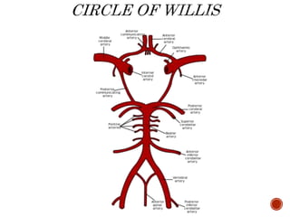
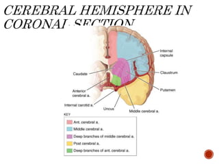
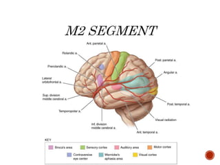
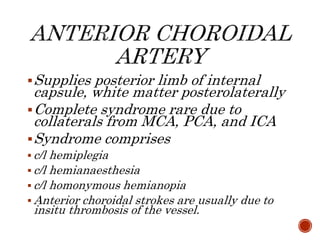
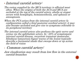
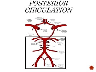
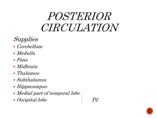
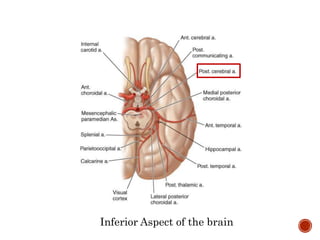
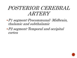
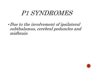
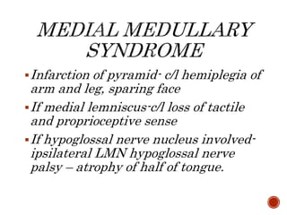
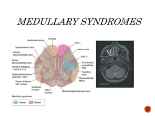
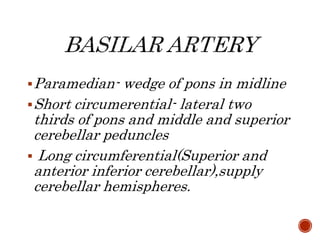
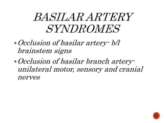
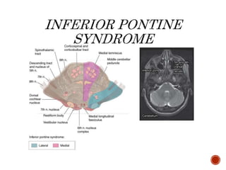
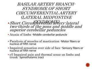
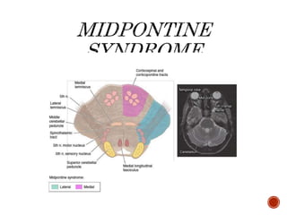
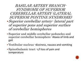
Cluster of signs and symptoms
produced due to the occlusion of an
artery(due to an atherothrombotic
lesion or an emboli or dissection )
supplying a particular region of the
brain
Large vessel stroke within the anterior
circulation
Large vessel stroke within the
posterior circulation
Small vessel disease of either vascular
bed
Anterior circulation- MCA, ACA, and
Anterior choroidal artery
Posterior circulation-Vertebral artery,
Basilar artery and Posterior cerebral
artery
Due to occlusion of Internal carotid
artery and its branches-Middle
cerebral artery, Anterior cerebral
artery and Anterior choroidal artery
M1 segment(proximal)- deep
penetrating or lenticulostriate
branches– Internal capsule, caudate
nuclues, putamen and outer pallidus
M2(distal)- superior and inferior
divisions- the entire superolateral
surface of frontal and parietal lobe
except frontal pole, strip along the
superomedial frontal and parietal
cortex, occipital lobe convolutions and
medial temporal cortex
Contralateral hemiplegia &
hemianaesthesia–Somatic motor area for face and arm and the fibres
descending from the leg area to enter the corona radiata & corresponding somatic
sensory system.
Contralateral homonymous hemianopia –Optic
radiation deep to second temporal convolution
Motor aphasia - Motor speech area of the dominant hemisphere
Conduction aphasia- Central speech area (parietal operculum)
Central aphasia, word deafness, anomia,
jargon speech, sensory agraphia, acalculia,
alexia, finger agnosia, right-left confusion-
Central, suprasylvian speech area and parietooccipital cortex of the dominant hemisphere
Gaze preference to the ipsilateral side
If dominant hemisphere involved-Global
aphasia
If non dominant hemisphere involved-
Hemispatial neglect, anasognosia and
constructional apraxia
Occlusion of lenticulostriate branches:
Lacunar stroke within the internal
capsule produces pure motor or sensory-
motor stroke contralateral to the side of
lesion; alternatively, the contralateral
hand may become ataxic and dysarthria
will be prominent (Clumsy hand
syndrome, dysarthria lacunar syndrome)
Lacunar infarct of putamen, globus
pallidus- predominantly parkinsonian
features
If superior division of M1 involved:
Brachial syndrome- weakness of hand
and arm
Frontal opercular syndrome-Broca’s
aphasia with facial weakness with or
without arm weakness
Proximal part of the superior division
involved- clinical features of motor
weakness, sensory disturbances and
broca’s aphasia
If inferior division of M2 involved:
If dominant hemisphere- Wernicke’s
aphasia without weakness with
contralateral homonymous superior
quadrantanopia
If non dominant hemisphere-
Hemineglect , spatial agonosia without
weakness
A1 segment- from internal carotid to
anterior communicating artery-
branches to anterior limb of internal
capsule, inferior part of caudate,
anterior hypothalamus
A2 segment- distal to anterior
communicating artery- supplies frontal
pole, entire medial part of cerebral
hemispheres
A1 segment occlusion rarely produces
clinical syndrome because collateral
flow through anterior communicating
artery and collaterals from MCA and
PCA
-c/l paralysis of foot and leg, paresis of
arm
-c/l cortical sensory loss of foot and leg
-urinary incontinence -Sensorimotor area in
paracentral lobule
-c/l grasp and suckling reflex
-abulia, slowness, delay
-gait apraxia
If both A2 segments arise from a single anterior cerebral
stem(c/l A1 segment atresia),it may affect both hemispheres-
Profound abulia, b/l pyramidal signs, paraparesis or
quadriparesis and urinary incontinence
Supplies posterior limb of internal
capsule, white matter posterolaterally
Complete syndrome rare due to
collaterals from MCA, PCA, and ICA
Syndrome comprises
 c/l hemiplegia
 c/l hemianaesthesia
 c/l homonymous hemianopia
 Anterior choroidal strokes are usually due to
insitu thrombosis of the vessel.
• Internal carotid artery:
The cortex supplied by the MCA territory is affected most
often. When the origins of both the ACA and MCA are
occluded at the top of the carotid artery, abulia or stupor
occurs with hemiplegia, hemianesthesia, and aphasia or
anosognosia.
When the PCA arises from the internal carotid artery (a
configuration called a fetal posterior cerebral artery), it may
also become occluded and give rise to symptoms referable to
its peripheral territory
The internal carotid artery also perfuses the optic nerve and
retina via the ophthalmic artery. In ~25% of symptomatic
internal carotid disease, recurrent transient monocular
blindness (amaurosis fugax) warns of the lesion-typically
describe a horizontal shade that sweeps down or up across
the field of vision
• Common carotid artery:
Jaw claudication may result from low flow in the external
carotid branches
 Paired Vertebral arteries
 Basilar artery
 Paired Posterior cerebral arteries
 Small penetrating branches and short
and long circumferential branches
Supplies
 Cerebellum
 Medulla
 Pons
 Midbrain
 Thalamus
 Subthalamus
 Hippocampus
 Medial part of temporal lobe
 Occipital lobe P2
Inferior Aspect of the brain
P1 segment-Precommunal- Midbrain,
thalamic and subthalamic
P2 segment-Temporal and occipital
cortex
Due to the involvement of ipsilateral
subthalamus, cerebral peduncles and
midbrain
Claude’s- 3rd nerve palsy with c/l ataxia- Red
nuclues
Weber’s- 3rd nerve palsy with c/l hemiplegia-
Cerebral peduncle
Subthalamus-c/l hemiballismus
Thalamus-
Thalamic ‘Dejerine Roussy syndrome’ -
c/l hemisensory loss followed later by
agonising,searing or burning pain in the
affected areas.
 Occlusion of the artery of Percheron produces paresis of upward gaze and
drowsiness and often abulia. Extensive infarction in the midbrain and
subthalamus occurring with bilateral proximal PCA occlusion presents as
coma, unreactive pupils, bilateral pyramidal signs, and decerebrate rigidity. `
 Infarction of medial temporal and occipital lobes
 Occipital lobe-c/l homonymous hemianopia with
macular sparing, if visual association area
spared, patient aware of visual defect
 Medial temporal lobe- Memory impairement
 Visual agnosia for faces, objects, mathematical
symbols, and colors and anomia with
paraphasic errors (amnestic aphasia) may also
occur
 Visual hallucinations of brightly coloured scenes
and objects.
Antons syndrome- bilateral occlusion in
distal PCAs – bilateral occipital lobe
infarction- cortical blindness and patient
often unaware and even deny it.
Rarely, only peripheral vision is lost and central vision is
spared-’gun-barrel’ vision
Balints syndrome- bilateral visual
association areas- disorder of orderly
visual scanning of the environment,
resulting from infarctions secondary to
low flow in watershed between distal
PCA and MCA-palinopsia and
asimultagnosia
Paramedian branches of upper basilar and
proximal posterior cerebral arteries
Eye “down and out” secondary to unopposed
action of fourth and sixth cranial nerves,
with dilated and unresponsive pupil: Third nerve
fibers
c/l Paralysis of face, arm, and leg: Corticobulbar and
corticospinal tract descending in crus cerebri.
Syndrome of small penetrating arteries
arising from posterior cerebral artery
 Eye “down and out” secondary to unopposed
action of fourth and sixth cranial nerves,
with dilated and unresponsive pupil: Third nerve
fibers
Hemiataxia, hyperkinesias, tremor: Red nucleus,
dentatorubrothalamic pathway
V1 and V4- prone for atherothrombosis
If occlusion is proximal to origin of
vertebral artery, reversal of blood flow-
subclavian steal syndrome
 Caused due to occlusion of V4 segment or PICA
 Pain, numbness and abnormal sensation over one
half of face -Descending tract and nucleus of trigeminal
nerve
 Vertigo, nausea, vomiting and diplopia -Vestibular
nucleus
 Dysphagia, hoarseness, palatal paralysis - Issuing
fibres of 9th and 10th nerve nucleus
 Ataxia of limbs -Restiform body, and cerebellar
hemispheres
 Horner’s syndrome -Descending sympathetic tract
 c/l loss of pain and temperature - Spinothalamic tract
Infarction of pyramid- c/l hemiplegia of
arm and leg, sparing face
If medial lemniscus-c/l loss of tactile
and proprioceptive sense
If hypoglossal nerve nucleus involved-
ipsilateral LMN hypoglossal nerve
palsy – atrophy of half of tongue.
Paramedian- wedge of pons in midline
Short circumerential- lateral two
thirds of pons and middle and superior
cerebellar peduncles
 Long circumferential(Superior and
anterior inferior cerebellar),supply
cerebellar hemispheres.
Occlusion of basilar artery- b/l
brainstem signs
Occlusion of basilar branch artery-
unilateral motor, sensory and cranial
nerves
• Complete basilar artery occlusion (Locked in
state)-
b/l long tract(sensory/motor) with cranial
nerve and cerebellar dysfunction- preserved
consciousness, quadriplegia and cranial nerve
signs
• TIAs in the proximal basilar distribution may
produce vertigo “swimming,” “swaying,”
“moving,” “unsteadiness,” or “light-headedness.
Other symptoms that warn of basilar
thrombosis include diplopia, dysarthria, facial or
circumoral numbness, and hemisensory
symptoms.
In general, symptoms of basilar branch
TIAs affect one side of the brainstem, whereas
symptoms of basilar artery TIAs usually affect
both sides
 TOP OF BASILAR SYNDROME:
Embolic occlusion of the top of the basilar
artery can produce any or all of the central or
peripheral PCA territory symptoms. The hallmark
is the sudden onset of bilateral signs, including
ptosis, pupillary asymmetry or lack of reaction to
light, and somnolence.
Anterior inferior cerebellar artery-
lateral part of inferior pons and
anterior part of inferior cerebellar
hemispheres
 Cerebellum- Ataxia of limb and gait
 7th nerve nuclues- Facial weakness
 8th nerve nucleus- Deafness, tinnitus, vertigo,
nausea, vomiting
 Spinothalamic tract- c/l loss of pain and
temperature
 Paralysis of conjugate gaze to side of lesion
(preservation of convergence): Center for conjugate
lateral gaze
 Nystagmus: Vestibular nucleus
 Ataxia of limbs and gait: Likely middle cerebellar
peduncle
 Diplopia on lateral gaze: Abducens nerve
 c/l Paralysis of face, arm, and leg: Corticobulbar
and corticospinal tract in lower pons
 Impaired tactile and proprioceptive sense over
c/l side of the body: Medial lemniscus
Short Circumferential artery-lateral
two-thirds of the pons and middle and
superior cerebellar peduncles
 Ataxia of limbs -Middle cerebellar peduncle
 Paralysis of muscles of mastication -Motor fibers or
nucleus of fifth nerve
 Impaired sensation over side of face -Sensory fibers or
nucleus of fifth nerve
 c/l Impaired pain and thermal sense on limbs and
trunk -Spinothalamic tract
Ataxia of limbs and gait : Pontine nuclei
c/l Paralysis of face, arm, and leg:
Corticobulbar and corticospinal tract
Variable impaired touch and proprioception
on c/l side: Medial lemniscus
Superior cerebellar artery- lateral part
of superior pons and superior surface
of cerebellar hemispheres
 Superior and middle cerebellar peduncles and
superior cerebellar hemisphere -Ataxia of limb and
gait
 Vestibular nucleus -dizziness, nausea and vomiting
 Spinothalamic tract -c/l loss of pain and
temperature
Cerebellar ataxia :Superior and/or middle
cerebellar peduncle
Internuclear ophthalmoplegia: Medial
longitudinal fasciculus
c/l Paralysis of face, arm, and leg:
Corticobulbar and corticospinal tract.
Rarely touch, vibration, and position are
affected on c/l side: Medial lemniscus
Reference: Harrison’s Principles Of Internal Medicine
19e

Clinical stroke syndromes

  • 2.
    Cluster of signsand symptoms produced due to the occlusion of an artery(due to an atherothrombotic lesion or an emboli or dissection ) supplying a particular region of the brain
  • 3.
    Large vessel strokewithin the anterior circulation Large vessel stroke within the posterior circulation Small vessel disease of either vascular bed
  • 4.
    Anterior circulation- MCA,ACA, and Anterior choroidal artery Posterior circulation-Vertebral artery, Basilar artery and Posterior cerebral artery
  • 6.
    Due to occlusionof Internal carotid artery and its branches-Middle cerebral artery, Anterior cerebral artery and Anterior choroidal artery
  • 7.
    M1 segment(proximal)- deep penetratingor lenticulostriate branches– Internal capsule, caudate nuclues, putamen and outer pallidus
  • 8.
    M2(distal)- superior andinferior divisions- the entire superolateral surface of frontal and parietal lobe except frontal pole, strip along the superomedial frontal and parietal cortex, occipital lobe convolutions and medial temporal cortex
  • 11.
    Contralateral hemiplegia & hemianaesthesia–Somaticmotor area for face and arm and the fibres descending from the leg area to enter the corona radiata & corresponding somatic sensory system. Contralateral homonymous hemianopia –Optic radiation deep to second temporal convolution Motor aphasia - Motor speech area of the dominant hemisphere Conduction aphasia- Central speech area (parietal operculum)
  • 12.
    Central aphasia, worddeafness, anomia, jargon speech, sensory agraphia, acalculia, alexia, finger agnosia, right-left confusion- Central, suprasylvian speech area and parietooccipital cortex of the dominant hemisphere Gaze preference to the ipsilateral side If dominant hemisphere involved-Global aphasia If non dominant hemisphere involved- Hemispatial neglect, anasognosia and constructional apraxia
  • 13.
    Occlusion of lenticulostriatebranches: Lacunar stroke within the internal capsule produces pure motor or sensory- motor stroke contralateral to the side of lesion; alternatively, the contralateral hand may become ataxic and dysarthria will be prominent (Clumsy hand syndrome, dysarthria lacunar syndrome) Lacunar infarct of putamen, globus pallidus- predominantly parkinsonian features
  • 14.
    If superior divisionof M1 involved: Brachial syndrome- weakness of hand and arm Frontal opercular syndrome-Broca’s aphasia with facial weakness with or without arm weakness Proximal part of the superior division involved- clinical features of motor weakness, sensory disturbances and broca’s aphasia
  • 15.
    If inferior divisionof M2 involved: If dominant hemisphere- Wernicke’s aphasia without weakness with contralateral homonymous superior quadrantanopia If non dominant hemisphere- Hemineglect , spatial agonosia without weakness
  • 16.
    A1 segment- frominternal carotid to anterior communicating artery- branches to anterior limb of internal capsule, inferior part of caudate, anterior hypothalamus A2 segment- distal to anterior communicating artery- supplies frontal pole, entire medial part of cerebral hemispheres
  • 19.
    A1 segment occlusionrarely produces clinical syndrome because collateral flow through anterior communicating artery and collaterals from MCA and PCA
  • 20.
    -c/l paralysis offoot and leg, paresis of arm -c/l cortical sensory loss of foot and leg -urinary incontinence -Sensorimotor area in paracentral lobule -c/l grasp and suckling reflex -abulia, slowness, delay -gait apraxia If both A2 segments arise from a single anterior cerebral stem(c/l A1 segment atresia),it may affect both hemispheres- Profound abulia, b/l pyramidal signs, paraparesis or quadriparesis and urinary incontinence
  • 21.
    Supplies posterior limbof internal capsule, white matter posterolaterally Complete syndrome rare due to collaterals from MCA, PCA, and ICA Syndrome comprises  c/l hemiplegia  c/l hemianaesthesia  c/l homonymous hemianopia  Anterior choroidal strokes are usually due to insitu thrombosis of the vessel.
  • 22.
    • Internal carotidartery: The cortex supplied by the MCA territory is affected most often. When the origins of both the ACA and MCA are occluded at the top of the carotid artery, abulia or stupor occurs with hemiplegia, hemianesthesia, and aphasia or anosognosia. When the PCA arises from the internal carotid artery (a configuration called a fetal posterior cerebral artery), it may also become occluded and give rise to symptoms referable to its peripheral territory The internal carotid artery also perfuses the optic nerve and retina via the ophthalmic artery. In ~25% of symptomatic internal carotid disease, recurrent transient monocular blindness (amaurosis fugax) warns of the lesion-typically describe a horizontal shade that sweeps down or up across the field of vision • Common carotid artery: Jaw claudication may result from low flow in the external carotid branches
  • 23.
     Paired Vertebralarteries  Basilar artery  Paired Posterior cerebral arteries  Small penetrating branches and short and long circumferential branches
  • 25.
    Supplies  Cerebellum  Medulla Pons  Midbrain  Thalamus  Subthalamus  Hippocampus  Medial part of temporal lobe  Occipital lobe P2
  • 26.
  • 27.
    P1 segment-Precommunal- Midbrain, thalamicand subthalamic P2 segment-Temporal and occipital cortex
  • 28.
    Due to theinvolvement of ipsilateral subthalamus, cerebral peduncles and midbrain
  • 29.
    Claude’s- 3rd nervepalsy with c/l ataxia- Red nuclues Weber’s- 3rd nerve palsy with c/l hemiplegia- Cerebral peduncle Subthalamus-c/l hemiballismus Thalamus- Thalamic ‘Dejerine Roussy syndrome’ - c/l hemisensory loss followed later by agonising,searing or burning pain in the affected areas.  Occlusion of the artery of Percheron produces paresis of upward gaze and drowsiness and often abulia. Extensive infarction in the midbrain and subthalamus occurring with bilateral proximal PCA occlusion presents as coma, unreactive pupils, bilateral pyramidal signs, and decerebrate rigidity. `
  • 30.
     Infarction ofmedial temporal and occipital lobes  Occipital lobe-c/l homonymous hemianopia with macular sparing, if visual association area spared, patient aware of visual defect  Medial temporal lobe- Memory impairement  Visual agnosia for faces, objects, mathematical symbols, and colors and anomia with paraphasic errors (amnestic aphasia) may also occur  Visual hallucinations of brightly coloured scenes and objects.
  • 31.
    Antons syndrome- bilateralocclusion in distal PCAs – bilateral occipital lobe infarction- cortical blindness and patient often unaware and even deny it. Rarely, only peripheral vision is lost and central vision is spared-’gun-barrel’ vision Balints syndrome- bilateral visual association areas- disorder of orderly visual scanning of the environment, resulting from infarctions secondary to low flow in watershed between distal PCA and MCA-palinopsia and asimultagnosia
  • 32.
    Paramedian branches ofupper basilar and proximal posterior cerebral arteries Eye “down and out” secondary to unopposed action of fourth and sixth cranial nerves, with dilated and unresponsive pupil: Third nerve fibers c/l Paralysis of face, arm, and leg: Corticobulbar and corticospinal tract descending in crus cerebri.
  • 33.
    Syndrome of smallpenetrating arteries arising from posterior cerebral artery  Eye “down and out” secondary to unopposed action of fourth and sixth cranial nerves, with dilated and unresponsive pupil: Third nerve fibers Hemiataxia, hyperkinesias, tremor: Red nucleus, dentatorubrothalamic pathway
  • 35.
    V1 and V4-prone for atherothrombosis If occlusion is proximal to origin of vertebral artery, reversal of blood flow- subclavian steal syndrome
  • 36.
     Caused dueto occlusion of V4 segment or PICA  Pain, numbness and abnormal sensation over one half of face -Descending tract and nucleus of trigeminal nerve  Vertigo, nausea, vomiting and diplopia -Vestibular nucleus  Dysphagia, hoarseness, palatal paralysis - Issuing fibres of 9th and 10th nerve nucleus  Ataxia of limbs -Restiform body, and cerebellar hemispheres  Horner’s syndrome -Descending sympathetic tract  c/l loss of pain and temperature - Spinothalamic tract
  • 37.
    Infarction of pyramid-c/l hemiplegia of arm and leg, sparing face If medial lemniscus-c/l loss of tactile and proprioceptive sense If hypoglossal nerve nucleus involved- ipsilateral LMN hypoglossal nerve palsy – atrophy of half of tongue.
  • 39.
    Paramedian- wedge ofpons in midline Short circumerential- lateral two thirds of pons and middle and superior cerebellar peduncles  Long circumferential(Superior and anterior inferior cerebellar),supply cerebellar hemispheres.
  • 40.
    Occlusion of basilarartery- b/l brainstem signs Occlusion of basilar branch artery- unilateral motor, sensory and cranial nerves
  • 41.
    • Complete basilarartery occlusion (Locked in state)- b/l long tract(sensory/motor) with cranial nerve and cerebellar dysfunction- preserved consciousness, quadriplegia and cranial nerve signs • TIAs in the proximal basilar distribution may produce vertigo “swimming,” “swaying,” “moving,” “unsteadiness,” or “light-headedness. Other symptoms that warn of basilar thrombosis include diplopia, dysarthria, facial or circumoral numbness, and hemisensory symptoms. In general, symptoms of basilar branch TIAs affect one side of the brainstem, whereas symptoms of basilar artery TIAs usually affect both sides
  • 42.
     TOP OFBASILAR SYNDROME: Embolic occlusion of the top of the basilar artery can produce any or all of the central or peripheral PCA territory symptoms. The hallmark is the sudden onset of bilateral signs, including ptosis, pupillary asymmetry or lack of reaction to light, and somnolence.
  • 43.
    Anterior inferior cerebellarartery- lateral part of inferior pons and anterior part of inferior cerebellar hemispheres  Cerebellum- Ataxia of limb and gait  7th nerve nuclues- Facial weakness  8th nerve nucleus- Deafness, tinnitus, vertigo, nausea, vomiting  Spinothalamic tract- c/l loss of pain and temperature
  • 44.
     Paralysis ofconjugate gaze to side of lesion (preservation of convergence): Center for conjugate lateral gaze  Nystagmus: Vestibular nucleus  Ataxia of limbs and gait: Likely middle cerebellar peduncle  Diplopia on lateral gaze: Abducens nerve  c/l Paralysis of face, arm, and leg: Corticobulbar and corticospinal tract in lower pons  Impaired tactile and proprioceptive sense over c/l side of the body: Medial lemniscus
  • 46.
    Short Circumferential artery-lateral two-thirdsof the pons and middle and superior cerebellar peduncles  Ataxia of limbs -Middle cerebellar peduncle  Paralysis of muscles of mastication -Motor fibers or nucleus of fifth nerve  Impaired sensation over side of face -Sensory fibers or nucleus of fifth nerve  c/l Impaired pain and thermal sense on limbs and trunk -Spinothalamic tract
  • 47.
    Ataxia of limbsand gait : Pontine nuclei c/l Paralysis of face, arm, and leg: Corticobulbar and corticospinal tract Variable impaired touch and proprioception on c/l side: Medial lemniscus
  • 49.
    Superior cerebellar artery-lateral part of superior pons and superior surface of cerebellar hemispheres  Superior and middle cerebellar peduncles and superior cerebellar hemisphere -Ataxia of limb and gait  Vestibular nucleus -dizziness, nausea and vomiting  Spinothalamic tract -c/l loss of pain and temperature
  • 50.
    Cerebellar ataxia :Superiorand/or middle cerebellar peduncle Internuclear ophthalmoplegia: Medial longitudinal fasciculus c/l Paralysis of face, arm, and leg: Corticobulbar and corticospinal tract. Rarely touch, vibration, and position are affected on c/l side: Medial lemniscus
  • 52.
    Reference: Harrison’s PrinciplesOf Internal Medicine 19e