SlideShare a Scribd company logo
1 of 11
Download to read offline
Sabancı University Program for Undergraduate Research (PURE) Summer 2017-2018
Synthesis and Characterization of Novel Nanomaterials for SERS
Biomedical/Environmental Application
Ammar Khan ammar_akhan@hotmail.com
Ziauddin University / Pharmacy / 2018
Finesa Xhibo finesaxhibo@gmail.com
Yildiz Technical University / Chemistry / 2018
Necla Yücel necla-ycl@hotmail.com
Bilgi University / Genetics and Bioengineering / 2018
Sena Turan s.turan@gtu.edu.tr
Gebze Technical University / Material Science and Engineering / 2018
Project Supervisors
Anjum Qureshi
Sabanci University SUNUM Nanotechnology Research Center, TR-34956 Istanbul, Turkey
(Functional nanomaterials, biosesnors)
Javed Kolkar
Sabanci University SUNUM Nanotechnology Research Center, TR-34956 Istanbul, Turkey
(Functional nanomaterials, biosesnors)
Abstract
In this study, a simple green method was employed to synthesize functionalized silver nanoparticles (Ag
NPs) as surface-enhanced Raman scattering (SERS) substrate for detection of dopamine (DA). In this
method, polyethylene glycol (PEG) was functionalized on silver nanoparticles to prepare the uniform and
controlled size of nanoparticles (NPs). The optical and structural properties of functionalized nanoparticles
were characterized. The Raman spectra of the prepared PEG-Ag SERS substrate clearly indicated an
enhancement in the SERS signal of dopamine. The developed functionalized SERS substrate can be
potentially used as a sensitive SERS substrate for detection of various neurotransmitters for biomedical
application.
Keywords: Surface-enhanced Raman scattering (SERS), silver nanoparticles (Ag NPs), polyethylene
glycol (PEG), dopamine (DA)
1 Introduction
Nanotechnology has received a great deal of attention in recent years, with the foundation being
laid by Feynman in 1959 and then consolidated a decade later by Taniguchi who came up with the
term nanotechnology (Benelmekki, M. (2015). A nanoparticle is the most central component in the
buildup of a nanostructure. A nanoparticle is bigger than an atom or a simple molecule that is
governed by quantum mechanics, and lies in the size range of between 1-100 nm (Al-Taa'y, Nabi
et al. 2014). Synthesis of these nanoparticles is carried out either through a bottom-up approach or
through the top-down approach. The bottom-up approach refers to how atoms are used to build up
to a nanomaterial, whereas in a top-down approach a larger material is broken down and eventually
reaches a smaller sized structure.
KHAN, XHIBO, YÜCEL, TURAN
2
Nanoparticles possess unique properties, especially optical, electronic and biological properties
(Ahmadpoor, Nateri et al. 2013, Lv, Li et al. 2013). As they have a high surface-to-volume ratio,
nanoparticles can be used as heterogeneous catalysts (Kusior, Klich-Kafel et al. 2013, Yang, Xie
et al. 2013). In addition, they are applied in many fields, including photonics, microelectronics,
lithography and surface-enhanced Raman spectroscopy (Bhui, Bar et al. 2009, Son, Ko et al. 2009).
The exceptional plasmonic properties of silver nanoparticles leads to a great enhancement of the
Raman signal. The surface-enhanced Raman scattering (SERS) is an advanced Raman technique
that enhances the vibrational spectrum of molecules adsorbed on or in the vicinity of metal particles.
According to previous studies, two main mechanisms, chemical and electromagnetic contribute to
SERS enhancement. (Aroca. 2007). In the electromagnetic mechanism, the laser light causes the
excitation of the localized surface plasmons which causes an enhancement of the local
electromagnetic field. The molecule which is near the metal surface or adsorbed to it experiences
this enhancement, and as a result, the intensity of the Raman scattered light is enhanced. In the
chemical mechanism, which typically occurs in those molecules with a lone pair of electrons, a
charge transfer takes place between the molecule and the surface of the metal. The chemical
mechanism is usually regarded as weaker than electromagnetic mechanism and occurs in concert
with it. Raman signal enhancement is because of both of these mechanisms. Due to its sensitivity,
readiness and minimum sample preparation requirements, SERS is being considered as a powerful
technique for detection of the wide variety of analytes at very low concentrations, even down to
the single molecule level (Kneipp, Wang et al. 1997).
A variety of materials are employed for SERS analysis. The aim while synthesizing these materials
is to prepare stable, reproducible and highly active SERS substrates. Metals such as gold and silver
are commonly used as part of metal hybrid nanostructures. The optical properties of silver and gold
depend on their size and shape which makes it important to control these parameters, and these
properties in turn influence the SERS effect. Nanoparticles can be synthesized according to various
methods depending on the shape of nanoparticle needed, as the shape affects SERS enhancement.
SERS substrates can be metal electrodes, colloidal metal solutions, or hybrid structures where the
metal nanoparticle is chemically or electrostatically attached to other inorganic materials such as
in metal island films or metal coated nanospheres (Fateixa, Nogueira et al. 2015). These methods
required complex and time consuming procedures. Therefore, in this study we opted to use easy
and simple method by employing PEG as an environmental friendly surface modifier and
stabilizing agent to prepare functionalized Ag NPs as SERS substrate for detection of a
neurotransmitter Dopamine (DA).
SERS provides a unique opportunity to detect a broader range of neurotransmitters in close proximity to
neurons. Dopamine is a catecholamine neurotransmitter that plays a significant role in the
functioning of central nervous, vascular, and hormonal systems (Zhang, Neumeyer et al. 2007,
Lussier, Brule et al. 2017). It is widely distributed in the brain tissues and body fluids of mammals.
The abnormal variation of the DA concentration in vivo has been linked to serious neurological,
renal, cardio disorders such as schizophrenia, Huntington’s disease, Alzheimer’s disease, and
Parkinson’s disease (Lotharius and Brundin 2002, Ji, Palui et al. 2012). Therefore, the aim of the
present work is to synthesize PEG functionalized Ag NPs, through a green method, which is
environmentally friendly, viable, and cost-effective to prepare an effective SERS substrate for the
detection of dopamine through Raman spectroscopy.
2 Experimental Section
2.1 Materials and Reagents
SYNTHESIS & CHARACTERIZATION OF NOVEL NANOMATERIALS FOR SERS BIOMEDICAL/ENVIRONMENTAL APPLICATION
3
All chemicals and reagents were of analytical grade and used as received. Silver nitrate (AgNO3),
sodium borohydride (NaBH4) and polyethylene glycol (PEG 6000) were obtained from Sigma
Aldrich. Distilled, deionized water was used throughout the experiments.
2.2 Apparatus
UV–Visible (UV-Vis) spectroscopy is a very important technique and is the simplest way to
confirm the formation of nanoparticles. The absorbance spectrum of the colloidal sample was
obtained using a UV–Vis spectrometer Nanodrop 2000 (200–800 nm). X-ray diffraction (XRD)
analysis was conducted using D8 ADVANCE using monochromatic Cu kα radiation (λ = 1.5406
Å) operated at a 2θ angle pattern. The scanning was done in the region of 20º–80º.Fourier-
Transform Infrared (FTIR) spectra of the samples was investigated using a Thermo Scientific™
Nicolet iS™10 FTIR Spectrometer). The characterization was in the wavenumber range from 4000
to 500 cm−1
accumulating 64 scans. The Raman spectra were recorded by Renishaw Invia Raman
microscope. Collection of the SERS spectra was performed through a 50 microscope objective
using low laser power (0.5%).
2.3 Methods
2.3.1 Synthesis of functionalized Ag NPs
Silver nanoparticles were prepared by the dropwise addition of aqueous AgNO3 solution (0.01 M)
to a mixture of 50 mL of PEG (0.1M) and 10 mL sodium borohydride (NaBH4) (0.01M) at room
temperature. The mixture was constantly stirred using a magnetic stirrer and kept in the laboratory
at room temperature. Three samples were prepared with differing volumes of AgNO3 in order to
determine the optimum volume of AgNO3 required to prepare the Ag NPs of adequate size. Three
volumes of AgNO3, 100, 200 and 300 μL were taken and added dropwise to the PEG and NaBH4
mixture. The non-functionalized Ag NPs were synthesized with same procedure without addition
of PEG.
2.3.2 Preparation of DA loaded SERS substrate
A volume of 100 μL of DA in phosphate buffer solution (pH 7.4, 0.05 mM) was added to the
functionalized Ag NP solution and the non-functionalized Ag NP solution, respectively. The
samples prepared for detection of dopamine (Table 1) were dropped to the silica substrate as shown
in Fig, 1. The samples were dried at room temperature and used to record the Raman spectra.
Sample
No
Sample Name
1-2
Non-functionalised Ag NPs without
dopamine
3-4
Non-functionalised Ag NPs with
dopamine
5-6
Functionalized Ag NPs without
dopamine
7-8 Functionalised Ag NPs with dopamine
9 dopamine
1-2
3-4
5-6
7-8
9
Figure 1: DA incubated synthesized PEG Ag
SERS substrate on silica plate.
Table 1: Showing DA incubated samples on silica
plate.
KHAN, XHIBO, YÜCEL, TURAN
4
2.4 Results and Discussion
Characterization
2.4.1 Uv-Vis spectroscopy
PEG functionalized Ag NPs were prepared by the dropwise addition of aqueous AgNO3 solution
(0.01 M) to a mixture of 50 mL of PEG (0.1M) and 10 mL of sodium borohydride (NaBH4) (0.01M)
at room temperature. Three different volumes of AgNO3 such as 100, 200 and 300 μL were taken
and added dropwise to the PEG and NaBH4 mixture and respective UV absorbance responses were
recorded and shown in Fig.2a. PEG Ag NPs showed a characteristic absorption peak at around 400
nm (Fig.2a). Increasing the volume of AgNO3 added in the reaction mixture, leads to the formation
of Ag NPs of a larger size, which is shown to increase UV absorption of light as shown in Fig.2.
For the synthesis of PEG-Ag NPs at room temperature, different volume/concentration of AgNO3
solution were considered and shown in Table 2. At 300 μL of AgNO3, a broader peak is observed
due to agglomeration of the Ag NPs (Fig.2).
Figure 2a: UV-Vis spectra of functionalized silver nanoparticles using different volumes of AgNO3.
SYNTHESIS & CHARACTERIZATION OF NOVEL NANOMATERIALS FOR SERS BIOMEDICAL/ENVIRONMENTAL APPLICATION
5
Figure 2b: The reaction procedure and color change with the addition of different volumes of AgNO3 to a
mixture of PEG and sodium borohydride. The figure shows the appearance of a light yellow color in the
reaction mixture which indicates the formation of PEG Ag NPs.
After the dropwise addition of AgNO3 to a mixture of PEG and sodium borohydride, a light yellow
color solution was obtained (Fig.2b) and it indicates the formation of Ag NPs. The change in color
of the solution was due to the surface plasmon resonance (SPR) and reduction of silver ions by
PEG and sodium borohydride. Further addition of AgNO3 resulted in a darker solution being
formed due to aggregation of the silver nanoparticles (Fig.2b).
Trial Number Quantity Of
AgNO3
Conc. Ag NO3 Absorbance Wavelength
(nm)
1 100 µL 0.01M 0.0952 388
2 200 µL 0.01M 0.1491 390
3 300 µL 0.01M 0.0130 408
Table 2: UV-Vis spectra results with optimizing parameters
2.4.2 XRD results
The X-ray diffraction (XRD) pattern of the prepared PEG Ag NPs was recorded. XRD profile was
taken from 2θ range of 20 to 80 o
with a step of 0.02 degree and shown in Fig.3.
KHAN, XHIBO, YÜCEL, TURAN
6
Figure 3: XRD profile of functionalized Ag NPs.
Three peaks at 2θ values of 38.03 o
, 44.22 o
, and 72.616 o
in the experimental diffractogram have
been identified and corresponding to (hkl) values - (111), (200), and (311) planes of silver,
respectively (Fig. 3). The XRD profile confirmed that the resultant particles in the prepared sample
are silver NPs and having a face-centered cubic crystal structure. There is one more unidentified
peak at 34.129 o
which may be due to AgNO3, which might not have been reduced and hence
remained in the sample in minute quantity.
2.4.3 FTIR results
The FTIR spectra of PEG and PEG Ag NPs were recoded and shown in Fig.4.
SYNTHESIS & CHARACTERIZATION OF NOVEL NANOMATERIALS FOR SERS BIOMEDICAL/ENVIRONMENTAL APPLICATION
7
Figure 4: FT-IR spectra of PEG and PEG- Ag NPs.
Intense absorption bands were observed at 2880, 1640, 1464, 1340, 1277, 1090, 945 and 842 cm−1
for PEG Ag sample (Fig.4). Aliphatic C–H stretching at 1464 and 1340 cm−1
due to C–H bending
vibrations (Shameli, Bin Ahmad et al. 2012), bands at 1277 cm−1
and 1090 cm−1
which were related
to the stretching vibrations of the alcoholic C–O bonds and C–O–C ether linkage ν(C–O–C) were
observed in FTIR spectrum of PEG functionalized Ag NPs. Further, bands at 945 cm−1
(ρ(CH2))
and 842 cm−1
(ρ(CH2)) were attributed to rocking vibrations and band at 3392 cm−1
was attributed
to the O–H stretching vibration in association with ν (CH3
s
) at ∼2880 cm−1
in case of PEG Ag NPs.
Finally, a new band was observed at ~1640 cm−1
which might correspond to a C=O stretching
vibration and this carbonyl group formation occurred probably due to the oxidation of hydroxyl
groups (Fahmy, El-Nasser et al. 2017) in FTIR spectra of PEG Ag NPs (Fig.4).
2.4.2 Detection of dopamine on synthesized SERS substrate
The Raman Spectra of Ag NPs and PEG functionalized Ag NPs (PEG Ag) were recorded (Fig.5).
The Raman spectra of PEG Ag showed enhanced Raman intensities at 813, 1035, 1340 and 1600
KHAN, XHIBO, YÜCEL, TURAN
8
cm-1
as compared with Ag NPs. These observations clearly indicated that PEG has stabilized silver
nanoparticles with controlled and uniform size distribution.
Figure 5: Raman spectra of PEG Ag and Ag NPs.
Furthermore, functionalized Ag NPs were utilized for detection of dopamine and SERS response
were recoded (Fig.6). The optical image of DA incubated PEG Ag NPs SERS substrate is shown
in Fig.7. The SERS spectra of PEG Ag NPs clearly showed enhanced peaks of dopamine (0.05
mM) as compared to the Ag NPs (Fig.6). The band at 1480 cm-1
corresponds to phenyl C=C
stretching has noticeably enhanced the peak signal as compared with dopamine peak signal. The
band at 1337 cm-1
can be assigned to ring stretching vibration of dopamine. The ring stretching
vibration mode of O-H bond is observed at 1395 cm-1
(Fig.6). The observations clearly indicated
that PEG functionalized Ag NPs can enhance the SERS peak signal of dopamine and can be used
as a sensitive SERS substrate for the detection of DA.
SYNTHESIS & CHARACTERIZATION OF NOVEL NANOMATERIALS FOR SERS BIOMEDICAL/ENVIRONMENTAL APPLICATION
9
Figure 6: Raman spectra of PEG Ag and Ag NPs incubated with DA (0.05 mM).
Figure 7: Raman optical image of functionalized Ag NPs incubated with dopamine (0.05 mM).
3 Conclusion and Future Work
In this study, a simple green method was employed to prepare PEG functionalized Ag NPs as a
sensitive SERS substrate for the detection of dopamine. The synthesized functionalized Ag NPs
were successfully characterized by UV-Vis, XRD and FTIR techniques. The synthesized PEG Ag
NPs showed enhanced Raman signal as compared to Ag NPs. Dopamine was successfully detected
by employing functionalized Ag NPs as a SERS substrate. The developed method to prepare
functionalized Ag SERS substrate is simple, low cost, environmentally friendly and easy to use.
The developed functionalized SERS substrate in this study can be potentially used as a sensitive
KHAN, XHIBO, YÜCEL, TURAN
10
SERS for the detection of a variety of neurotransmitters for biomedical applications. The next step
would be to study the dopamine concentration in human cerebrospinal fluid (CSF) samples to see
if this method can quantitatively measure the amount of dopamine in a reliable and accurate
manner. Additional investigations can be made regarding altering the chain length of PEG or using
PEG as the sole reducing agent instead of NaBH4.
References
Benelmekki, M. (2015). Designing hybrid nanoparticle. San Rafael, Calif.: Morgan et Claypool
Al-Taa'Y, W., Abdul Nabi, M., Yusop, R. M., Yousif, E., Abdullah, B. M., Salimon, J., ... Zubairi,
S. I. (2014). Effect of nano ZnO on the optical properties of poly(vinyl chloride)
films. International Journal of Polymer Science, 2014, [697809]. DOI: 10.1155/2014/697809
Arole, V., & Munde, S. (2014). FABRICATION OF NANOMATERIALS BY TOP-DOWN AND
BOTTOM-UP APPROACHES – AN OVERVIEW. Journal Of Advances In Applied Sciences
And Technology, 1(2), 89-93.
Ahmadpoor, P., et al. (2013). "The optical properties of PVA/TiO2 composite nanofibers." Journal
of Applied Polymer Science 130(1): 78-85.
Al-Taa'y, W., et al. (2014). "Effect of Nano ZnO on the Optical Properties of Poly(vinyl chloride)
Films." International Journal of Polymer Science.
Bhui, D. K., et al. (2009). "Synthesis and UV-vis spectroscopic study of silver nanoparticles in
aqueous SDS solution." Journal of Molecular Liquids 145(1): 33-37.
Fahmy, A., et al. (2017). "Tuned interactions of silver nanoparticles with ZSM-5 zeolite by
adhesion-promoting poly(acrylic acid) deposited by electrospray ionization (ESI)." Journal of
Adhesion Science and Technology 31(24): 2641-2656.
Fateixa, S., et al. (2015). "Hybrid nanostructures for SERS: materials development and chemical
detection." Physical Chemistry Chemical Physics 17(33): 21046-21071.
Ji, X., et al. (2012). "On the pH-Dependent Quenching of Quantum Dot Photoluminescence by
Redox Active Dopamine." Journal of the American Chemical Society 134(13): 6006-6017.
Kneipp, K., et al. (1997). "Single molecule detection using surface-enhanced Raman scattering
(SERS)." Physical Review Letters 78(9): 1667-1670.
Kusior, A., et al. (2013). "TiO2-SnO2 nanomaterials for gas sensing and photocatalysis." Journal
of the European Ceramic Society 33(12): 2285-2290.
Lotharius, J. and P. Brundin (2002). "Pathogenesis of Parkinson's disease: Dopamine, vesicles and
alpha-synuclein." Nature Reviews Neuroscience 3(12): 932-942.
SYNTHESIS & CHARACTERIZATION OF NOVEL NANOMATERIALS FOR SERS BIOMEDICAL/ENVIRONMENTAL APPLICATION
11
Lussier, F., et al. (2017). "Dynamic SERS nanosensor for neurotransmitter sensing near neurons."
Faraday Discussions 205: 387-407.
Lv, Y., et al. (2013). "Surface modification of quantum dots and magnetic nanoparticles with PEG-
conjugated chitosan derivatives for biological applications." Chemical Papers 67(11): 1404-1413.
Muzamil, M., et al. (2014). "Synthesis of silver nanoparticles by silver salt reduction and its
characterization." 13th International Symposium on Advanced Materials (Isam 2013) 60.
Shameli, K., et al. (2012). "Synthesis and Characterization of Polyethylene Glycol Mediated Silver
Nanoparticles by the Green Method." International Journal of Molecular Sciences 13(6): 6639-
6650.
Son, H. S., et al. (2009). "Kinetics and mechanism of photolysis and TiO2 photocatalysis of
triclosan." Journal of Hazardous Materials 166(2-3): 954-960.
Yang, W. S., et al. (2013). "Facile preparation of Ag2S nanoparticles with broad photoelectric
response region." Colloids and Surfaces a-Physicochemical and Engineering Aspects 433: 55-58.
ctor. Crown Copyright (C) 2013 Published by Elsevier B.V. All rights reserved.
Zhang, A., et al. (2007). "Recent progress in development of dopamine receptor subtype-selective
agents: Potential therapeutics for neurological and psychiatric disorders." Chemical Reviews
107(1): 274-302.

More Related Content

What's hot

Enginneered nanoparticles and microbial activity- Dinesh et al (2012)
Enginneered nanoparticles and microbial activity- Dinesh et al (2012)Enginneered nanoparticles and microbial activity- Dinesh et al (2012)
Enginneered nanoparticles and microbial activity- Dinesh et al (2012)Raghavan Dinesh
 
nanotechnology
nanotechnologynanotechnology
nanotechnologyPriti Pal
 
Green synthesis of zinc oxide nano particles using flower extract cassia dens...
Green synthesis of zinc oxide nano particles using flower extract cassia dens...Green synthesis of zinc oxide nano particles using flower extract cassia dens...
Green synthesis of zinc oxide nano particles using flower extract cassia dens...IJERD Editor
 
Nanoparticles for magnetic resonance imaging
Nanoparticles for magnetic resonance imagingNanoparticles for magnetic resonance imaging
Nanoparticles for magnetic resonance imagingAlex Chris
 
SYNTHESIS AND CHARACTERIZATION OF ZNO NANO STRUCTURES BY MICROWAVE-ASSISTED T...
SYNTHESIS AND CHARACTERIZATION OF ZNO NANO STRUCTURES BY MICROWAVE-ASSISTED T...SYNTHESIS AND CHARACTERIZATION OF ZNO NANO STRUCTURES BY MICROWAVE-ASSISTED T...
SYNTHESIS AND CHARACTERIZATION OF ZNO NANO STRUCTURES BY MICROWAVE-ASSISTED T...Mathankumar S
 
BIOSYNTHESIS AND CHARACTERIZATION OF ZINC OXIDE NANOPARTICLE USING FICUS RELI...
BIOSYNTHESIS AND CHARACTERIZATION OF ZINC OXIDE NANOPARTICLE USING FICUS RELI...BIOSYNTHESIS AND CHARACTERIZATION OF ZINC OXIDE NANOPARTICLE USING FICUS RELI...
BIOSYNTHESIS AND CHARACTERIZATION OF ZINC OXIDE NANOPARTICLE USING FICUS RELI...Arvind Singh Heer
 
Magnetic nanoparticles applications and bioavailability for cancer therapy
Magnetic nanoparticles applications and bioavailability for cancer therapyMagnetic nanoparticles applications and bioavailability for cancer therapy
Magnetic nanoparticles applications and bioavailability for cancer therapyPravin Chinchole
 
Scintillating Metal Organic Frameworks
Scintillating Metal Organic FrameworksScintillating Metal Organic Frameworks
Scintillating Metal Organic Frameworksfpdoty
 
Structural and Surface Morphology Studies of Zinc Sulphide Nanopowders
Structural and Surface Morphology Studies of Zinc Sulphide NanopowdersStructural and Surface Morphology Studies of Zinc Sulphide Nanopowders
Structural and Surface Morphology Studies of Zinc Sulphide NanopowdersIRJET Journal
 
Optical Studies of Ni and Fe Doped Tin Oxide Nanoparticles by Co-Precipitatio...
Optical Studies of Ni and Fe Doped Tin Oxide Nanoparticles by Co-Precipitatio...Optical Studies of Ni and Fe Doped Tin Oxide Nanoparticles by Co-Precipitatio...
Optical Studies of Ni and Fe Doped Tin Oxide Nanoparticles by Co-Precipitatio...IRJET Journal
 
Composites of nano zincoxide for efficientphotocatalytic activity
Composites of nano zincoxide for efficientphotocatalytic activityComposites of nano zincoxide for efficientphotocatalytic activity
Composites of nano zincoxide for efficientphotocatalytic activityMuhammad Mudassir
 
Ag doped zno
Ag doped znoAg doped zno
Ag doped znoalisanim
 
Drug delivery and nanomedicine with nanomagnetic material
Drug delivery and nanomedicine with nanomagnetic materialDrug delivery and nanomedicine with nanomagnetic material
Drug delivery and nanomedicine with nanomagnetic materialshashank chetty
 
Nanostructured Electromaterials for Energy Applications
Nanostructured Electromaterials for Energy ApplicationsNanostructured Electromaterials for Energy Applications
Nanostructured Electromaterials for Energy ApplicationsTrinity College Dublin
 
magnetic nano particles in bio medical applications
magnetic nano particles in bio medical applicationsmagnetic nano particles in bio medical applications
magnetic nano particles in bio medical applicationshassan7777777
 
Plant Mediated Synthesis of ZnO and Mn Doped ZnO Nanoparticles Using Carica P...
Plant Mediated Synthesis of ZnO and Mn Doped ZnO Nanoparticles Using Carica P...Plant Mediated Synthesis of ZnO and Mn Doped ZnO Nanoparticles Using Carica P...
Plant Mediated Synthesis of ZnO and Mn Doped ZnO Nanoparticles Using Carica P...IIJSRJournal
 
Preliminary study of poly (tetrahydrofurturyl acrylate) thin film as a potent...
Preliminary study of poly (tetrahydrofurturyl acrylate) thin film as a potent...Preliminary study of poly (tetrahydrofurturyl acrylate) thin film as a potent...
Preliminary study of poly (tetrahydrofurturyl acrylate) thin film as a potent...Journal Papers
 

What's hot (20)

Enginneered nanoparticles and microbial activity- Dinesh et al (2012)
Enginneered nanoparticles and microbial activity- Dinesh et al (2012)Enginneered nanoparticles and microbial activity- Dinesh et al (2012)
Enginneered nanoparticles and microbial activity- Dinesh et al (2012)
 
nanotechnology
nanotechnologynanotechnology
nanotechnology
 
Green synthesis of zinc oxide nano particles using flower extract cassia dens...
Green synthesis of zinc oxide nano particles using flower extract cassia dens...Green synthesis of zinc oxide nano particles using flower extract cassia dens...
Green synthesis of zinc oxide nano particles using flower extract cassia dens...
 
Nanoparticles for magnetic resonance imaging
Nanoparticles for magnetic resonance imagingNanoparticles for magnetic resonance imaging
Nanoparticles for magnetic resonance imaging
 
SYNTHESIS AND CHARACTERIZATION OF ZNO NANO STRUCTURES BY MICROWAVE-ASSISTED T...
SYNTHESIS AND CHARACTERIZATION OF ZNO NANO STRUCTURES BY MICROWAVE-ASSISTED T...SYNTHESIS AND CHARACTERIZATION OF ZNO NANO STRUCTURES BY MICROWAVE-ASSISTED T...
SYNTHESIS AND CHARACTERIZATION OF ZNO NANO STRUCTURES BY MICROWAVE-ASSISTED T...
 
BIOSYNTHESIS AND CHARACTERIZATION OF ZINC OXIDE NANOPARTICLE USING FICUS RELI...
BIOSYNTHESIS AND CHARACTERIZATION OF ZINC OXIDE NANOPARTICLE USING FICUS RELI...BIOSYNTHESIS AND CHARACTERIZATION OF ZINC OXIDE NANOPARTICLE USING FICUS RELI...
BIOSYNTHESIS AND CHARACTERIZATION OF ZINC OXIDE NANOPARTICLE USING FICUS RELI...
 
Od3424682473
Od3424682473Od3424682473
Od3424682473
 
Magnetic nanoparticles applications and bioavailability for cancer therapy
Magnetic nanoparticles applications and bioavailability for cancer therapyMagnetic nanoparticles applications and bioavailability for cancer therapy
Magnetic nanoparticles applications and bioavailability for cancer therapy
 
Nanoparticle
NanoparticleNanoparticle
Nanoparticle
 
Scintillating Metal Organic Frameworks
Scintillating Metal Organic FrameworksScintillating Metal Organic Frameworks
Scintillating Metal Organic Frameworks
 
Structural and Surface Morphology Studies of Zinc Sulphide Nanopowders
Structural and Surface Morphology Studies of Zinc Sulphide NanopowdersStructural and Surface Morphology Studies of Zinc Sulphide Nanopowders
Structural and Surface Morphology Studies of Zinc Sulphide Nanopowders
 
Optical Studies of Ni and Fe Doped Tin Oxide Nanoparticles by Co-Precipitatio...
Optical Studies of Ni and Fe Doped Tin Oxide Nanoparticles by Co-Precipitatio...Optical Studies of Ni and Fe Doped Tin Oxide Nanoparticles by Co-Precipitatio...
Optical Studies of Ni and Fe Doped Tin Oxide Nanoparticles by Co-Precipitatio...
 
Composites of nano zincoxide for efficientphotocatalytic activity
Composites of nano zincoxide for efficientphotocatalytic activityComposites of nano zincoxide for efficientphotocatalytic activity
Composites of nano zincoxide for efficientphotocatalytic activity
 
Ag doped zno
Ag doped znoAg doped zno
Ag doped zno
 
Drug delivery and nanomedicine with nanomagnetic material
Drug delivery and nanomedicine with nanomagnetic materialDrug delivery and nanomedicine with nanomagnetic material
Drug delivery and nanomedicine with nanomagnetic material
 
Nanostructured Electromaterials for Energy Applications
Nanostructured Electromaterials for Energy ApplicationsNanostructured Electromaterials for Energy Applications
Nanostructured Electromaterials for Energy Applications
 
magnetic nano particles in bio medical applications
magnetic nano particles in bio medical applicationsmagnetic nano particles in bio medical applications
magnetic nano particles in bio medical applications
 
Plant Mediated Synthesis of ZnO and Mn Doped ZnO Nanoparticles Using Carica P...
Plant Mediated Synthesis of ZnO and Mn Doped ZnO Nanoparticles Using Carica P...Plant Mediated Synthesis of ZnO and Mn Doped ZnO Nanoparticles Using Carica P...
Plant Mediated Synthesis of ZnO and Mn Doped ZnO Nanoparticles Using Carica P...
 
Preliminary study of poly (tetrahydrofurturyl acrylate) thin film as a potent...
Preliminary study of poly (tetrahydrofurturyl acrylate) thin film as a potent...Preliminary study of poly (tetrahydrofurturyl acrylate) thin film as a potent...
Preliminary study of poly (tetrahydrofurturyl acrylate) thin film as a potent...
 
7 imaging
7 imaging7 imaging
7 imaging
 

Similar to Synthesis and Characterization of Novel Nanomaterials for SERS Biomedical/Environmental Application

Passivative effect of polyethylene glycol and carboxylmethly cellulose as cap...
Passivative effect of polyethylene glycol and carboxylmethly cellulose as cap...Passivative effect of polyethylene glycol and carboxylmethly cellulose as cap...
Passivative effect of polyethylene glycol and carboxylmethly cellulose as cap...Alexander Decker
 
Synthesis, Characterization of ZnS nanoparticles by Coprecipitation method us...
Synthesis, Characterization of ZnS nanoparticles by Coprecipitation method us...Synthesis, Characterization of ZnS nanoparticles by Coprecipitation method us...
Synthesis, Characterization of ZnS nanoparticles by Coprecipitation method us...IOSR Journals
 
Silver Nano Dots as Effective Biosensors in the Field of Medicine
Silver Nano Dots as Effective Biosensors in the Field of MedicineSilver Nano Dots as Effective Biosensors in the Field of Medicine
Silver Nano Dots as Effective Biosensors in the Field of MedicineIRJET Journal
 
Laser Ablation Molecular Isotopic Spectrometry for rare isotopes of the light...
Laser Ablation Molecular Isotopic Spectrometry for rare isotopes of the light...Laser Ablation Molecular Isotopic Spectrometry for rare isotopes of the light...
Laser Ablation Molecular Isotopic Spectrometry for rare isotopes of the light...Alexander Bolshakov
 
2016_Appl Spectrosc_Long-Standing Stability of Silver Nanorod Substrate Funct...
2016_Appl Spectrosc_Long-Standing Stability of Silver Nanorod Substrate Funct...2016_Appl Spectrosc_Long-Standing Stability of Silver Nanorod Substrate Funct...
2016_Appl Spectrosc_Long-Standing Stability of Silver Nanorod Substrate Funct...Ranjit De
 
Biosynthesis and characterization of silver nanoparticles using ficus benghal...
Biosynthesis and characterization of silver nanoparticles using ficus benghal...Biosynthesis and characterization of silver nanoparticles using ficus benghal...
Biosynthesis and characterization of silver nanoparticles using ficus benghal...eSAT Publishing House
 
3 Rapid and selective detection of dopamine in human serum using an.pdf
3 Rapid and selective detection of dopamine in human serum using an.pdf3 Rapid and selective detection of dopamine in human serum using an.pdf
3 Rapid and selective detection of dopamine in human serum using an.pdfVictorHugo214270
 
Design and development of nanomaterials for biomolecular detection and cancer...
Design and development of nanomaterials for biomolecular detection and cancer...Design and development of nanomaterials for biomolecular detection and cancer...
Design and development of nanomaterials for biomolecular detection and cancer...Arun kumar
 
progress SEMINAR 7th JulyUUUUUUUUUUU.pptx
progress SEMINAR 7th JulyUUUUUUUUUUU.pptxprogress SEMINAR 7th JulyUUUUUUUUUUU.pptx
progress SEMINAR 7th JulyUUUUUUUUUUU.pptxAvik Paul
 
Structural characterization of chloride salt of conducting pani by xrd, saxd,...
Structural characterization of chloride salt of conducting pani by xrd, saxd,...Structural characterization of chloride salt of conducting pani by xrd, saxd,...
Structural characterization of chloride salt of conducting pani by xrd, saxd,...Grupo de Pesquisa em Nanoneurobiofisica
 
Synthesis and study of structural and magnetic properties of superparamagneti...
Synthesis and study of structural and magnetic properties of superparamagneti...Synthesis and study of structural and magnetic properties of superparamagneti...
Synthesis and study of structural and magnetic properties of superparamagneti...Nanomedicine Journal (NMJ)
 
nanobiotechnology QD in bioimaging
nanobiotechnology QD in bioimagingnanobiotechnology QD in bioimaging
nanobiotechnology QD in bioimagingYong Zhang
 
silver nano.pptx
silver nano.pptxsilver nano.pptx
silver nano.pptxArpanKotal1
 

Similar to Synthesis and Characterization of Novel Nanomaterials for SERS Biomedical/Environmental Application (20)

Passivative effect of polyethylene glycol and carboxylmethly cellulose as cap...
Passivative effect of polyethylene glycol and carboxylmethly cellulose as cap...Passivative effect of polyethylene glycol and carboxylmethly cellulose as cap...
Passivative effect of polyethylene glycol and carboxylmethly cellulose as cap...
 
Synthesis, Characterization of ZnS nanoparticles by Coprecipitation method us...
Synthesis, Characterization of ZnS nanoparticles by Coprecipitation method us...Synthesis, Characterization of ZnS nanoparticles by Coprecipitation method us...
Synthesis, Characterization of ZnS nanoparticles by Coprecipitation method us...
 
Silver Nano Dots as Effective Biosensors in the Field of Medicine
Silver Nano Dots as Effective Biosensors in the Field of MedicineSilver Nano Dots as Effective Biosensors in the Field of Medicine
Silver Nano Dots as Effective Biosensors in the Field of Medicine
 
Laser Ablation Molecular Isotopic Spectrometry for rare isotopes of the light...
Laser Ablation Molecular Isotopic Spectrometry for rare isotopes of the light...Laser Ablation Molecular Isotopic Spectrometry for rare isotopes of the light...
Laser Ablation Molecular Isotopic Spectrometry for rare isotopes of the light...
 
2016_Appl Spectrosc_Long-Standing Stability of Silver Nanorod Substrate Funct...
2016_Appl Spectrosc_Long-Standing Stability of Silver Nanorod Substrate Funct...2016_Appl Spectrosc_Long-Standing Stability of Silver Nanorod Substrate Funct...
2016_Appl Spectrosc_Long-Standing Stability of Silver Nanorod Substrate Funct...
 
7 murugan pccp 2017
7 murugan pccp 20177 murugan pccp 2017
7 murugan pccp 2017
 
Austin Biomolecules: open access
Austin Biomolecules: open accessAustin Biomolecules: open access
Austin Biomolecules: open access
 
Biosynthesis and characterization of silver nanoparticles using ficus benghal...
Biosynthesis and characterization of silver nanoparticles using ficus benghal...Biosynthesis and characterization of silver nanoparticles using ficus benghal...
Biosynthesis and characterization of silver nanoparticles using ficus benghal...
 
3 Rapid and selective detection of dopamine in human serum using an.pdf
3 Rapid and selective detection of dopamine in human serum using an.pdf3 Rapid and selective detection of dopamine in human serum using an.pdf
3 Rapid and selective detection of dopamine in human serum using an.pdf
 
Design and development of nanomaterials for biomolecular detection and cancer...
Design and development of nanomaterials for biomolecular detection and cancer...Design and development of nanomaterials for biomolecular detection and cancer...
Design and development of nanomaterials for biomolecular detection and cancer...
 
progress SEMINAR 7th JulyUUUUUUUUUUU.pptx
progress SEMINAR 7th JulyUUUUUUUUUUU.pptxprogress SEMINAR 7th JulyUUUUUUUUUUU.pptx
progress SEMINAR 7th JulyUUUUUUUUUUU.pptx
 
O046038489
O046038489O046038489
O046038489
 
Structural characterization of chloride salt of conducting pani by xrd, saxd,...
Structural characterization of chloride salt of conducting pani by xrd, saxd,...Structural characterization of chloride salt of conducting pani by xrd, saxd,...
Structural characterization of chloride salt of conducting pani by xrd, saxd,...
 
Synthesis and study of structural and magnetic properties of superparamagneti...
Synthesis and study of structural and magnetic properties of superparamagneti...Synthesis and study of structural and magnetic properties of superparamagneti...
Synthesis and study of structural and magnetic properties of superparamagneti...
 
nanobiotechnology QD in bioimaging
nanobiotechnology QD in bioimagingnanobiotechnology QD in bioimaging
nanobiotechnology QD in bioimaging
 
Gdt 2-136 (1)
Gdt 2-136 (1)Gdt 2-136 (1)
Gdt 2-136 (1)
 
Gdt 2-136
Gdt 2-136Gdt 2-136
Gdt 2-136
 
Nanotechnology15
Nanotechnology15Nanotechnology15
Nanotechnology15
 
silver nano.pptx
silver nano.pptxsilver nano.pptx
silver nano.pptx
 
1-s2.0-S1386142514009287-main
1-s2.0-S1386142514009287-main1-s2.0-S1386142514009287-main
1-s2.0-S1386142514009287-main
 

More from Necla YÜCEL

Kanserin tanı ve tedavisinde kullanılan nanoteknolojik yaklaşımlar
Kanserin tanı ve tedavisinde kullanılan nanoteknolojik yaklaşımlarKanserin tanı ve tedavisinde kullanılan nanoteknolojik yaklaşımlar
Kanserin tanı ve tedavisinde kullanılan nanoteknolojik yaklaşımlarNecla YÜCEL
 
Synthesis and characterization of ciprofloxacin imprinted polymer nanoparticl...
Synthesis and characterization of ciprofloxacin imprinted polymer nanoparticl...Synthesis and characterization of ciprofloxacin imprinted polymer nanoparticl...
Synthesis and characterization of ciprofloxacin imprinted polymer nanoparticl...Necla YÜCEL
 
Çevresel Radyoaktivite
Çevresel RadyoaktiviteÇevresel Radyoaktivite
Çevresel RadyoaktiviteNecla YÜCEL
 
Yara Örtü Uygulaması için çinko-oksit (ZnO) Nanopartiküllerinin heparinize PV...
Yara Örtü Uygulaması için çinko-oksit (ZnO) Nanopartiküllerinin heparinize PV...Yara Örtü Uygulaması için çinko-oksit (ZnO) Nanopartiküllerinin heparinize PV...
Yara Örtü Uygulaması için çinko-oksit (ZnO) Nanopartiküllerinin heparinize PV...Necla YÜCEL
 
Necla yucel ytu_muk_rapor
Necla yucel ytu_muk_raporNecla yucel ytu_muk_rapor
Necla yucel ytu_muk_raporNecla YÜCEL
 
Bi̇oremediation presentation v.1
Bi̇oremediation presentation v.1Bi̇oremediation presentation v.1
Bi̇oremediation presentation v.1Necla YÜCEL
 
Internship notebook
Internship notebookInternship notebook
Internship notebookNecla YÜCEL
 
Drosophila - Laboratory Report 2
Drosophila  - Laboratory Report 2Drosophila  - Laboratory Report 2
Drosophila - Laboratory Report 2Necla YÜCEL
 
Drosophila - Laboratory Report 1
Drosophila  - Laboratory Report 1Drosophila  - Laboratory Report 1
Drosophila - Laboratory Report 1Necla YÜCEL
 

More from Necla YÜCEL (11)

Kanserin tanı ve tedavisinde kullanılan nanoteknolojik yaklaşımlar
Kanserin tanı ve tedavisinde kullanılan nanoteknolojik yaklaşımlarKanserin tanı ve tedavisinde kullanılan nanoteknolojik yaklaşımlar
Kanserin tanı ve tedavisinde kullanılan nanoteknolojik yaklaşımlar
 
Synthesis and characterization of ciprofloxacin imprinted polymer nanoparticl...
Synthesis and characterization of ciprofloxacin imprinted polymer nanoparticl...Synthesis and characterization of ciprofloxacin imprinted polymer nanoparticl...
Synthesis and characterization of ciprofloxacin imprinted polymer nanoparticl...
 
Çevresel Radyoaktivite
Çevresel RadyoaktiviteÇevresel Radyoaktivite
Çevresel Radyoaktivite
 
Gene gun+cfd ny
Gene gun+cfd nyGene gun+cfd ny
Gene gun+cfd ny
 
Yara Örtü Uygulaması için çinko-oksit (ZnO) Nanopartiküllerinin heparinize PV...
Yara Örtü Uygulaması için çinko-oksit (ZnO) Nanopartiküllerinin heparinize PV...Yara Örtü Uygulaması için çinko-oksit (ZnO) Nanopartiküllerinin heparinize PV...
Yara Örtü Uygulaması için çinko-oksit (ZnO) Nanopartiküllerinin heparinize PV...
 
Necla y poster
Necla y posterNecla y poster
Necla y poster
 
Necla yucel ytu_muk_rapor
Necla yucel ytu_muk_raporNecla yucel ytu_muk_rapor
Necla yucel ytu_muk_rapor
 
Bi̇oremediation presentation v.1
Bi̇oremediation presentation v.1Bi̇oremediation presentation v.1
Bi̇oremediation presentation v.1
 
Internship notebook
Internship notebookInternship notebook
Internship notebook
 
Drosophila - Laboratory Report 2
Drosophila  - Laboratory Report 2Drosophila  - Laboratory Report 2
Drosophila - Laboratory Report 2
 
Drosophila - Laboratory Report 1
Drosophila  - Laboratory Report 1Drosophila  - Laboratory Report 1
Drosophila - Laboratory Report 1
 

Recently uploaded

Cot curve, melting temperature, unique and repetitive DNA
Cot curve, melting temperature, unique and repetitive DNACot curve, melting temperature, unique and repetitive DNA
Cot curve, melting temperature, unique and repetitive DNACherry
 
FAIRSpectra - Enabling the FAIRification of Spectroscopy and Spectrometry
FAIRSpectra - Enabling the FAIRification of Spectroscopy and SpectrometryFAIRSpectra - Enabling the FAIRification of Spectroscopy and Spectrometry
FAIRSpectra - Enabling the FAIRification of Spectroscopy and SpectrometryAlex Henderson
 
FS P2 COMBO MSTA LAST PUSH past exam papers.
FS P2 COMBO MSTA LAST PUSH past exam papers.FS P2 COMBO MSTA LAST PUSH past exam papers.
FS P2 COMBO MSTA LAST PUSH past exam papers.takadzanijustinmaime
 
CURRENT SCENARIO OF POULTRY PRODUCTION IN INDIA
CURRENT SCENARIO OF POULTRY PRODUCTION IN INDIACURRENT SCENARIO OF POULTRY PRODUCTION IN INDIA
CURRENT SCENARIO OF POULTRY PRODUCTION IN INDIADr. TATHAGAT KHOBRAGADE
 
Role of AI in seed science Predictive modelling and Beyond.pptx
Role of AI in seed science  Predictive modelling and  Beyond.pptxRole of AI in seed science  Predictive modelling and  Beyond.pptx
Role of AI in seed science Predictive modelling and Beyond.pptxArvind Kumar
 
ONLINE VOTING SYSTEM SE Project for vote
ONLINE VOTING SYSTEM SE Project for voteONLINE VOTING SYSTEM SE Project for vote
ONLINE VOTING SYSTEM SE Project for voteRaunakRastogi4
 
Cyathodium bryophyte: morphology, anatomy, reproduction etc.
Cyathodium bryophyte: morphology, anatomy, reproduction etc.Cyathodium bryophyte: morphology, anatomy, reproduction etc.
Cyathodium bryophyte: morphology, anatomy, reproduction etc.Cherry
 
module for grade 9 for distance learning
module for grade 9 for distance learningmodule for grade 9 for distance learning
module for grade 9 for distance learninglevieagacer
 
CYTOGENETIC MAP................ ppt.pptx
CYTOGENETIC MAP................ ppt.pptxCYTOGENETIC MAP................ ppt.pptx
CYTOGENETIC MAP................ ppt.pptxCherry
 
FAIRSpectra - Enabling the FAIRification of Analytical Science
FAIRSpectra - Enabling the FAIRification of Analytical ScienceFAIRSpectra - Enabling the FAIRification of Analytical Science
FAIRSpectra - Enabling the FAIRification of Analytical ScienceAlex Henderson
 
Understanding Partial Differential Equations: Types and Solution Methods
Understanding Partial Differential Equations: Types and Solution MethodsUnderstanding Partial Differential Equations: Types and Solution Methods
Understanding Partial Differential Equations: Types and Solution Methodsimroshankoirala
 
Digital Dentistry.Digital Dentistryvv.pptx
Digital Dentistry.Digital Dentistryvv.pptxDigital Dentistry.Digital Dentistryvv.pptx
Digital Dentistry.Digital Dentistryvv.pptxMohamedFarag457087
 
Human & Veterinary Respiratory Physilogy_DR.E.Muralinath_Associate Professor....
Human & Veterinary Respiratory Physilogy_DR.E.Muralinath_Associate Professor....Human & Veterinary Respiratory Physilogy_DR.E.Muralinath_Associate Professor....
Human & Veterinary Respiratory Physilogy_DR.E.Muralinath_Associate Professor....muralinath2
 
Bhiwandi Bhiwandi ❤CALL GIRL 7870993772 ❤CALL GIRLS ESCORT SERVICE In Bhiwan...
Bhiwandi Bhiwandi ❤CALL GIRL 7870993772 ❤CALL GIRLS  ESCORT SERVICE In Bhiwan...Bhiwandi Bhiwandi ❤CALL GIRL 7870993772 ❤CALL GIRLS  ESCORT SERVICE In Bhiwan...
Bhiwandi Bhiwandi ❤CALL GIRL 7870993772 ❤CALL GIRLS ESCORT SERVICE In Bhiwan...Monika Rani
 
(May 9, 2024) Enhanced Ultrafast Vector Flow Imaging (VFI) Using Multi-Angle ...
(May 9, 2024) Enhanced Ultrafast Vector Flow Imaging (VFI) Using Multi-Angle ...(May 9, 2024) Enhanced Ultrafast Vector Flow Imaging (VFI) Using Multi-Angle ...
(May 9, 2024) Enhanced Ultrafast Vector Flow Imaging (VFI) Using Multi-Angle ...Scintica Instrumentation
 
Genome sequencing,shotgun sequencing.pptx
Genome sequencing,shotgun sequencing.pptxGenome sequencing,shotgun sequencing.pptx
Genome sequencing,shotgun sequencing.pptxCherry
 
GBSN - Microbiology (Unit 3)Defense Mechanism of the body
GBSN - Microbiology (Unit 3)Defense Mechanism of the body GBSN - Microbiology (Unit 3)Defense Mechanism of the body
GBSN - Microbiology (Unit 3)Defense Mechanism of the body Areesha Ahmad
 
LUNULARIA -features, morphology, anatomy ,reproduction etc.
LUNULARIA -features, morphology, anatomy ,reproduction etc.LUNULARIA -features, morphology, anatomy ,reproduction etc.
LUNULARIA -features, morphology, anatomy ,reproduction etc.Cherry
 
The Mariana Trench remarkable geological features on Earth.pptx
The Mariana Trench remarkable geological features on Earth.pptxThe Mariana Trench remarkable geological features on Earth.pptx
The Mariana Trench remarkable geological features on Earth.pptxseri bangash
 

Recently uploaded (20)

Cot curve, melting temperature, unique and repetitive DNA
Cot curve, melting temperature, unique and repetitive DNACot curve, melting temperature, unique and repetitive DNA
Cot curve, melting temperature, unique and repetitive DNA
 
FAIRSpectra - Enabling the FAIRification of Spectroscopy and Spectrometry
FAIRSpectra - Enabling the FAIRification of Spectroscopy and SpectrometryFAIRSpectra - Enabling the FAIRification of Spectroscopy and Spectrometry
FAIRSpectra - Enabling the FAIRification of Spectroscopy and Spectrometry
 
FS P2 COMBO MSTA LAST PUSH past exam papers.
FS P2 COMBO MSTA LAST PUSH past exam papers.FS P2 COMBO MSTA LAST PUSH past exam papers.
FS P2 COMBO MSTA LAST PUSH past exam papers.
 
CURRENT SCENARIO OF POULTRY PRODUCTION IN INDIA
CURRENT SCENARIO OF POULTRY PRODUCTION IN INDIACURRENT SCENARIO OF POULTRY PRODUCTION IN INDIA
CURRENT SCENARIO OF POULTRY PRODUCTION IN INDIA
 
Role of AI in seed science Predictive modelling and Beyond.pptx
Role of AI in seed science  Predictive modelling and  Beyond.pptxRole of AI in seed science  Predictive modelling and  Beyond.pptx
Role of AI in seed science Predictive modelling and Beyond.pptx
 
ONLINE VOTING SYSTEM SE Project for vote
ONLINE VOTING SYSTEM SE Project for voteONLINE VOTING SYSTEM SE Project for vote
ONLINE VOTING SYSTEM SE Project for vote
 
Cyathodium bryophyte: morphology, anatomy, reproduction etc.
Cyathodium bryophyte: morphology, anatomy, reproduction etc.Cyathodium bryophyte: morphology, anatomy, reproduction etc.
Cyathodium bryophyte: morphology, anatomy, reproduction etc.
 
module for grade 9 for distance learning
module for grade 9 for distance learningmodule for grade 9 for distance learning
module for grade 9 for distance learning
 
PATNA CALL GIRLS 8617370543 LOW PRICE ESCORT SERVICE
PATNA CALL GIRLS 8617370543 LOW PRICE ESCORT SERVICEPATNA CALL GIRLS 8617370543 LOW PRICE ESCORT SERVICE
PATNA CALL GIRLS 8617370543 LOW PRICE ESCORT SERVICE
 
CYTOGENETIC MAP................ ppt.pptx
CYTOGENETIC MAP................ ppt.pptxCYTOGENETIC MAP................ ppt.pptx
CYTOGENETIC MAP................ ppt.pptx
 
FAIRSpectra - Enabling the FAIRification of Analytical Science
FAIRSpectra - Enabling the FAIRification of Analytical ScienceFAIRSpectra - Enabling the FAIRification of Analytical Science
FAIRSpectra - Enabling the FAIRification of Analytical Science
 
Understanding Partial Differential Equations: Types and Solution Methods
Understanding Partial Differential Equations: Types and Solution MethodsUnderstanding Partial Differential Equations: Types and Solution Methods
Understanding Partial Differential Equations: Types and Solution Methods
 
Digital Dentistry.Digital Dentistryvv.pptx
Digital Dentistry.Digital Dentistryvv.pptxDigital Dentistry.Digital Dentistryvv.pptx
Digital Dentistry.Digital Dentistryvv.pptx
 
Human & Veterinary Respiratory Physilogy_DR.E.Muralinath_Associate Professor....
Human & Veterinary Respiratory Physilogy_DR.E.Muralinath_Associate Professor....Human & Veterinary Respiratory Physilogy_DR.E.Muralinath_Associate Professor....
Human & Veterinary Respiratory Physilogy_DR.E.Muralinath_Associate Professor....
 
Bhiwandi Bhiwandi ❤CALL GIRL 7870993772 ❤CALL GIRLS ESCORT SERVICE In Bhiwan...
Bhiwandi Bhiwandi ❤CALL GIRL 7870993772 ❤CALL GIRLS  ESCORT SERVICE In Bhiwan...Bhiwandi Bhiwandi ❤CALL GIRL 7870993772 ❤CALL GIRLS  ESCORT SERVICE In Bhiwan...
Bhiwandi Bhiwandi ❤CALL GIRL 7870993772 ❤CALL GIRLS ESCORT SERVICE In Bhiwan...
 
(May 9, 2024) Enhanced Ultrafast Vector Flow Imaging (VFI) Using Multi-Angle ...
(May 9, 2024) Enhanced Ultrafast Vector Flow Imaging (VFI) Using Multi-Angle ...(May 9, 2024) Enhanced Ultrafast Vector Flow Imaging (VFI) Using Multi-Angle ...
(May 9, 2024) Enhanced Ultrafast Vector Flow Imaging (VFI) Using Multi-Angle ...
 
Genome sequencing,shotgun sequencing.pptx
Genome sequencing,shotgun sequencing.pptxGenome sequencing,shotgun sequencing.pptx
Genome sequencing,shotgun sequencing.pptx
 
GBSN - Microbiology (Unit 3)Defense Mechanism of the body
GBSN - Microbiology (Unit 3)Defense Mechanism of the body GBSN - Microbiology (Unit 3)Defense Mechanism of the body
GBSN - Microbiology (Unit 3)Defense Mechanism of the body
 
LUNULARIA -features, morphology, anatomy ,reproduction etc.
LUNULARIA -features, morphology, anatomy ,reproduction etc.LUNULARIA -features, morphology, anatomy ,reproduction etc.
LUNULARIA -features, morphology, anatomy ,reproduction etc.
 
The Mariana Trench remarkable geological features on Earth.pptx
The Mariana Trench remarkable geological features on Earth.pptxThe Mariana Trench remarkable geological features on Earth.pptx
The Mariana Trench remarkable geological features on Earth.pptx
 

Synthesis and Characterization of Novel Nanomaterials for SERS Biomedical/Environmental Application

  • 1. Sabancı University Program for Undergraduate Research (PURE) Summer 2017-2018 Synthesis and Characterization of Novel Nanomaterials for SERS Biomedical/Environmental Application Ammar Khan ammar_akhan@hotmail.com Ziauddin University / Pharmacy / 2018 Finesa Xhibo finesaxhibo@gmail.com Yildiz Technical University / Chemistry / 2018 Necla Yücel necla-ycl@hotmail.com Bilgi University / Genetics and Bioengineering / 2018 Sena Turan s.turan@gtu.edu.tr Gebze Technical University / Material Science and Engineering / 2018 Project Supervisors Anjum Qureshi Sabanci University SUNUM Nanotechnology Research Center, TR-34956 Istanbul, Turkey (Functional nanomaterials, biosesnors) Javed Kolkar Sabanci University SUNUM Nanotechnology Research Center, TR-34956 Istanbul, Turkey (Functional nanomaterials, biosesnors) Abstract In this study, a simple green method was employed to synthesize functionalized silver nanoparticles (Ag NPs) as surface-enhanced Raman scattering (SERS) substrate for detection of dopamine (DA). In this method, polyethylene glycol (PEG) was functionalized on silver nanoparticles to prepare the uniform and controlled size of nanoparticles (NPs). The optical and structural properties of functionalized nanoparticles were characterized. The Raman spectra of the prepared PEG-Ag SERS substrate clearly indicated an enhancement in the SERS signal of dopamine. The developed functionalized SERS substrate can be potentially used as a sensitive SERS substrate for detection of various neurotransmitters for biomedical application. Keywords: Surface-enhanced Raman scattering (SERS), silver nanoparticles (Ag NPs), polyethylene glycol (PEG), dopamine (DA) 1 Introduction Nanotechnology has received a great deal of attention in recent years, with the foundation being laid by Feynman in 1959 and then consolidated a decade later by Taniguchi who came up with the term nanotechnology (Benelmekki, M. (2015). A nanoparticle is the most central component in the buildup of a nanostructure. A nanoparticle is bigger than an atom or a simple molecule that is governed by quantum mechanics, and lies in the size range of between 1-100 nm (Al-Taa'y, Nabi et al. 2014). Synthesis of these nanoparticles is carried out either through a bottom-up approach or through the top-down approach. The bottom-up approach refers to how atoms are used to build up to a nanomaterial, whereas in a top-down approach a larger material is broken down and eventually reaches a smaller sized structure.
  • 2. KHAN, XHIBO, YÜCEL, TURAN 2 Nanoparticles possess unique properties, especially optical, electronic and biological properties (Ahmadpoor, Nateri et al. 2013, Lv, Li et al. 2013). As they have a high surface-to-volume ratio, nanoparticles can be used as heterogeneous catalysts (Kusior, Klich-Kafel et al. 2013, Yang, Xie et al. 2013). In addition, they are applied in many fields, including photonics, microelectronics, lithography and surface-enhanced Raman spectroscopy (Bhui, Bar et al. 2009, Son, Ko et al. 2009). The exceptional plasmonic properties of silver nanoparticles leads to a great enhancement of the Raman signal. The surface-enhanced Raman scattering (SERS) is an advanced Raman technique that enhances the vibrational spectrum of molecules adsorbed on or in the vicinity of metal particles. According to previous studies, two main mechanisms, chemical and electromagnetic contribute to SERS enhancement. (Aroca. 2007). In the electromagnetic mechanism, the laser light causes the excitation of the localized surface plasmons which causes an enhancement of the local electromagnetic field. The molecule which is near the metal surface or adsorbed to it experiences this enhancement, and as a result, the intensity of the Raman scattered light is enhanced. In the chemical mechanism, which typically occurs in those molecules with a lone pair of electrons, a charge transfer takes place between the molecule and the surface of the metal. The chemical mechanism is usually regarded as weaker than electromagnetic mechanism and occurs in concert with it. Raman signal enhancement is because of both of these mechanisms. Due to its sensitivity, readiness and minimum sample preparation requirements, SERS is being considered as a powerful technique for detection of the wide variety of analytes at very low concentrations, even down to the single molecule level (Kneipp, Wang et al. 1997). A variety of materials are employed for SERS analysis. The aim while synthesizing these materials is to prepare stable, reproducible and highly active SERS substrates. Metals such as gold and silver are commonly used as part of metal hybrid nanostructures. The optical properties of silver and gold depend on their size and shape which makes it important to control these parameters, and these properties in turn influence the SERS effect. Nanoparticles can be synthesized according to various methods depending on the shape of nanoparticle needed, as the shape affects SERS enhancement. SERS substrates can be metal electrodes, colloidal metal solutions, or hybrid structures where the metal nanoparticle is chemically or electrostatically attached to other inorganic materials such as in metal island films or metal coated nanospheres (Fateixa, Nogueira et al. 2015). These methods required complex and time consuming procedures. Therefore, in this study we opted to use easy and simple method by employing PEG as an environmental friendly surface modifier and stabilizing agent to prepare functionalized Ag NPs as SERS substrate for detection of a neurotransmitter Dopamine (DA). SERS provides a unique opportunity to detect a broader range of neurotransmitters in close proximity to neurons. Dopamine is a catecholamine neurotransmitter that plays a significant role in the functioning of central nervous, vascular, and hormonal systems (Zhang, Neumeyer et al. 2007, Lussier, Brule et al. 2017). It is widely distributed in the brain tissues and body fluids of mammals. The abnormal variation of the DA concentration in vivo has been linked to serious neurological, renal, cardio disorders such as schizophrenia, Huntington’s disease, Alzheimer’s disease, and Parkinson’s disease (Lotharius and Brundin 2002, Ji, Palui et al. 2012). Therefore, the aim of the present work is to synthesize PEG functionalized Ag NPs, through a green method, which is environmentally friendly, viable, and cost-effective to prepare an effective SERS substrate for the detection of dopamine through Raman spectroscopy. 2 Experimental Section 2.1 Materials and Reagents
  • 3. SYNTHESIS & CHARACTERIZATION OF NOVEL NANOMATERIALS FOR SERS BIOMEDICAL/ENVIRONMENTAL APPLICATION 3 All chemicals and reagents were of analytical grade and used as received. Silver nitrate (AgNO3), sodium borohydride (NaBH4) and polyethylene glycol (PEG 6000) were obtained from Sigma Aldrich. Distilled, deionized water was used throughout the experiments. 2.2 Apparatus UV–Visible (UV-Vis) spectroscopy is a very important technique and is the simplest way to confirm the formation of nanoparticles. The absorbance spectrum of the colloidal sample was obtained using a UV–Vis spectrometer Nanodrop 2000 (200–800 nm). X-ray diffraction (XRD) analysis was conducted using D8 ADVANCE using monochromatic Cu kα radiation (λ = 1.5406 Å) operated at a 2θ angle pattern. The scanning was done in the region of 20º–80º.Fourier- Transform Infrared (FTIR) spectra of the samples was investigated using a Thermo Scientific™ Nicolet iS™10 FTIR Spectrometer). The characterization was in the wavenumber range from 4000 to 500 cm−1 accumulating 64 scans. The Raman spectra were recorded by Renishaw Invia Raman microscope. Collection of the SERS spectra was performed through a 50 microscope objective using low laser power (0.5%). 2.3 Methods 2.3.1 Synthesis of functionalized Ag NPs Silver nanoparticles were prepared by the dropwise addition of aqueous AgNO3 solution (0.01 M) to a mixture of 50 mL of PEG (0.1M) and 10 mL sodium borohydride (NaBH4) (0.01M) at room temperature. The mixture was constantly stirred using a magnetic stirrer and kept in the laboratory at room temperature. Three samples were prepared with differing volumes of AgNO3 in order to determine the optimum volume of AgNO3 required to prepare the Ag NPs of adequate size. Three volumes of AgNO3, 100, 200 and 300 μL were taken and added dropwise to the PEG and NaBH4 mixture. The non-functionalized Ag NPs were synthesized with same procedure without addition of PEG. 2.3.2 Preparation of DA loaded SERS substrate A volume of 100 μL of DA in phosphate buffer solution (pH 7.4, 0.05 mM) was added to the functionalized Ag NP solution and the non-functionalized Ag NP solution, respectively. The samples prepared for detection of dopamine (Table 1) were dropped to the silica substrate as shown in Fig, 1. The samples were dried at room temperature and used to record the Raman spectra. Sample No Sample Name 1-2 Non-functionalised Ag NPs without dopamine 3-4 Non-functionalised Ag NPs with dopamine 5-6 Functionalized Ag NPs without dopamine 7-8 Functionalised Ag NPs with dopamine 9 dopamine 1-2 3-4 5-6 7-8 9 Figure 1: DA incubated synthesized PEG Ag SERS substrate on silica plate. Table 1: Showing DA incubated samples on silica plate.
  • 4. KHAN, XHIBO, YÜCEL, TURAN 4 2.4 Results and Discussion Characterization 2.4.1 Uv-Vis spectroscopy PEG functionalized Ag NPs were prepared by the dropwise addition of aqueous AgNO3 solution (0.01 M) to a mixture of 50 mL of PEG (0.1M) and 10 mL of sodium borohydride (NaBH4) (0.01M) at room temperature. Three different volumes of AgNO3 such as 100, 200 and 300 μL were taken and added dropwise to the PEG and NaBH4 mixture and respective UV absorbance responses were recorded and shown in Fig.2a. PEG Ag NPs showed a characteristic absorption peak at around 400 nm (Fig.2a). Increasing the volume of AgNO3 added in the reaction mixture, leads to the formation of Ag NPs of a larger size, which is shown to increase UV absorption of light as shown in Fig.2. For the synthesis of PEG-Ag NPs at room temperature, different volume/concentration of AgNO3 solution were considered and shown in Table 2. At 300 μL of AgNO3, a broader peak is observed due to agglomeration of the Ag NPs (Fig.2). Figure 2a: UV-Vis spectra of functionalized silver nanoparticles using different volumes of AgNO3.
  • 5. SYNTHESIS & CHARACTERIZATION OF NOVEL NANOMATERIALS FOR SERS BIOMEDICAL/ENVIRONMENTAL APPLICATION 5 Figure 2b: The reaction procedure and color change with the addition of different volumes of AgNO3 to a mixture of PEG and sodium borohydride. The figure shows the appearance of a light yellow color in the reaction mixture which indicates the formation of PEG Ag NPs. After the dropwise addition of AgNO3 to a mixture of PEG and sodium borohydride, a light yellow color solution was obtained (Fig.2b) and it indicates the formation of Ag NPs. The change in color of the solution was due to the surface plasmon resonance (SPR) and reduction of silver ions by PEG and sodium borohydride. Further addition of AgNO3 resulted in a darker solution being formed due to aggregation of the silver nanoparticles (Fig.2b). Trial Number Quantity Of AgNO3 Conc. Ag NO3 Absorbance Wavelength (nm) 1 100 µL 0.01M 0.0952 388 2 200 µL 0.01M 0.1491 390 3 300 µL 0.01M 0.0130 408 Table 2: UV-Vis spectra results with optimizing parameters 2.4.2 XRD results The X-ray diffraction (XRD) pattern of the prepared PEG Ag NPs was recorded. XRD profile was taken from 2θ range of 20 to 80 o with a step of 0.02 degree and shown in Fig.3.
  • 6. KHAN, XHIBO, YÜCEL, TURAN 6 Figure 3: XRD profile of functionalized Ag NPs. Three peaks at 2θ values of 38.03 o , 44.22 o , and 72.616 o in the experimental diffractogram have been identified and corresponding to (hkl) values - (111), (200), and (311) planes of silver, respectively (Fig. 3). The XRD profile confirmed that the resultant particles in the prepared sample are silver NPs and having a face-centered cubic crystal structure. There is one more unidentified peak at 34.129 o which may be due to AgNO3, which might not have been reduced and hence remained in the sample in minute quantity. 2.4.3 FTIR results The FTIR spectra of PEG and PEG Ag NPs were recoded and shown in Fig.4.
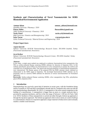
  • 7. SYNTHESIS & CHARACTERIZATION OF NOVEL NANOMATERIALS FOR SERS BIOMEDICAL/ENVIRONMENTAL APPLICATION 7 Figure 4: FT-IR spectra of PEG and PEG- Ag NPs. Intense absorption bands were observed at 2880, 1640, 1464, 1340, 1277, 1090, 945 and 842 cm−1 for PEG Ag sample (Fig.4). Aliphatic C–H stretching at 1464 and 1340 cm−1 due to C–H bending vibrations (Shameli, Bin Ahmad et al. 2012), bands at 1277 cm−1 and 1090 cm−1 which were related to the stretching vibrations of the alcoholic C–O bonds and C–O–C ether linkage ν(C–O–C) were observed in FTIR spectrum of PEG functionalized Ag NPs. Further, bands at 945 cm−1 (ρ(CH2)) and 842 cm−1 (ρ(CH2)) were attributed to rocking vibrations and band at 3392 cm−1 was attributed to the O–H stretching vibration in association with ν (CH3 s ) at ∼2880 cm−1 in case of PEG Ag NPs. Finally, a new band was observed at ~1640 cm−1 which might correspond to a C=O stretching vibration and this carbonyl group formation occurred probably due to the oxidation of hydroxyl groups (Fahmy, El-Nasser et al. 2017) in FTIR spectra of PEG Ag NPs (Fig.4). 2.4.2 Detection of dopamine on synthesized SERS substrate The Raman Spectra of Ag NPs and PEG functionalized Ag NPs (PEG Ag) were recorded (Fig.5). The Raman spectra of PEG Ag showed enhanced Raman intensities at 813, 1035, 1340 and 1600
  • 8. KHAN, XHIBO, YÜCEL, TURAN 8 cm-1 as compared with Ag NPs. These observations clearly indicated that PEG has stabilized silver nanoparticles with controlled and uniform size distribution. Figure 5: Raman spectra of PEG Ag and Ag NPs. Furthermore, functionalized Ag NPs were utilized for detection of dopamine and SERS response were recoded (Fig.6). The optical image of DA incubated PEG Ag NPs SERS substrate is shown in Fig.7. The SERS spectra of PEG Ag NPs clearly showed enhanced peaks of dopamine (0.05 mM) as compared to the Ag NPs (Fig.6). The band at 1480 cm-1 corresponds to phenyl C=C stretching has noticeably enhanced the peak signal as compared with dopamine peak signal. The band at 1337 cm-1 can be assigned to ring stretching vibration of dopamine. The ring stretching vibration mode of O-H bond is observed at 1395 cm-1 (Fig.6). The observations clearly indicated that PEG functionalized Ag NPs can enhance the SERS peak signal of dopamine and can be used as a sensitive SERS substrate for the detection of DA.
  • 9. SYNTHESIS & CHARACTERIZATION OF NOVEL NANOMATERIALS FOR SERS BIOMEDICAL/ENVIRONMENTAL APPLICATION 9 Figure 6: Raman spectra of PEG Ag and Ag NPs incubated with DA (0.05 mM). Figure 7: Raman optical image of functionalized Ag NPs incubated with dopamine (0.05 mM). 3 Conclusion and Future Work In this study, a simple green method was employed to prepare PEG functionalized Ag NPs as a sensitive SERS substrate for the detection of dopamine. The synthesized functionalized Ag NPs were successfully characterized by UV-Vis, XRD and FTIR techniques. The synthesized PEG Ag NPs showed enhanced Raman signal as compared to Ag NPs. Dopamine was successfully detected by employing functionalized Ag NPs as a SERS substrate. The developed method to prepare functionalized Ag SERS substrate is simple, low cost, environmentally friendly and easy to use. The developed functionalized SERS substrate in this study can be potentially used as a sensitive
  • 10. KHAN, XHIBO, YÜCEL, TURAN 10 SERS for the detection of a variety of neurotransmitters for biomedical applications. The next step would be to study the dopamine concentration in human cerebrospinal fluid (CSF) samples to see if this method can quantitatively measure the amount of dopamine in a reliable and accurate manner. Additional investigations can be made regarding altering the chain length of PEG or using PEG as the sole reducing agent instead of NaBH4. References Benelmekki, M. (2015). Designing hybrid nanoparticle. San Rafael, Calif.: Morgan et Claypool Al-Taa'Y, W., Abdul Nabi, M., Yusop, R. M., Yousif, E., Abdullah, B. M., Salimon, J., ... Zubairi, S. I. (2014). Effect of nano ZnO on the optical properties of poly(vinyl chloride) films. International Journal of Polymer Science, 2014, [697809]. DOI: 10.1155/2014/697809 Arole, V., & Munde, S. (2014). FABRICATION OF NANOMATERIALS BY TOP-DOWN AND BOTTOM-UP APPROACHES – AN OVERVIEW. Journal Of Advances In Applied Sciences And Technology, 1(2), 89-93. Ahmadpoor, P., et al. (2013). "The optical properties of PVA/TiO2 composite nanofibers." Journal of Applied Polymer Science 130(1): 78-85. Al-Taa'y, W., et al. (2014). "Effect of Nano ZnO on the Optical Properties of Poly(vinyl chloride) Films." International Journal of Polymer Science. Bhui, D. K., et al. (2009). "Synthesis and UV-vis spectroscopic study of silver nanoparticles in aqueous SDS solution." Journal of Molecular Liquids 145(1): 33-37. Fahmy, A., et al. (2017). "Tuned interactions of silver nanoparticles with ZSM-5 zeolite by adhesion-promoting poly(acrylic acid) deposited by electrospray ionization (ESI)." Journal of Adhesion Science and Technology 31(24): 2641-2656. Fateixa, S., et al. (2015). "Hybrid nanostructures for SERS: materials development and chemical detection." Physical Chemistry Chemical Physics 17(33): 21046-21071. Ji, X., et al. (2012). "On the pH-Dependent Quenching of Quantum Dot Photoluminescence by Redox Active Dopamine." Journal of the American Chemical Society 134(13): 6006-6017. Kneipp, K., et al. (1997). "Single molecule detection using surface-enhanced Raman scattering (SERS)." Physical Review Letters 78(9): 1667-1670. Kusior, A., et al. (2013). "TiO2-SnO2 nanomaterials for gas sensing and photocatalysis." Journal of the European Ceramic Society 33(12): 2285-2290. Lotharius, J. and P. Brundin (2002). "Pathogenesis of Parkinson's disease: Dopamine, vesicles and alpha-synuclein." Nature Reviews Neuroscience 3(12): 932-942.
  • 11. SYNTHESIS & CHARACTERIZATION OF NOVEL NANOMATERIALS FOR SERS BIOMEDICAL/ENVIRONMENTAL APPLICATION 11 Lussier, F., et al. (2017). "Dynamic SERS nanosensor for neurotransmitter sensing near neurons." Faraday Discussions 205: 387-407. Lv, Y., et al. (2013). "Surface modification of quantum dots and magnetic nanoparticles with PEG- conjugated chitosan derivatives for biological applications." Chemical Papers 67(11): 1404-1413. Muzamil, M., et al. (2014). "Synthesis of silver nanoparticles by silver salt reduction and its characterization." 13th International Symposium on Advanced Materials (Isam 2013) 60. Shameli, K., et al. (2012). "Synthesis and Characterization of Polyethylene Glycol Mediated Silver Nanoparticles by the Green Method." International Journal of Molecular Sciences 13(6): 6639- 6650. Son, H. S., et al. (2009). "Kinetics and mechanism of photolysis and TiO2 photocatalysis of triclosan." Journal of Hazardous Materials 166(2-3): 954-960. Yang, W. S., et al. (2013). "Facile preparation of Ag2S nanoparticles with broad photoelectric response region." Colloids and Surfaces a-Physicochemical and Engineering Aspects 433: 55-58. ctor. Crown Copyright (C) 2013 Published by Elsevier B.V. All rights reserved. Zhang, A., et al. (2007). "Recent progress in development of dopamine receptor subtype-selective agents: Potential therapeutics for neurological and psychiatric disorders." Chemical Reviews 107(1): 274-302.