The Sac Fungi: An Introduction to Ascomycota
•
0 likes•245 views

The document provides an overview of Ascomycota, or sac fungi, which accounts for approximately 75% of all described fungi. Key points include: - Ascomycota is defined by the ascus, where nuclear fusion and meiosis take place to form ascospores. - It includes many important and well-known fungi, from baker's yeast to morels and penicillin producer Penicillium. - Ascomycota obtain nutrients as saprotrophs, biotrophs, and parasites and can be found worldwide. - They reproduce both sexually via ascospores and asexually via conidia and have complex life cycles involving haploid and/or diplo

Recommended
More Related Content
What's hot
What's hot (20)
Similar to The Sac Fungi: An Introduction to Ascomycota
Similar to The Sac Fungi: An Introduction to Ascomycota (20)
Recently uploaded
Recently uploaded (20)
The Sac Fungi: An Introduction to Ascomycota
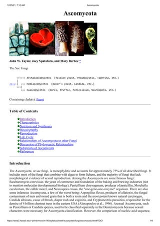
- 1. 12/25/21, 7:10 AM Ascomycota https://www2.hawaii.edu/~johnb/micro/m140/syllabus/week/eucaryotes/fungi/ascomycota.html#TOC1 1/6 Ascomycota John W. Taylor, Joey Spatafora, and Mary Berbee * The Sac Fungi ====== Archaeascomycetes (fission yeast, Pneumocystis, Taphrina, etc.) | <<===| === Hemiascomycetes (baker's yeast, Candida, etc.) ===| === Euascomycetes (morel, truffle, Penicillium, Neurospora, etc.) Containing clade(s): Fungi Table of Contents Introduction Characteristics Nutrition and Symbioses Biogeography Reproduction Life Cycle Relationships of Ascomycota to other Fungi Discussion of Phylogenetic Relationships Subgroups of Ascomycota References Introduction The Ascomycota, or sac fungi, is monophyletic and accounts for approximately 75% of all described fungi. It includes most of the fungi that combine with algae to form lichens, and the majority of fungi that lack morphological evidence of sexual reproduction. Among the Ascomycota are some famous fungi: Saccharomyces cerevisiae, the yeast of commerce and foundation of the baking and brewing industries (not to mention molecular developmental biology), Penicillium chrysogenum, producer of penicillin, Morchella esculentum, the edible morel, and Neurospora crassa, the "one-gene-one-enzyme" organism. There are also some infamous Ascomycota, a few of the worst being: Aspergillus flavus, producer of aflatoxin, the fungal contaminant of nuts and stored grain that is both a toxin and the most potent known natural carcinogen, Candida albicans, cause of thrush, diaper rash and vaginitis, and Cryphonectria parasitica, responsible for the demise of 4 billion chestnut trees in the eastern USA (Alexopoulos et al., 1996). Asexual Ascomycota, such as Penicillium or Candida species, used to be classified separately in the Deuteromycota because sexual characters were necessary for Ascomycota classification. However, the comparison of nucleic acid sequence,
- 2. 12/25/21, 7:10 AM Ascomycota https://www2.hawaii.edu/~johnb/micro/m140/syllabus/week/eucaryotes/fungi/ascomycota.html#TOC1 2/6 as well as nonsexual phenotypic characters, have permitted the integration of asexual fungi into the Ascomycota (Taylor, 1995). Characteristics The shared derived character that defines the Ascomycota is the ascus. It is within the ascus that nuclear fusion and meiosis take place. In the ascus, one round of mitosis typically follows meiosis to leave eight nuclei, and eventually eight ascospores. Ascospores are formed within the ascus by an enveloping membrane system, which packages each nucleus with its adjacent cytoplasm and provides the site for ascospore wall formation. These membranes apparently are derived from the ascus plasma membrane in the Euascomycetes and the nuclear membrane in the Hemiascomycetes (Wu and Kimbrough, 1992; Raju, 1992). In hyphal Ascomycota (left), the youngest, terminal hyphal segments develop into 8-spored asci. In yeasts (right) a single cell simply becomes the ascus, often with just 4 spores. At the time they are released from the ascus, the thick-walled haploid ascospores are resistant to adverse environments. But, given the right conditions, they will germinate to form a new haploid fungus. The body of Ascomycota is shared by other fungi and consists of a typical eukaryotic cell surrounded by a wall. The body can be a single cell, as in yeasts, or a long tubular filament divided into cellular segments, which is called a hypha (plural, hyphae). Both yeasts and hyphae have cell walls made of varying proportions of chitin and beta glucans (Wessels, 1994). Nutrition and Symbioses Like other fungi, Ascomycota are heterotrophs and obtain nutrients from dead or living organisms (Griffin, 1994; Carroll and Wicklow, 1992). If water is present, as saprotrophs they can consume almost any carbonaceous substrate, including jet fuel (Amorphotheca resinae) and wall paint (Aureobasidium pullulans), and play their biggest role in recycling dead plant material. As biotrophs, they may form symbioses with algae (lichens), plant roots (mycorrhizae) or the leaves and stems of plants (endophytes). Other Ascomycota (Ceratocystis and Ophiostoma) form symbiotic associations with an array of arthropods, where they can line beetle galleries and provide nutrition for the developing larvae. In return, the beetles maintain a pure culture of the fungus and transport it to newly established galleries. As parasites, ascomycetes account for most of the animal and plant pathogens including Pneumocystis carinii, responsible for pneumonia of humans with compromised immune systems and Ophiostoma ulmi, the Dutch elm disease fungus that is responsible for the demise of elm trees in North America and Europe (Agrios, 1988) Biogeography
- 3. 12/25/21, 7:10 AM Ascomycota https://www2.hawaii.edu/~johnb/micro/m140/syllabus/week/eucaryotes/fungi/ascomycota.html#TOC1 3/6 Ascomycota can be found on all continents and many genera and species display a cosmopolitan distribution (Candida albicans or Aspergillus flavus). Others are found on more than one continent (Ophiostoma ulmi, or Cryphonectria parasitica), but many are known from only one narrowly restricted location. For example, the White Piedmont Truffle (Tuber magnatum) is known from only one provence of Northern Italy. Reproduction From a human perspective, the most unusual aspect of all fungi is that they have more than one reproductive option. The textbook Ascomycota can make spores sexually (ascospores or meiospores) and asexually (condia or mitospores). Following meiosis,the ascospores take shape inside the ascus when new cell walls surround each nucleus as can be seen in the electron micrograph above (Wu and Kimbrough, 1992). Conidia contain mitotic nuclei, and their cell wall is simply a modified hyphal or yeast wall. Ascospores may or may not be shot by water pressure from the ascus and although wind is the primary dispersal agent once the spores have been released from the ascus, Ascomycota also use splashing or running water or animals to disperse their spores (Ingold, 1965). Conidial diversity reaches its climax with the ascomycetes, with forms ranging from single spores hardly different from hyphae(Geotrichum candidum) to elaborate heads of ornamented condida (Aspergillus niger) and beyond (Cole and Kendrick, 1981). Life Cycle Ascomycota are either single-celled (yeasts) or filamentous (hyphal) or both (dimorphic). Yeasts grow by budding or fission and hyphae grow apically and branch laterally. Most yeasts and filamentous Ascomycota are haploid, but some species, Saccharomyces cerevisiae for example, can also be diploid. Mitospores may simply reproduce the parent, or may also act as gametes to fertilize a compatible partner. Some Ascomycota must outbreed (heterothallic), others can also self, and some can only self (homothallic) (Alexopoulos et al. 1996). Genetic regulation of sex expression and mating is well-understood in some model Ascomycota such as yeast, where there are two sexes and mating is coordinated by oligopeptide pheromones (Marsh, 1991; Glass
- 4. 12/25/21, 7:10 AM Ascomycota https://www2.hawaii.edu/~johnb/micro/m140/syllabus/week/eucaryotes/fungi/ascomycota.html#TOC1 4/6 and Lorimer, 1991). In hyphal species, cytoplasmic fusion may not be immediately followed by nuclear fusion, leading to a short dikaryotic phase. The dikaryotic hyphae may be protected and nourished by differentiated haploid hyphae which form a fruiting body (the ascoma; plural ascomata). Ascomata may be closed (cleistothecium), open by a narrow orifice (perithecium), or broadly open like a cup (apothecium). Ascospores are released from the ascoma and germinate to form a new haploid mycelium. Relationships of Ascomycota to other Fungi The Ascomycota is a sister group to the Basidiomycota. This relationship is supported by the presence in members of both phyla of cross-walls (septa) that divide the hypahe into segments, and pairs of unfused nuclei in these segments after mating and before nuclear fusion (dikaryons). Further support comes from the apparent homology between structures that coordinate simultaneous mitosis of the two dikaryotic nucli (Ascomycota croziers and Basidiomycota clamp-connections). Discussion of Phylogenetic Relationships Sexual Ascomycota all have asci. Comparison of nuclear small subunit ribosomal RNA gene sequence demonstrates a monophyletic Ascomycota, although support for the basal branch is not strong (Berbee and Taylor, 1993; Bruns et al., 1992). Early diverging Ascomycota have been grouped into the Archaeascomycetes, although support for the monophyly of this group is not strong (Nishida and Sugiyama, 1994). The placement of Neolecta among the Archaeascomycetes is surprising because of the presence of an ascoma, a feature not found in other Archaeascomycetes or in any Hemiascomycetes (Landvik et al. 1992). However, there is no reason that the Hemiascomycetes could not have lost ascomata as hyphal growth became suppressed in favor of yeasts. The Hemiascomycetes form a well-supported monophyletic taxon, as do the Euascomycetes (Gargas et al., 1995). Asexual fungi sharing morphological or molecular characters of sexual Ascomycota are classified in the Ascomycota; examples include Candida albicans (Hemiascomycetes) and Pencillium chrysogenum (Euascomycetes). By comparing nucleic acid sequences, the timing of Ascomycota evolution has been estimated (Berbee and Taylor, 1993). The Archaeascomycetes, Hemiascomycetes and Euascomycetes all became established in the coal age, a bit more than 300 million years ago. Fossils of these early Ascomycota are not going to be easy to recognize, because they probably lacked ascoma and their spores were not distinctive. Fungal-like fossils claimed to be older than 1.0 to 1.2 billion years are probably artifactual. The earliest ascomycete fossil ascomata and spores are controversial because their age of deposition significantly predates molecular estimates of their time of origin. The fruiting bodies may be zygomycetous, and the spores may have washed into older sediments, or the molecular estimates may be erroneous. Subgroups of Ascomycota Archaeascomycetes is a class recently discovered from comparison of nucleic acid sequences and contains species previously thought to be Hemiascomycetes. Some species, such as the fission yeast, Schizosaccharomyces pombe, are unicellular, but others grow as hyphae as well as single cells (for example, Taphrina species). The genera are distantly related to each other, possibly remnants of an early radiation of Ascomycota. Archaeascomycetes lack ascomata (Nishida and Sugiyama, 1994). The Hemiascomycetes comprises the yeasts and is home to the most famous fungus, Saccharomyces cerevisiae, better known as the baker's yeast. Although most members are primarily unicellular, the basal taxa make abundant hyphae. Hemiascomycetes lack ascomata (Barnett et al., 1990). Euascomycetes contain well over 90% of Ascomycota, and the species are hyphal, with almost all of the sexually reproducing forms possessing ascomata. Most of the recent molecular phylogenetic effort has been directed at this class (e.g., Berbee and Taylor 1992a, b; Spatafora and Blackwell, 1993, Spatafora, 1995).
- 5. 12/25/21, 7:10 AM Ascomycota https://www2.hawaii.edu/~johnb/micro/m140/syllabus/week/eucaryotes/fungi/ascomycota.html#TOC1 5/6 References Agrios, G. N. 1988. Plant Pathology, third edition. Academic Press, San Diego. Alexopoulos, C. J., C. W. Mims, and M. Blackwell. 1996. Introductory Mycology. John Wiley and Sons, New York. 868p. Barnett, J. A., R. W. Payne, and D. Yarrow. 1990. Yeasts: characteristics and identification. Cambridge University Press, Cambridge. Berbee, M. L., and J. W. Taylor. 1992a. Convergence in ascospore discharge mechanism among Pyrenomycete fungi based on 18S ribosomal RNA gene sequence. Mol. Phylog. Evol. 1:59-71. Berbee, M. L., and J. W. Taylor. 1992b. Two ascomycete classes based on fruiting-body characters and ribosomal DNA sequence. Mol. Biol. Evol. 9:278-284. Berbee, M. L., and J. W. Taylor. 1993. Dating the evolutionary radiations of the true fungi. Can. J. Bot. 71:1114-1127. Bruns, T. D., R. Vilgalys, S. M. Barns, D. Gonzalez, D. S. Hibbett, D. J. Lane, L. Simon, S. Stickel, T. M. Szaro, W. G. Weisburg, and M. L. Sogin. 1992. Evolutionary relationships within the fungi: analyses of nuclear small subunit rRNA sequences. Mol. Phylog. Evol. 1:231-241. Carroll, G.C. and D. T. Wicklow, 1992. The Fungal Community: Its Organization and Role in the Ecosystem. Marcel Dekker, Inc., New York. Cole, G. T., and B. Kendrick. 1981. Biology of conidial fungi. Academic Press, New York. Gargas, A., P. T. DePriest, M. Grube, and A. Tehler. 1995. Multiple origins of lichen symbioses in fungi suggested by SSU rDNA phylogeny. Science 268:1492-1495. Glass, N. L., and I. A. J. Lorimer. 1991. Ascomycete mating types. Pages 193-216. in More gene manipulations in fungi (J. W. Bennett and L. L. Lasure, eds.). Academic Press, Orlando. Griffin, D. H. 1994. Fungal Physiology. 2nd. Wiley-Liss, New York. Ingold, C. T. 1965. Spore Liberation. Clarendon Press, Oxford. Landvik, S., O. E. Eriksson, A. Gargas, and P. Gustafsson. 1993. Relationships of the genus Neolecta (Neolectales ordo nov., Ascomycotina) inferred from 18s rDNA sequences. Syst. Ascomycetum 11:107-118. Marsh, L. 1991. Signal transduction during pheromone response in yeast. Annu. Rev. Cell biol. 7:699-728. Nishida, H., and J. Sugiyama. 1994. Archiascomycetes: Detection of a major new linage within the Ascomycota. Mycoscience 35:361-366. Raju, N. B. 1992. Genetic control of the sexual cycle in Neurospora. Mycol. Res. 96:241-262,. Spatafora, J. W. 1995a. Ascomal evolution among filamentous ascomycetes: evidence from molecular data. Can. J. Bot. S811-S815. Spatafora, J., and M. Blackwell. 1993. Molecular systematics of unitunicate perithecial Ascomycetes. The Clavicipitales - Hypocreales connection. Mycologia 85:912-922. Taylor, J. W. 1995. Making the Deuteromycota redundant: a practical integration of mitosporic and meiosporic fungi. Can. J. Bot. 73 (suppl.):s754-s759. Taylor, J. W., B. Bowman, M. L. Berbee, and T. J. White. 1993. Fungal model organisms: phylogenetics of Saccharomyces, Aspergillus and Neurospora. Syst. Biol. 42:440-457. Wessels, J. G. H. 1994. Developmental regulation of fungal cell wall formation. Ann. Rev. Phytopathol. 32:413-437. Wu, C. G., and J. W. Kimbrough. 1992. Ultrastructural studies of ascosporogenesis in Ascobolus immersus. Mycologia 84:459-466. About this page
- 6. 12/25/21, 7:10 AM Ascomycota https://www2.hawaii.edu/~johnb/micro/m140/syllabus/week/eucaryotes/fungi/ascomycota.html#TOC1 6/6 Many thanks to Dave Carmean, Soren Rosendahl for scanning photos and David Maddison for page design advice. John W. Taylor E-mail: jtaylor@violet.berkeley.edu. Department of Plant and Microbial Biology, 111 Koshland Hall, University of California, Berkeley, CA 94720-3120, USA Joey Spatafora E-mail: spatafora@UO.edu. Department of Botany and Plant Pathology, 2082 Cordley Hall, Oregon State University, Corvallis, OR 97330-2902, USA Mary Berbee E-mail: berbee@unix.ubc.ca. Department of Botany, University of British Columbia, Vancouver, BC V6T 2B1, CANADA Correspondence regarding this page should be directed to John Taylor, at jtaylor@violet.berkeley.edu. Page copyright © 1996 John Taylor, Joey Spatafora, Mary Berbee First online 11 March 1996 Last saved 27 May 1996 Title Illustrations From left to right: Asci of Taphrina (Peach leaf curl fungus) atop a peach leaf, © K. Wells 1996. Budding cells of Saccharomyces (the baker's and brewer's yeast), © K. Wells 1996. Two fruiting bodies (ascomata) of Morchella (the edible morel) with one sliced open, © J. Taylor 1996. Mitospores (conidia) of Penicillium, one of the asexual Ascomycota, © K. Wells 1996. Text Illustrations From top to bottom: Asci of a hyphal Ascomycota (Euascomycetes), Podospora , © R. Vilgalys 1996. Ascus of a yeast (Hemiascomycetes), Saccharomyces, © J. Taylor 1996. Ascus of a hyphal ascomycete (Euascomycetes) as viewed by the electron microscope, © R. Vilgalys 1996. Life cycle of Ascomycota, © J. Taylor 1996 Information on the Internet Mycologists Online Virtual Library - Yeast Fungal Genetics Stock Center Yahoo Mycology Taxon name: Ascomycota Query GenBank Tree of Life design and icons copyright © 1996 David Maddison and Wayne Maddison.