SlideShare a Scribd company logo
1 of 42
PHYLUM: ASCOMYCOTA
• Phylum Ascomycota and Basidiomycota are sometimes called “higher fungi” considerably
more complex in structure; share no. of characters such as septate mycelium, having
simple pores and the presence of woronin bodies (while concentric bodies are present in
the lichen-forming ascomycetes) and a dikaryotic stage in life cycle having either short
lived (Ascomycota) and long lived (Basidiomycota) dikaryotic stage interspersed between
plasmogamy and karyogamy unlike other fungi in which karyogamy follows immediately
after plasmogamy.
• Phylum Ascomycota are distinguished from other fungi in terms of formation of ascus (pl.
asci Gr. askos=goat skin, sac), a sac like cell containing the ascospores (Gr. askos+ spora
=seed, spore), cleaved from within by free-cell formation after karyogamy and meiosis.
Eight ascospores are typically formed within the ascus, but this number may vary from
one to over a thousand according to the species.
General characteristics: As in Zygomycota ascomycetes may have distinct
reproductive phases
(i) Sexual involving formation of asci and ascospores and
(ii) Asexual with spore production occurring at different times on the same
mycelium
(iii) Some Ascomycota have only a sexual state while vast numbers of ascomycetes
are known by their asexual stages. These taxa are placed in artificial group
called Deuteromycota or Fungi Imperfecti.
Somatic structure:
• Except for some yeasts ascomycetes fungi possess well developed and profusely
branched mycelium with one or several nuclei. They may be single celled, mycelial
or dimorphic.
• The cell wall largely contains chitin and cellulose is generally absent except few
species.
• The septa are produced towards the center of hypha from the hyphal wall/periphery
and a pore (1.0 to 1.5) is left in the centre. The pore allows for cytoplasmic
continuity and occurrence of nuclear and mitochondrial migration throughout the
hypha.
• In many yeasts that produce a mycelium a closure line demarking the complete
inward growth of the septum or micropores analogous to the plasmodesmata of plants
are present.
• Dowding (1958) reported that the nuclei in Gelasinospora tetrasperma are carried at
the rate of 40 mm/hour. The actual mechanism of nuclear migration probably depends
on cytoplasmic streaming (Snider, 1965).
• Woronin bodies highly refractive bodies are found associated with pores (both in ascomycetes and
deuteromycetes fungi) which are rounded or elongated oval, spherical or hexagonal or rectangular
with a crystalline protein matrix that are usually associated with septum often known to plug the
septal pores of hyphae although they are believed to serve to separate aging or damaged hyphae from
rest of mycelium.
• Hyphal compartments often are uninucleate, but mycelia consisting of multinucleate cells are well
known. Three kinds of nuclei may exist within ascomycetes hyphae:
• A homokaryotic mycelium that results from germination of a uninucleate haploid ascospore. A
homokaryotic multinucleate condition arises by mitotic duplication of nucleus and subsequent
migration of daughter nuclei.
• The heterokaryotic condition may arise as a result of the mutation of nuclei in an originally
homokaryotic mycelium; or by the fusion of genetically distinct entities that results in dikaryon;
or a multinucleate cell in which the nuclei are unpaired and not daughter cells of the same parent
cell.
• These various multinucleate situations are possible because the pores in the septa allow nuclear
migration.
• Aggregation of modified vegetative hyphae may lead to the production of sclerotia.
• In certain members the aggregation of mycelial strands produces rhizomorphs.
• Somatic structures known as stroma (pl. stromata Gr. Stroma= mattress) are
commonly produced in many ascomycetes members. Stroma consists of a compact
mass of vegetative hyphae with or without tissue of the host or substratum,
sometimes sclerotium–like in form, often bearing internal or external fructifications.
These are especially produced in the order Sphaeriales.
• Anatomically, it may be prosenchymatous or pseudoparenchymatous and has two
distinct regions.
• The outer narrow ectostroma which has mechanical function and inner massive
endostroma that gives rise to the sporophores or ascocarps. [Ascomycetes mycelium
may be organized into fungal tissues (plectenchyma).
• Plectenchyma differs from the parenchyma of plants because it arises from apical
growth of hyphae that become closely associated only secondarily.
• If such a tissue is loosely woven and the mycelial strands are more or less evident, it is
known as prosenchyma.
• If however, the hyphae have lost their individuality and cells are more or less
isodiametric, closely resembling the parenchyma of plants it is known as
pseudoparenchyma.
• Prosenchymatous and pseudoparenchymatous tissues are associated primarily with
reproductive and resting structures of ascomycetes as well as basidiomycetes.
Ascomycetes may produce specialized structures; appressoria and haustoria some of
which are associated with host infection by pathogens.
• In addition certain ascomycetes form structures known as hyphopodia (sing.
Hyphopodium) which are two types: Capitate hyphopodia produced by some plant
pathogenic ascomycetes considered to be specialized form of appressorium. These are
stalked, thick-walled, lobed cells that stick to the host surface; certain stalked
hyphopodia may have additional function i.e. initiation of ascocarp development upon
their fusion.
Melanized hyphopodia of Gaeumannomyces graminis arising from an equally strongly
melanized runner hypha formed on the hydrophobic surface of a plastic coverslip.
• Pointed mucronate hyphopodia best known in Meliolales are not associated with
infection structures, reported to produce conidia that function as spermatia (Hughes,
1981).
• Some of the specializations of ascomycetes mycelia are the nooses, traps, coils and
sticky pegs that trap nematodes. These systems have not only developed in
ascomycetes, but in zygomycetes and basidiomycetes as well. Trap formation in
Arthrobotrys may be induced by nematode presence or by addition of proteinaceous
material into a culture.
• The fungus mycelium is more attractive to nematodes when traps are present, and
the modified trap hyphae produce lectins (carbohydrate binding proteins) that bind
to specific sites on the nematode surface; for this reason the traps may also attach to
other organisms, including fungi with similar cell surface carbohydrates.
Reproduction: These fungi reproduce both sexually and asexually. The ascus or sexual
stage is often called the ascigerous or perfect stage (or state), and the conidial or asexual
stage is often designated as imperfect stage. No flagellated structures are produced
while some fungi reproduce both sexually and asexually, others multiply exclusively by
sexual reproduction through ascospores. Hennebert and Weresub (1977) used the term
teleomorph for the sexual form; anamorph for the asexual form and holomorph for
the whole fungus in all its forms and phases. Synanamorph is applied to any one of the
two or more anamorphs (asexual states) which have the same teleomorph. Classification
of ascomycetes is based on ascigerous or perfect stage. The conidia and other methods
of asexual reproduction are secondary in importance.
Asexual reproduction:
• Asexual reproduction in ascomycetes may be carried out by fission, fragmentation, or
formation of chlamydospores or conidia according to species and environmental
conditions.
• Budding and fission are characteristic of the yeasts and some dimorphic ascomycetes, the
spores produced by budding are known as blastospores etymologically means bud spores.
Fission occurs in yeast genus Schizosaccharomyces only.
• Fragmentation of hyphae and chlamydospore formation are other means of asexual
reproduction. Because all living portions of the thallus potentially are capabale of growth,
fragmentation (including laboratory transfer of cultured mycelium) may result in as many
new individuals as there are fragments.
• Although some ascomycetes produce only sterile mycelia, many are known for their
conidium formation.
• Conidia are the chief reproductive organs of majority of organisms and are produced
from or within a conidiogenous cell, which is borne on a simple or branched hypha.
Conidia are may arise directly from somatic hyphae or from specialized
conidiogenous cells, often borne on hyphal branches known as conidiophores.
• Conidiophores vary from short hyphal branches to those that are long and intricately
branched.
• In some species the conidiophores may be free from each other without evident
organization while in others they are joined togeey often their to form complex
structures such as synnemata, sporodochia, pycnidia and acervuli which are found
in Deuteromycota fungi as these fungi are the imperfect states of ascomycetes fungi
whose perfect states have not been discovered so far or extinct.
• Conidia are often important in propagating and disseminating fungal species
throughout the spring and summer with several generations produced in a season.
The conidia vary greatly in size, shape, colour and number and arrangement of cells,
if multicellular, often determined by dispersal mechanisms.
• The conidia may be produced successively at the tip, separating from conidiophore as
soon as formed, or clinging together in a mucilaginous drop or remaining attached in
chains.
• A chain may be acogenously (acropetal) formed with the basal conidium being the oldest
and youngest conidium at the apex, or it may be bassigenous (basipetal) in which the
oldest conidium is at the apex of chain.
• In another type almost simultaneous rounding off of the cells of simple or branched
hyphae into catenulate conidia (oidia) which then fall apart almost simultaneously.
• Among many ascomycetes other types of ‘resistant spores/structures’ which serve the
fungus to survive unfavourable condtions include sclerotia and chlamydospores which are
characterized by thick walls.
• Stromata have a similar function but rather than initiating mycelial growth they give rise
to conidia or ascocarps.
Life cycle:
• As there is so much diversity in ascomycetes, no typical life cycle exists. However,
life cycle of Pyronema omphaloides generally illustrates both sexual and asexual
reproduction in a single life cycle.
• An ascospore germinates by one or more germ tubes to produce a mycelium. As germ
tubes issue from a spore, the spore nucleus divides and nuclear progeny are
distributed within growing hyphae. Soon after hyphal growth septa are laid down to
delimit uninuclate compartments and hyphal branching takes place. The vigourously
growing mycelium may begin to produce conidia almost immediately which are
continuous and abundant sources of inoculum for localized dispersal of fungus for a
period of months.
• Many crops of conidia may be borne in a single season. The conidia produced in
enormous numbers germinate by germ tube to produce mycelium similar to ascospore
germination.
• At some time in appropriate conditions the mycelium ceases conidial production and
begins to undergo a complex series of physiological changes that initiate sexual
reproduction.
• The presence of second mating type in heterothallic fungi is required. The ascogonia are
differentiated from somatic hyphae (may be uni- or multinucleate).
• Compatible nuclei are brought to the ascogonium by one or more methods of
copulations briefed earlier. The contact of antheridium and ascogonium and passage of
antheridial nuclei into the trichogyne and eventually into the ascogonial base (Fig. A to
C) takes place.
• After plasmogamy the ascogonium produces a number of papillae just opposite groups
of nuclei located in the periphery of ascogonium. The ascogonial walls appear thinner at
these points. As these papillae enlarge, nuclei from the ascogonium begin to pass into
them one by one. Eventually the papillae elongate into ascogenous hyphae (Fig. D) in
which a leading pair of nuclei can be detected, followed often by second pair.
• The nuclei in the ascogenous hyphae and those still in ascogonium soon undergo
simultaneous mitosis known as conjugate divisions.
• Primary septa are formed between daughter nuclei in such a way that the tip cell of
ascogenous hypha is uninucleate and followed by a series of binucleate cells that contain
pairs of non-sister nuclei.
• It is probable that one nucleus in each binucleate cell of ascogenous hyphae is acogonial
in origin and other antheridial, indicating dikaryotic phase of life cycle. In a large number
of ascomycetes including P. omphaloides, one of the binucleate cells of ascogenous hypha
elongates and bends over to form a hook or crozier (Fig. E).
• The two nuclei in this hooked cell (ascus mother cell AMC) divide in such a way that
their spindles are oriented more or less vertically and parallel to one another(Fig. F), so
that two of the daughter nuclei –one from each spindle and therefore of different origin-
are close to one another at the bend of the hook. Two septa are laid down, separating the
hook into three cells (Fig. G).
• The tip and basal cells are uninucleate, one containing an antheridial nucleus and the other
an ascogonial nucleus; the crook cell is binucleate which is the young ascus where
eventual nuclear fusion (Fig. H) takes place.
• Karyogamy takes place in the young ascus soon after septa are formed in the
hook thus ending the dikaryotic phase. The ascus with its diploid zygote
nucleus begins to elongate (Fig. I to J) and the zygote nucleus undergoes
meiosis.
• The four haploid (Fig. K) nuclei (products of meiosis) each undergo a mitotic
division so that a total of eight nuclei are formed.
• These will become incorporated in the ascospores during ascosporogenesis or
free cell formation.
Ascosporogenesis:
The ascus mother cell gets transformed into an ascus generally containing eight
ascospores which develop around the nuclei by free cell formation. It involves two
mechanisms:
(1) Portions of cytoplasm each typically consisting a single nucleus are delimited by an
envelope consisting of two closely appressed unit membranes and
(2) The ascospore wall is deposited between these two membranes to separate them from
one another as spore matures which are present in a discontinuous cylindrical ascus
vesicle or enveloping membrane system (EMS) which invaginates and fragments into
sheets and then cleave out the young ascospore.
• The portion of cytoplasm that remains outside the ascospore is called epiplasm,
possibly serves to nourish the developing spore and deposit external ornamentation.
The inner membrane of EMS becomes spore plasma membrane.
• Commonly the number of asscopores per ascus in most species is eight however,
some species routinely produce asci containing smaller numbers of spore including
one, two, three or four while large numbers of spores up to possibly as many as
thousand are produced in few species.
• Ascospores come in variety of sizes and shape (Fig.) ranging from long, thin and
threadlike to globose and even hat shaped in appearance.
• Ascospore may be one celled or septate, also may be arranged in uni- or biserate
manner in an ascus.
Ascospore:
• They are “spores produced as a result of meiosis and enclosed within a specialized
cover” (Sussman and Halvorson, 1966).
• After nuclear divisions, ascospore boundaries become delineated around the clear
areas enclosing the asci by a process of “free cell formation” which involves the
plasmalemma differently from conidiogenesis.
• Ascospores vary in size, shape and colour. Variations in the shape of mature
ascospores provide a good criterion for differentiation in yeast systematics (Kreger-
van Rij, 1969).
Asci:
• The asci are typically elongated, cylindrical, ovoid, club shaped/clavate (Gnomonia) or
globose form (Eremascus), septate (Dothidea), broadly ovate with stalk (Taphrina).
Spherical asci are produced by some yeasts (Hansenula anomala) and may also be
produced directly on hyphae (Endomycopsis fibuliger) or in ascocarps (Emericella
variecolor) where they are generally arranged in a scattered fashion.
• Elongated asci usually are formed in a hymenium as component of ascocarp. Asci may
be stalked or sessile and may arise at various levels within the ascocarps
(Plectomycetes) or from a single level formed naked (Hemiascomcetes) or arranged in a
palisade like manner forming a hymenial layer (Pyrenomyectes and Discomycetes). A
definite layer of asci whether naked or enclosed in an ascocarp is called a hymenium
(pl. hymenia; Gr. hymen= membrane). Studies have shown that sometimes asci may
develop in hymenium but become rearranged at maturity so that they appear to be
scattered.
• In the ascus morphology three basic types of asci such as prototunicate, unitunicate and
bitunicate can be distinguished on the basis of their wall structure and mode of dehiscence
used as important criterion in taxonomy of ascomycetes.
• Prototunicate asci, a thin delicate wall and release their spores either by breaking apart or
deliquescing.
• The walls of both unitunicate and bitunicate ascus is said to consist of two layers:
exotunica/ectotunica (exoascus), the outer layer and endotunica (endoascus), the inner
layer.
• In the unitunicate ascus these layers adhere closely throughout the life of ascus, the asci
are thickened at the apex and characteristically perforated by a pore for ascospore
discharge (a terminal pore), or spores are released through slit or hinged cap (operculum).
• In the bitunicate ascus the endotunica usually expands up to twice or more its original
length separating from the ruptured exotunica at the time of spore release. The outer wall
ruptures during dehiscence and enables elastic inner wall to expand to form a cylindric sac
and ascospores are successively discharged through elastic pore in the expanded sac or
endotunica. Due to this behaviour of bitunicate ascus has been called the Jack-in-the-box
(Ingold, 1933) or the fissitunicate ascus (Henssen and Jahns, 1974).
• In the unitunicate ascus may have a pore, slit, operculum or tear (formed in the lateral wall) for
liberation of ascospores before the discharge occurs; the pore is plugged with a substance that stains
blue with iodine and is different from wall material (or Pot. iodide called as Melzier’s reagent used
for this; unitunicate blue stain while in bitunicate no blue stain).
• In many ascomycetes the ascal tip has distinct characters called apical apparatus are of great
taxanomic importance.
• It can be stained with writing ink, congo-red and janus green. In the discomycetes the tip of the
ascus has a cap or operculum or may be perforated by a pore without an operculum.
• These two types of asci are termed as operculate and inoperculate respectively and the presence or
absence of an operculum is an important feature in classification.
Ascospore release, dispersal and germination: Eriksson (1981) distinguished eight
types of dehiscence in bitunicate asci with ectotunica and endotunica; (1) fissitunicate
(e.g. Pleospora); (2) rostrate (e.g. Melaspilea); (3) semifissitunicate (e.g. Peltigera); (4)
pseudofissitunicate (e.g. Diatrypiella); (5) bilabiate (e.g. Pertusaria); (6) poricidal (e.g.
Porina); (7) operculate and (8) explosive (e.g. Erysiphe).
Asci contained in cleistothecia are exposed for release of ascospore only after the
ascocarp disintegrates and ascospore release is non-explosive. In majority of ascomycetes
however, the asci are cylindrical and ascospore release is explosive. In the Erysiphales
the cleistothecial wall is non-disintegrating; the ascus enlarges and protrudes through
confining ascocarp wall. The wall which is forced open is under some strain and
suddenly snaps shut, ejecting the entire acsus to a distance of several centimeters. The
ascus then forcibly ejects its ascospores. Water uptake is possibly responsible for increase
in turgor which causes the explosive release. After the delimitation of ascospores in the
ascus the epiplasm continues to line the ascus wall that surrounds a large central vacuole
containing ascus sap. The ascospores are suspended in the vacuole.
Ascocarp: Except for a few filamentous ascomycetes (Hemiascomycetes- asci are naked) that do not
produce their asci/ascus in fruiting bodies called ascocarp (Gr. askos =sac + karpos=fruit). In general
there are five ways that ascomycetes can be separated according to the way they bear asci:
(1) Those bear naked asci without any fruiting body; thallus mycelial or yeast like [Order
Endomycetales (yeast-Saccharomyces), Protomycetales (Protomyces macrospores-stem gall of
coriander) and Taphrinales (Taphrina deformans-peach leaf curl; T. maculans-turmeric).
(2) Those that produce their asci inside a completely closed and usually globular ascocarp called a
cleistothecium (pl. cleistothecia, Gr. kleistos = closed+ theke = case) without any opening for
liberation of acsospores. By disintegration or breaking of cleistothecium liberates asci. Ex:
Sphaerotheca, Erysiphe, Meliola, Order Eurotiales- Eurotium (conidial state: Aspergillus) and
Talaromyces (conidial state: Penicillium).
(3) Those with an ascocarp the perithecium (pl. perithecia, Gr. peri = around + theke= case) that is
more or less closed (flask shaped ascocarp) but at maturity is provided with a pore (ostiole)
through which ascospores escape. Ex: Pyrenomycetes Order Sphaeriales [Family: Clavicipitaceae
(Claviceps), Ophiostomataceae (Ceratocystis), Nectria, Gibberella etc.]
(4) Those that produce their asci in an open /cup or saucer shaped ascocarp called an
apothecium (pl. apothecia, Gr. apotheke = store house) bearing an exposed
hymenium Ex: Peziza, Monilinia, Sclerotinia, Pyronema.
(5) Those that form their asci directly in a cavity (locule) within a stroma. Stroma
may be likened to a cushion of closely woven somatic hyphae forms the wall of the
ascocarp called an ascostroma (pl. ascostromata, ascus + stroma) or
pseudothecium. Ex: Mycosphaerella, Elsinoe, Guignardia etc.
• In addition, various modifications of ascocarps are known to occur in few
members: hysterothecium (pl.hysterothecia, Gr. hysteros = womb + theke = case)
which is elongated, boat shaped with a longitudinal slit (Order Hysteriales and
lichenized Order Arthoniales) and sclerotoid ascocarps with tough peridia (as in
some species of Penicillium).
10 Ascus development.pptx

More Related Content

Similar to 10 Ascus development.pptx (20)

General characters of fungi
General characters of  fungiGeneral characters of  fungi
General characters of fungi
 
Mycology 1st Year B.Sc. Hons.ppt
Mycology 1st Year B.Sc. Hons.pptMycology 1st Year B.Sc. Hons.ppt
Mycology 1st Year B.Sc. Hons.ppt
 
Loculoascomycetes^.^.pptx
Loculoascomycetes^.^.pptxLoculoascomycetes^.^.pptx
Loculoascomycetes^.^.pptx
 
Zygomycotina
ZygomycotinaZygomycotina
Zygomycotina
 
Ppt of fungi
Ppt of fungiPpt of fungi
Ppt of fungi
 
PRESENTATION ON FUNGI.
PRESENTATION ON FUNGI.PRESENTATION ON FUNGI.
PRESENTATION ON FUNGI.
 
Deuteromycotina
Deuteromycotina Deuteromycotina
Deuteromycotina
 
Lec 9. Intro myco1.pptx
Lec 9. Intro myco1.pptxLec 9. Intro myco1.pptx
Lec 9. Intro myco1.pptx
 
1) Strategies and structuresIn Protozoans the method of movement .pdf
1) Strategies and structuresIn Protozoans the method of movement .pdf1) Strategies and structuresIn Protozoans the method of movement .pdf
1) Strategies and structuresIn Protozoans the method of movement .pdf
 
Morphologyoffungi 170226153026
Morphologyoffungi 170226153026Morphologyoffungi 170226153026
Morphologyoffungi 170226153026
 
Morphology of fungi
Morphology of fungiMorphology of fungi
Morphology of fungi
 
Class xi ch 2
Class xi ch   2Class xi ch   2
Class xi ch 2
 
Mycology Introduction
Mycology IntroductionMycology Introduction
Mycology Introduction
 
Asexual reproduction in ASCOMYCOTA.pptx
Asexual reproduction in ASCOMYCOTA.pptxAsexual reproduction in ASCOMYCOTA.pptx
Asexual reproduction in ASCOMYCOTA.pptx
 
Fungi 1195182837648559-3
Fungi 1195182837648559-3Fungi 1195182837648559-3
Fungi 1195182837648559-3
 
Fungi- Cell Wall & Thallus Structure.pdf
Fungi- Cell Wall & Thallus Structure.pdfFungi- Cell Wall & Thallus Structure.pdf
Fungi- Cell Wall & Thallus Structure.pdf
 
Myxomycota
MyxomycotaMyxomycota
Myxomycota
 
Algae.pptx
Algae.pptxAlgae.pptx
Algae.pptx
 
Fungi.
Fungi.Fungi.
Fungi.
 
Fungi
FungiFungi
Fungi
 

More from Lavanya943804

Lecture-6 Reproduction in bacteria.ppt
Lecture-6 Reproduction  in bacteria.pptLecture-6 Reproduction  in bacteria.ppt
Lecture-6 Reproduction in bacteria.pptLavanya943804
 
plant quarantine in plant health mngt.ppt
plant quarantine in plant health mngt.pptplant quarantine in plant health mngt.ppt
plant quarantine in plant health mngt.pptLavanya943804
 
advances in chilli.pptx
advances in chilli.pptxadvances in chilli.pptx
advances in chilli.pptxLavanya943804
 
Presentation reading .ppt
Presentation reading .pptPresentation reading .ppt
Presentation reading .pptLavanya943804
 
Stress PhysiologyL#3.ppt
Stress PhysiologyL#3.pptStress PhysiologyL#3.ppt
Stress PhysiologyL#3.pptLavanya943804
 
CLIIMATE CHANGE AND BIODIVERSITY.pptx
CLIIMATE CHANGE AND BIODIVERSITY.pptxCLIIMATE CHANGE AND BIODIVERSITY.pptx
CLIIMATE CHANGE AND BIODIVERSITY.pptxLavanya943804
 
Clamate change introduction class 1.pptx
Clamate change introduction class 1.pptxClamate change introduction class 1.pptx
Clamate change introduction class 1.pptxLavanya943804
 
International Convension, PPT - 1.pptx
International Convension, PPT - 1.pptxInternational Convension, PPT - 1.pptx
International Convension, PPT - 1.pptxLavanya943804
 

More from Lavanya943804 (10)

Lecture-6 Reproduction in bacteria.ppt
Lecture-6 Reproduction  in bacteria.pptLecture-6 Reproduction  in bacteria.ppt
Lecture-6 Reproduction in bacteria.ppt
 
plant quarantine in plant health mngt.ppt
plant quarantine in plant health mngt.pptplant quarantine in plant health mngt.ppt
plant quarantine in plant health mngt.ppt
 
advances in chilli.pptx
advances in chilli.pptxadvances in chilli.pptx
advances in chilli.pptx
 
Presentation reading .ppt
Presentation reading .pptPresentation reading .ppt
Presentation reading .ppt
 
endophyte.pptx
endophyte.pptxendophyte.pptx
endophyte.pptx
 
Stress PhysiologyL#3.ppt
Stress PhysiologyL#3.pptStress PhysiologyL#3.ppt
Stress PhysiologyL#3.ppt
 
CLIIMATE CHANGE AND BIODIVERSITY.pptx
CLIIMATE CHANGE AND BIODIVERSITY.pptxCLIIMATE CHANGE AND BIODIVERSITY.pptx
CLIIMATE CHANGE AND BIODIVERSITY.pptx
 
Clamate change introduction class 1.pptx
Clamate change introduction class 1.pptxClamate change introduction class 1.pptx
Clamate change introduction class 1.pptx
 
International Convension, PPT - 1.pptx
International Convension, PPT - 1.pptxInternational Convension, PPT - 1.pptx
International Convension, PPT - 1.pptx
 
virology.pptx
virology.pptxvirology.pptx
virology.pptx
 

Recently uploaded

BAG TECHNIQUE Bag technique-a tool making use of public health bag through wh...
BAG TECHNIQUE Bag technique-a tool making use of public health bag through wh...BAG TECHNIQUE Bag technique-a tool making use of public health bag through wh...
BAG TECHNIQUE Bag technique-a tool making use of public health bag through wh...Sapna Thakur
 
fourth grading exam for kindergarten in writing
fourth grading exam for kindergarten in writingfourth grading exam for kindergarten in writing
fourth grading exam for kindergarten in writingTeacherCyreneCayanan
 
Q4-W6-Restating Informational Text Grade 3
Q4-W6-Restating Informational Text Grade 3Q4-W6-Restating Informational Text Grade 3
Q4-W6-Restating Informational Text Grade 3JemimahLaneBuaron
 
social pharmacy d-pharm 1st year by Pragati K. Mahajan
social pharmacy d-pharm 1st year by Pragati K. Mahajansocial pharmacy d-pharm 1st year by Pragati K. Mahajan
social pharmacy d-pharm 1st year by Pragati K. Mahajanpragatimahajan3
 
Nutritional Needs Presentation - HLTH 104
Nutritional Needs Presentation - HLTH 104Nutritional Needs Presentation - HLTH 104
Nutritional Needs Presentation - HLTH 104misteraugie
 
Kisan Call Centre - To harness potential of ICT in Agriculture by answer farm...
Kisan Call Centre - To harness potential of ICT in Agriculture by answer farm...Kisan Call Centre - To harness potential of ICT in Agriculture by answer farm...
Kisan Call Centre - To harness potential of ICT in Agriculture by answer farm...Krashi Coaching
 
The Most Excellent Way | 1 Corinthians 13
The Most Excellent Way | 1 Corinthians 13The Most Excellent Way | 1 Corinthians 13
The Most Excellent Way | 1 Corinthians 13Steve Thomason
 
9548086042 for call girls in Indira Nagar with room service
9548086042  for call girls in Indira Nagar  with room service9548086042  for call girls in Indira Nagar  with room service
9548086042 for call girls in Indira Nagar with room servicediscovermytutordmt
 
Explore beautiful and ugly buildings. Mathematics helps us create beautiful d...
Explore beautiful and ugly buildings. Mathematics helps us create beautiful d...Explore beautiful and ugly buildings. Mathematics helps us create beautiful d...
Explore beautiful and ugly buildings. Mathematics helps us create beautiful d...christianmathematics
 
Presentation by Andreas Schleicher Tackling the School Absenteeism Crisis 30 ...
Presentation by Andreas Schleicher Tackling the School Absenteeism Crisis 30 ...Presentation by Andreas Schleicher Tackling the School Absenteeism Crisis 30 ...
Presentation by Andreas Schleicher Tackling the School Absenteeism Crisis 30 ...EduSkills OECD
 
General AI for Medical Educators April 2024
General AI for Medical Educators April 2024General AI for Medical Educators April 2024
General AI for Medical Educators April 2024Janet Corral
 
Arihant handbook biology for class 11 .pdf
Arihant handbook biology for class 11 .pdfArihant handbook biology for class 11 .pdf
Arihant handbook biology for class 11 .pdfchloefrazer622
 
Key note speaker Neum_Admir Softic_ENG.pdf
Key note speaker Neum_Admir Softic_ENG.pdfKey note speaker Neum_Admir Softic_ENG.pdf
Key note speaker Neum_Admir Softic_ENG.pdfAdmir Softic
 
The basics of sentences session 2pptx copy.pptx
The basics of sentences session 2pptx copy.pptxThe basics of sentences session 2pptx copy.pptx
The basics of sentences session 2pptx copy.pptxheathfieldcps1
 
Disha NEET Physics Guide for classes 11 and 12.pdf
Disha NEET Physics Guide for classes 11 and 12.pdfDisha NEET Physics Guide for classes 11 and 12.pdf
Disha NEET Physics Guide for classes 11 and 12.pdfchloefrazer622
 
Beyond the EU: DORA and NIS 2 Directive's Global Impact
Beyond the EU: DORA and NIS 2 Directive's Global ImpactBeyond the EU: DORA and NIS 2 Directive's Global Impact
Beyond the EU: DORA and NIS 2 Directive's Global ImpactPECB
 
Russian Escort Service in Delhi 11k Hotel Foreigner Russian Call Girls in Delhi
Russian Escort Service in Delhi 11k Hotel Foreigner Russian Call Girls in DelhiRussian Escort Service in Delhi 11k Hotel Foreigner Russian Call Girls in Delhi
Russian Escort Service in Delhi 11k Hotel Foreigner Russian Call Girls in Delhikauryashika82
 

Recently uploaded (20)

BAG TECHNIQUE Bag technique-a tool making use of public health bag through wh...
BAG TECHNIQUE Bag technique-a tool making use of public health bag through wh...BAG TECHNIQUE Bag technique-a tool making use of public health bag through wh...
BAG TECHNIQUE Bag technique-a tool making use of public health bag through wh...
 
fourth grading exam for kindergarten in writing
fourth grading exam for kindergarten in writingfourth grading exam for kindergarten in writing
fourth grading exam for kindergarten in writing
 
Q4-W6-Restating Informational Text Grade 3
Q4-W6-Restating Informational Text Grade 3Q4-W6-Restating Informational Text Grade 3
Q4-W6-Restating Informational Text Grade 3
 
social pharmacy d-pharm 1st year by Pragati K. Mahajan
social pharmacy d-pharm 1st year by Pragati K. Mahajansocial pharmacy d-pharm 1st year by Pragati K. Mahajan
social pharmacy d-pharm 1st year by Pragati K. Mahajan
 
Nutritional Needs Presentation - HLTH 104
Nutritional Needs Presentation - HLTH 104Nutritional Needs Presentation - HLTH 104
Nutritional Needs Presentation - HLTH 104
 
Kisan Call Centre - To harness potential of ICT in Agriculture by answer farm...
Kisan Call Centre - To harness potential of ICT in Agriculture by answer farm...Kisan Call Centre - To harness potential of ICT in Agriculture by answer farm...
Kisan Call Centre - To harness potential of ICT in Agriculture by answer farm...
 
The Most Excellent Way | 1 Corinthians 13
The Most Excellent Way | 1 Corinthians 13The Most Excellent Way | 1 Corinthians 13
The Most Excellent Way | 1 Corinthians 13
 
9548086042 for call girls in Indira Nagar with room service
9548086042  for call girls in Indira Nagar  with room service9548086042  for call girls in Indira Nagar  with room service
9548086042 for call girls in Indira Nagar with room service
 
Mattingly "AI & Prompt Design: The Basics of Prompt Design"
Mattingly "AI & Prompt Design: The Basics of Prompt Design"Mattingly "AI & Prompt Design: The Basics of Prompt Design"
Mattingly "AI & Prompt Design: The Basics of Prompt Design"
 
Explore beautiful and ugly buildings. Mathematics helps us create beautiful d...
Explore beautiful and ugly buildings. Mathematics helps us create beautiful d...Explore beautiful and ugly buildings. Mathematics helps us create beautiful d...
Explore beautiful and ugly buildings. Mathematics helps us create beautiful d...
 
Presentation by Andreas Schleicher Tackling the School Absenteeism Crisis 30 ...
Presentation by Andreas Schleicher Tackling the School Absenteeism Crisis 30 ...Presentation by Andreas Schleicher Tackling the School Absenteeism Crisis 30 ...
Presentation by Andreas Schleicher Tackling the School Absenteeism Crisis 30 ...
 
General AI for Medical Educators April 2024
General AI for Medical Educators April 2024General AI for Medical Educators April 2024
General AI for Medical Educators April 2024
 
Arihant handbook biology for class 11 .pdf
Arihant handbook biology for class 11 .pdfArihant handbook biology for class 11 .pdf
Arihant handbook biology for class 11 .pdf
 
Key note speaker Neum_Admir Softic_ENG.pdf
Key note speaker Neum_Admir Softic_ENG.pdfKey note speaker Neum_Admir Softic_ENG.pdf
Key note speaker Neum_Admir Softic_ENG.pdf
 
The basics of sentences session 2pptx copy.pptx
The basics of sentences session 2pptx copy.pptxThe basics of sentences session 2pptx copy.pptx
The basics of sentences session 2pptx copy.pptx
 
Advance Mobile Application Development class 07
Advance Mobile Application Development class 07Advance Mobile Application Development class 07
Advance Mobile Application Development class 07
 
Disha NEET Physics Guide for classes 11 and 12.pdf
Disha NEET Physics Guide for classes 11 and 12.pdfDisha NEET Physics Guide for classes 11 and 12.pdf
Disha NEET Physics Guide for classes 11 and 12.pdf
 
Beyond the EU: DORA and NIS 2 Directive's Global Impact
Beyond the EU: DORA and NIS 2 Directive's Global ImpactBeyond the EU: DORA and NIS 2 Directive's Global Impact
Beyond the EU: DORA and NIS 2 Directive's Global Impact
 
Código Creativo y Arte de Software | Unidad 1
Código Creativo y Arte de Software | Unidad 1Código Creativo y Arte de Software | Unidad 1
Código Creativo y Arte de Software | Unidad 1
 
Russian Escort Service in Delhi 11k Hotel Foreigner Russian Call Girls in Delhi
Russian Escort Service in Delhi 11k Hotel Foreigner Russian Call Girls in DelhiRussian Escort Service in Delhi 11k Hotel Foreigner Russian Call Girls in Delhi
Russian Escort Service in Delhi 11k Hotel Foreigner Russian Call Girls in Delhi
 

10 Ascus development.pptx

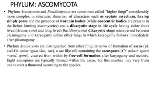
  • 1. PHYLUM: ASCOMYCOTA • Phylum Ascomycota and Basidiomycota are sometimes called “higher fungi” considerably more complex in structure; share no. of characters such as septate mycelium, having simple pores and the presence of woronin bodies (while concentric bodies are present in the lichen-forming ascomycetes) and a dikaryotic stage in life cycle having either short lived (Ascomycota) and long lived (Basidiomycota) dikaryotic stage interspersed between plasmogamy and karyogamy unlike other fungi in which karyogamy follows immediately after plasmogamy. • Phylum Ascomycota are distinguished from other fungi in terms of formation of ascus (pl. asci Gr. askos=goat skin, sac), a sac like cell containing the ascospores (Gr. askos+ spora =seed, spore), cleaved from within by free-cell formation after karyogamy and meiosis. Eight ascospores are typically formed within the ascus, but this number may vary from one to over a thousand according to the species.
  • 2.
  • 3. General characteristics: As in Zygomycota ascomycetes may have distinct reproductive phases (i) Sexual involving formation of asci and ascospores and (ii) Asexual with spore production occurring at different times on the same mycelium (iii) Some Ascomycota have only a sexual state while vast numbers of ascomycetes are known by their asexual stages. These taxa are placed in artificial group called Deuteromycota or Fungi Imperfecti.
  • 4. Somatic structure: • Except for some yeasts ascomycetes fungi possess well developed and profusely branched mycelium with one or several nuclei. They may be single celled, mycelial or dimorphic. • The cell wall largely contains chitin and cellulose is generally absent except few species. • The septa are produced towards the center of hypha from the hyphal wall/periphery and a pore (1.0 to 1.5) is left in the centre. The pore allows for cytoplasmic continuity and occurrence of nuclear and mitochondrial migration throughout the hypha. • In many yeasts that produce a mycelium a closure line demarking the complete inward growth of the septum or micropores analogous to the plasmodesmata of plants are present. • Dowding (1958) reported that the nuclei in Gelasinospora tetrasperma are carried at the rate of 40 mm/hour. The actual mechanism of nuclear migration probably depends on cytoplasmic streaming (Snider, 1965).
  • 5. • Woronin bodies highly refractive bodies are found associated with pores (both in ascomycetes and deuteromycetes fungi) which are rounded or elongated oval, spherical or hexagonal or rectangular with a crystalline protein matrix that are usually associated with septum often known to plug the septal pores of hyphae although they are believed to serve to separate aging or damaged hyphae from rest of mycelium. • Hyphal compartments often are uninucleate, but mycelia consisting of multinucleate cells are well known. Three kinds of nuclei may exist within ascomycetes hyphae: • A homokaryotic mycelium that results from germination of a uninucleate haploid ascospore. A homokaryotic multinucleate condition arises by mitotic duplication of nucleus and subsequent migration of daughter nuclei. • The heterokaryotic condition may arise as a result of the mutation of nuclei in an originally homokaryotic mycelium; or by the fusion of genetically distinct entities that results in dikaryon; or a multinucleate cell in which the nuclei are unpaired and not daughter cells of the same parent cell. • These various multinucleate situations are possible because the pores in the septa allow nuclear migration.
  • 6. • Aggregation of modified vegetative hyphae may lead to the production of sclerotia. • In certain members the aggregation of mycelial strands produces rhizomorphs. • Somatic structures known as stroma (pl. stromata Gr. Stroma= mattress) are commonly produced in many ascomycetes members. Stroma consists of a compact mass of vegetative hyphae with or without tissue of the host or substratum, sometimes sclerotium–like in form, often bearing internal or external fructifications. These are especially produced in the order Sphaeriales. • Anatomically, it may be prosenchymatous or pseudoparenchymatous and has two distinct regions. • The outer narrow ectostroma which has mechanical function and inner massive endostroma that gives rise to the sporophores or ascocarps. [Ascomycetes mycelium may be organized into fungal tissues (plectenchyma).
  • 7. • Plectenchyma differs from the parenchyma of plants because it arises from apical growth of hyphae that become closely associated only secondarily. • If such a tissue is loosely woven and the mycelial strands are more or less evident, it is known as prosenchyma. • If however, the hyphae have lost their individuality and cells are more or less isodiametric, closely resembling the parenchyma of plants it is known as pseudoparenchyma. • Prosenchymatous and pseudoparenchymatous tissues are associated primarily with reproductive and resting structures of ascomycetes as well as basidiomycetes. Ascomycetes may produce specialized structures; appressoria and haustoria some of which are associated with host infection by pathogens. • In addition certain ascomycetes form structures known as hyphopodia (sing. Hyphopodium) which are two types: Capitate hyphopodia produced by some plant pathogenic ascomycetes considered to be specialized form of appressorium. These are stalked, thick-walled, lobed cells that stick to the host surface; certain stalked hyphopodia may have additional function i.e. initiation of ascocarp development upon their fusion.
  • 8. Melanized hyphopodia of Gaeumannomyces graminis arising from an equally strongly melanized runner hypha formed on the hydrophobic surface of a plastic coverslip.
  • 9. • Pointed mucronate hyphopodia best known in Meliolales are not associated with infection structures, reported to produce conidia that function as spermatia (Hughes, 1981). • Some of the specializations of ascomycetes mycelia are the nooses, traps, coils and sticky pegs that trap nematodes. These systems have not only developed in ascomycetes, but in zygomycetes and basidiomycetes as well. Trap formation in Arthrobotrys may be induced by nematode presence or by addition of proteinaceous material into a culture. • The fungus mycelium is more attractive to nematodes when traps are present, and the modified trap hyphae produce lectins (carbohydrate binding proteins) that bind to specific sites on the nematode surface; for this reason the traps may also attach to other organisms, including fungi with similar cell surface carbohydrates.
  • 10. Reproduction: These fungi reproduce both sexually and asexually. The ascus or sexual stage is often called the ascigerous or perfect stage (or state), and the conidial or asexual stage is often designated as imperfect stage. No flagellated structures are produced while some fungi reproduce both sexually and asexually, others multiply exclusively by sexual reproduction through ascospores. Hennebert and Weresub (1977) used the term teleomorph for the sexual form; anamorph for the asexual form and holomorph for the whole fungus in all its forms and phases. Synanamorph is applied to any one of the two or more anamorphs (asexual states) which have the same teleomorph. Classification of ascomycetes is based on ascigerous or perfect stage. The conidia and other methods of asexual reproduction are secondary in importance.
  • 11. Asexual reproduction: • Asexual reproduction in ascomycetes may be carried out by fission, fragmentation, or formation of chlamydospores or conidia according to species and environmental conditions. • Budding and fission are characteristic of the yeasts and some dimorphic ascomycetes, the spores produced by budding are known as blastospores etymologically means bud spores. Fission occurs in yeast genus Schizosaccharomyces only. • Fragmentation of hyphae and chlamydospore formation are other means of asexual reproduction. Because all living portions of the thallus potentially are capabale of growth, fragmentation (including laboratory transfer of cultured mycelium) may result in as many new individuals as there are fragments. • Although some ascomycetes produce only sterile mycelia, many are known for their conidium formation.
  • 12.
  • 13.
  • 14. • Conidia are the chief reproductive organs of majority of organisms and are produced from or within a conidiogenous cell, which is borne on a simple or branched hypha. Conidia are may arise directly from somatic hyphae or from specialized conidiogenous cells, often borne on hyphal branches known as conidiophores. • Conidiophores vary from short hyphal branches to those that are long and intricately branched. • In some species the conidiophores may be free from each other without evident organization while in others they are joined togeey often their to form complex structures such as synnemata, sporodochia, pycnidia and acervuli which are found in Deuteromycota fungi as these fungi are the imperfect states of ascomycetes fungi whose perfect states have not been discovered so far or extinct. • Conidia are often important in propagating and disseminating fungal species throughout the spring and summer with several generations produced in a season. The conidia vary greatly in size, shape, colour and number and arrangement of cells, if multicellular, often determined by dispersal mechanisms.
  • 15. • The conidia may be produced successively at the tip, separating from conidiophore as soon as formed, or clinging together in a mucilaginous drop or remaining attached in chains. • A chain may be acogenously (acropetal) formed with the basal conidium being the oldest and youngest conidium at the apex, or it may be bassigenous (basipetal) in which the oldest conidium is at the apex of chain. • In another type almost simultaneous rounding off of the cells of simple or branched hyphae into catenulate conidia (oidia) which then fall apart almost simultaneously. • Among many ascomycetes other types of ‘resistant spores/structures’ which serve the fungus to survive unfavourable condtions include sclerotia and chlamydospores which are characterized by thick walls. • Stromata have a similar function but rather than initiating mycelial growth they give rise to conidia or ascocarps.
  • 16. Life cycle: • As there is so much diversity in ascomycetes, no typical life cycle exists. However, life cycle of Pyronema omphaloides generally illustrates both sexual and asexual reproduction in a single life cycle. • An ascospore germinates by one or more germ tubes to produce a mycelium. As germ tubes issue from a spore, the spore nucleus divides and nuclear progeny are distributed within growing hyphae. Soon after hyphal growth septa are laid down to delimit uninuclate compartments and hyphal branching takes place. The vigourously growing mycelium may begin to produce conidia almost immediately which are continuous and abundant sources of inoculum for localized dispersal of fungus for a period of months. • Many crops of conidia may be borne in a single season. The conidia produced in enormous numbers germinate by germ tube to produce mycelium similar to ascospore germination.
  • 17. • At some time in appropriate conditions the mycelium ceases conidial production and begins to undergo a complex series of physiological changes that initiate sexual reproduction. • The presence of second mating type in heterothallic fungi is required. The ascogonia are differentiated from somatic hyphae (may be uni- or multinucleate). • Compatible nuclei are brought to the ascogonium by one or more methods of copulations briefed earlier. The contact of antheridium and ascogonium and passage of antheridial nuclei into the trichogyne and eventually into the ascogonial base (Fig. A to C) takes place. • After plasmogamy the ascogonium produces a number of papillae just opposite groups of nuclei located in the periphery of ascogonium. The ascogonial walls appear thinner at these points. As these papillae enlarge, nuclei from the ascogonium begin to pass into them one by one. Eventually the papillae elongate into ascogenous hyphae (Fig. D) in which a leading pair of nuclei can be detected, followed often by second pair. • The nuclei in the ascogenous hyphae and those still in ascogonium soon undergo simultaneous mitosis known as conjugate divisions.
  • 18.
  • 19.
  • 20. • Primary septa are formed between daughter nuclei in such a way that the tip cell of ascogenous hypha is uninucleate and followed by a series of binucleate cells that contain pairs of non-sister nuclei. • It is probable that one nucleus in each binucleate cell of ascogenous hyphae is acogonial in origin and other antheridial, indicating dikaryotic phase of life cycle. In a large number of ascomycetes including P. omphaloides, one of the binucleate cells of ascogenous hypha elongates and bends over to form a hook or crozier (Fig. E). • The two nuclei in this hooked cell (ascus mother cell AMC) divide in such a way that their spindles are oriented more or less vertically and parallel to one another(Fig. F), so that two of the daughter nuclei –one from each spindle and therefore of different origin- are close to one another at the bend of the hook. Two septa are laid down, separating the hook into three cells (Fig. G). • The tip and basal cells are uninucleate, one containing an antheridial nucleus and the other an ascogonial nucleus; the crook cell is binucleate which is the young ascus where eventual nuclear fusion (Fig. H) takes place.
  • 21.
  • 22.
  • 23. • Karyogamy takes place in the young ascus soon after septa are formed in the hook thus ending the dikaryotic phase. The ascus with its diploid zygote nucleus begins to elongate (Fig. I to J) and the zygote nucleus undergoes meiosis. • The four haploid (Fig. K) nuclei (products of meiosis) each undergo a mitotic division so that a total of eight nuclei are formed. • These will become incorporated in the ascospores during ascosporogenesis or free cell formation.
  • 24.
  • 25. Ascosporogenesis: The ascus mother cell gets transformed into an ascus generally containing eight ascospores which develop around the nuclei by free cell formation. It involves two mechanisms: (1) Portions of cytoplasm each typically consisting a single nucleus are delimited by an envelope consisting of two closely appressed unit membranes and (2) The ascospore wall is deposited between these two membranes to separate them from one another as spore matures which are present in a discontinuous cylindrical ascus vesicle or enveloping membrane system (EMS) which invaginates and fragments into sheets and then cleave out the young ascospore.
  • 26.
  • 27.
  • 28. • The portion of cytoplasm that remains outside the ascospore is called epiplasm, possibly serves to nourish the developing spore and deposit external ornamentation. The inner membrane of EMS becomes spore plasma membrane. • Commonly the number of asscopores per ascus in most species is eight however, some species routinely produce asci containing smaller numbers of spore including one, two, three or four while large numbers of spores up to possibly as many as thousand are produced in few species. • Ascospores come in variety of sizes and shape (Fig.) ranging from long, thin and threadlike to globose and even hat shaped in appearance. • Ascospore may be one celled or septate, also may be arranged in uni- or biserate manner in an ascus.
  • 29.
  • 30. Ascospore: • They are “spores produced as a result of meiosis and enclosed within a specialized cover” (Sussman and Halvorson, 1966). • After nuclear divisions, ascospore boundaries become delineated around the clear areas enclosing the asci by a process of “free cell formation” which involves the plasmalemma differently from conidiogenesis. • Ascospores vary in size, shape and colour. Variations in the shape of mature ascospores provide a good criterion for differentiation in yeast systematics (Kreger- van Rij, 1969).
  • 31.
  • 32. Asci: • The asci are typically elongated, cylindrical, ovoid, club shaped/clavate (Gnomonia) or globose form (Eremascus), septate (Dothidea), broadly ovate with stalk (Taphrina). Spherical asci are produced by some yeasts (Hansenula anomala) and may also be produced directly on hyphae (Endomycopsis fibuliger) or in ascocarps (Emericella variecolor) where they are generally arranged in a scattered fashion. • Elongated asci usually are formed in a hymenium as component of ascocarp. Asci may be stalked or sessile and may arise at various levels within the ascocarps (Plectomycetes) or from a single level formed naked (Hemiascomcetes) or arranged in a palisade like manner forming a hymenial layer (Pyrenomyectes and Discomycetes). A definite layer of asci whether naked or enclosed in an ascocarp is called a hymenium (pl. hymenia; Gr. hymen= membrane). Studies have shown that sometimes asci may develop in hymenium but become rearranged at maturity so that they appear to be scattered.
  • 33.
  • 34. • In the ascus morphology three basic types of asci such as prototunicate, unitunicate and bitunicate can be distinguished on the basis of their wall structure and mode of dehiscence used as important criterion in taxonomy of ascomycetes. • Prototunicate asci, a thin delicate wall and release their spores either by breaking apart or deliquescing. • The walls of both unitunicate and bitunicate ascus is said to consist of two layers: exotunica/ectotunica (exoascus), the outer layer and endotunica (endoascus), the inner layer. • In the unitunicate ascus these layers adhere closely throughout the life of ascus, the asci are thickened at the apex and characteristically perforated by a pore for ascospore discharge (a terminal pore), or spores are released through slit or hinged cap (operculum). • In the bitunicate ascus the endotunica usually expands up to twice or more its original length separating from the ruptured exotunica at the time of spore release. The outer wall ruptures during dehiscence and enables elastic inner wall to expand to form a cylindric sac and ascospores are successively discharged through elastic pore in the expanded sac or endotunica. Due to this behaviour of bitunicate ascus has been called the Jack-in-the-box (Ingold, 1933) or the fissitunicate ascus (Henssen and Jahns, 1974).
  • 35.
  • 36. • In the unitunicate ascus may have a pore, slit, operculum or tear (formed in the lateral wall) for liberation of ascospores before the discharge occurs; the pore is plugged with a substance that stains blue with iodine and is different from wall material (or Pot. iodide called as Melzier’s reagent used for this; unitunicate blue stain while in bitunicate no blue stain). • In many ascomycetes the ascal tip has distinct characters called apical apparatus are of great taxanomic importance. • It can be stained with writing ink, congo-red and janus green. In the discomycetes the tip of the ascus has a cap or operculum or may be perforated by a pore without an operculum. • These two types of asci are termed as operculate and inoperculate respectively and the presence or absence of an operculum is an important feature in classification.
  • 37. Ascospore release, dispersal and germination: Eriksson (1981) distinguished eight types of dehiscence in bitunicate asci with ectotunica and endotunica; (1) fissitunicate (e.g. Pleospora); (2) rostrate (e.g. Melaspilea); (3) semifissitunicate (e.g. Peltigera); (4) pseudofissitunicate (e.g. Diatrypiella); (5) bilabiate (e.g. Pertusaria); (6) poricidal (e.g. Porina); (7) operculate and (8) explosive (e.g. Erysiphe). Asci contained in cleistothecia are exposed for release of ascospore only after the ascocarp disintegrates and ascospore release is non-explosive. In majority of ascomycetes however, the asci are cylindrical and ascospore release is explosive. In the Erysiphales the cleistothecial wall is non-disintegrating; the ascus enlarges and protrudes through confining ascocarp wall. The wall which is forced open is under some strain and suddenly snaps shut, ejecting the entire acsus to a distance of several centimeters. The ascus then forcibly ejects its ascospores. Water uptake is possibly responsible for increase in turgor which causes the explosive release. After the delimitation of ascospores in the ascus the epiplasm continues to line the ascus wall that surrounds a large central vacuole containing ascus sap. The ascospores are suspended in the vacuole.
  • 38. Ascocarp: Except for a few filamentous ascomycetes (Hemiascomycetes- asci are naked) that do not produce their asci/ascus in fruiting bodies called ascocarp (Gr. askos =sac + karpos=fruit). In general there are five ways that ascomycetes can be separated according to the way they bear asci: (1) Those bear naked asci without any fruiting body; thallus mycelial or yeast like [Order Endomycetales (yeast-Saccharomyces), Protomycetales (Protomyces macrospores-stem gall of coriander) and Taphrinales (Taphrina deformans-peach leaf curl; T. maculans-turmeric). (2) Those that produce their asci inside a completely closed and usually globular ascocarp called a cleistothecium (pl. cleistothecia, Gr. kleistos = closed+ theke = case) without any opening for liberation of acsospores. By disintegration or breaking of cleistothecium liberates asci. Ex: Sphaerotheca, Erysiphe, Meliola, Order Eurotiales- Eurotium (conidial state: Aspergillus) and Talaromyces (conidial state: Penicillium). (3) Those with an ascocarp the perithecium (pl. perithecia, Gr. peri = around + theke= case) that is more or less closed (flask shaped ascocarp) but at maturity is provided with a pore (ostiole) through which ascospores escape. Ex: Pyrenomycetes Order Sphaeriales [Family: Clavicipitaceae (Claviceps), Ophiostomataceae (Ceratocystis), Nectria, Gibberella etc.]
  • 39. (4) Those that produce their asci in an open /cup or saucer shaped ascocarp called an apothecium (pl. apothecia, Gr. apotheke = store house) bearing an exposed hymenium Ex: Peziza, Monilinia, Sclerotinia, Pyronema. (5) Those that form their asci directly in a cavity (locule) within a stroma. Stroma may be likened to a cushion of closely woven somatic hyphae forms the wall of the ascocarp called an ascostroma (pl. ascostromata, ascus + stroma) or pseudothecium. Ex: Mycosphaerella, Elsinoe, Guignardia etc. • In addition, various modifications of ascocarps are known to occur in few members: hysterothecium (pl.hysterothecia, Gr. hysteros = womb + theke = case) which is elongated, boat shaped with a longitudinal slit (Order Hysteriales and lichenized Order Arthoniales) and sclerotoid ascocarps with tough peridia (as in some species of Penicillium).

Editor's Notes

  1. hyphopodium is defined as an appressorium produced from a vegetative hypha rather than a germinating spore (Fig. 12.49).