How to Send Pro Forma Invoice to Your Customers in Odoo 17
1)Digestive system, Nervous system, circulatory system self made note-- Summary type.pdf
1. Insect Digestive system Self-made note
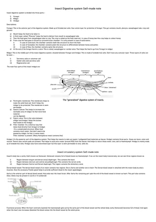
Insect digestive system is divided into three parts—
1) Foregut
2) Midgut
3) Hindgut
Descriptions:
Foregut: This is the anterior part of the digestive system. Made up of Ectodermal cells. Has cuticle layer for protection of foregut. This gut contains mouth, pharynx, oesophageal tube, crop and
gizzard.
a) Mouth helps the food to be taken in.
b) A thick layer called; “Pharynx” helps the food to deliver from mouth to oesophageal tube.
c) Then the food goes from oesophageal tube to crop. The crop is called as the food reservoir. In case of honey bee this crop helps to collect honey.
d) From crop the food goes to Gizzard or proventricular chamber. It is only present in case of solid eaters.
i) In case of cockroach, the chamber contains teeth like structure to churn the food.
ii) In case of honeybee, the chamber contains plate like structure to differentiate between honey and pollen.
iii) In case of flies, the chamber contains spine like structure.
e) At the end of the foregut there’s a valve. This is called, oesophageal or cardial valve. That helps the food to go from Foregut to midgut.
Midgut: This is the middle part of the insect digestive system. situated between Foregut and hindgut. This is made of endodermal cells. Don’t have any cuticular layer. Three layers of cells can
be seen —
i) Secretory cells or columnar cell
ii) Goblet cells (old secretive cell)
iii) Regenerative cell.
The main four parts of the insect midgut are
a) Peritrophic membrane: This membrane helps to
make the solid food wet. And it helps the
midgut to be protected. This membrane is semi
permeable.
b) Gastric Caecae: This helps to increase the
workable area of midgut. So that more food
material
can be digested.
c) Pyloric valve: This is the valve between
midgut and hindgut. Helps the excess
food waste to the hindgut.
d) Filter chamber: It’s a shortcut way that
takes food directly from foregut to hindgut.
It’s a complicated structure. When food
contains much water, that don’t need to be
passed by midgut. So that, in some insect,
Filter chamber is present. (Many Homopteran insect contains this)
Hindgut: It’s the posterior part of the digestive system. That helps the insects to take out waste / undigested food materials as faeces. Hindgut contains three parts- those are ileum, colon and
rectum. There’s also rectum pads are present. That helps to suck extra water (around 90%) from the faeces. And helps to return those water, ions, salt to Haemolymph. Hindgut is mainly made
up of endodermal cells. Hindgut also have sclerotised layer but this layer is semi permeable to ions, salts etc.
Insect circulatory system Self-made note
Insect’s body has a cavity; that’s known as Hemocoel. Hemocoel remains full of blood known as Haemolymph. If we cut the insect body transversely, we can see three regions known as
i) Region between tergum and dorsal sinus/ diaphragm : This contains the heart.
ii) Region between sternum and ventral sinus/diaphragm: This contains the nerval cords.
iii) Region between Ventral and Dorsal diaphragm: This region contains the alimentary canal.
Dorsal blood vessel: The dorsal blood vessel of an insect contains two regions. One is Aorta and another one is heart. The Dorsal blood vessel in attached with the insect body by allary
muscles. Also the accessory Pulsite gland helps to provide sufficient blood to the insect appendages.
Aorta is the anterior part of dorsal blood vessel, that ends near the insect brain. After Aorta the remaining part upto the end of the blood vessel is known as heart. This part also contains
Ostia. (Ostia may be present in 4,6,10 or 12 numbered)
Functional process: When the heart contracts (systole) the haemolymph gets out by the aorta part of the blood vessel and the whole body cavity (hemocoel) becomes full of blood. And again
when the heart size increases (diastole) the blood comes into the blood vessel by the ostial pores.
2. Characteristics of insect blood:
a) Insect blood does not carry Oxygen.
b) Insect blood has no colours as it does not contain haemoglobin.
c) Insect blood have the specific gravity of 1.1 to 1.6
d) Insect blood contains lipid, protein, phospholipids etc.
e) Phytophagous insect contains more K than Carnivorous insect. Which contains more Na.
f) Insect blood have no Vitamin K.
g) Insect blood contains sugar called Trehalose.
Functions of insect blood:
a) This helps to carry water in insect’s body.
b) Blood helps to prevent various organisms and microorganisms from attacking the insect.
c) Blood helps to regenerate injured place of body system of insect.
d) Blood helps during moulting.
e) Sometimes some insects can actually show false bleeding to protect themselves from their predators.
Nervous system of insect body self-made note
The main component of any nerval system is neuron. On the basis of structure Neuron can be divided into three parts. These are-
i) Monopolar neuron: It doesn’t contain any dendrite. It contains only the main nucleus body with an axon.
ii) Bipolar : This contains the main nucleus body with an axon and a long dendrite.
iii) Multipolar : This contains the main nucleus body with many dendrites and a axon.
On the basis of functions, neurones can be divided into three classes.
i) Sensory: This helps to connect the signal from sensory organs to CNS (Central Nervous System)
ii) Motor: This helps to connect the signal from CNS to other organs that will react for respective sense.
iii) Inter: This helps to connect between Sensory and Motor neurons.
# The whole neural system is divided into three parts. 1) CNS (Central Nervous system) ; 2) VNS (Visceral Nervous system) ; 3) Peripheral Nervous system. (PNS)
Central Nervous system:
Central nervous system is made of—Brain, Ventral nerval canal, Thoracic nerve, abdominal nerve and Thoraco-Abdominal nerve.
a) Brain: This is made of three neuromeres.
i) Protocerebrum: This helps to sense the visual organs including complex eyes and ocelli.
ii) Deutocerebrum: This is situated beneath protocerebrum. Helps to sense antenna.
iii) Tritocerebrum: This helps to sense the labrum.
b) Ventral nerval canal: Situated under the oesophageal canal.
c) Thoracic ganglia: 3 thoracic segments. Each of them contains one pair of ganglia. This helps to sense the muscles and legs.
d) Abdominal ganglia: Maximum 8 ganglia (Plural of ganglion). Helps to sense the Spiracles.
e) Thoraco-Abdominal ganglia: These ganglia is made up of mixed abdominal and thoracic ganglia. These help to sense reproductive/ genital organs and also cerci.
(Imp dif between Cerci and ovipositor: Cerci is used as sensory organ, reproductive organ but Ovipositor is used for egg laying organ in insect)
How neuron carries the impulses?
- Neuron can carry the impulses by two methods:
i) From a neuron’s dendrite to axon: This impulse can be flowed as an electrical charge by using the difference of charges (K+, Na+) in the inner and outer part
of an neuron.
ii) From a neuron to another neuron: This impulse can be flowed through neurotransmitters (eg: Acytile CoA) by synaptic nobs.